AO/PI双染试剂盒是一种基于荧光核酸染料的检测系统,能快速可靠地区分生物样本中的活细胞、凋亡细胞和坏死细胞。其核心原理在于利用吖啶橙(Acridine Orange, AO)和碘化丙啶(Propidium Iodide, PI)这两种染料对细胞膜通透性的差异及其与核酸结合后的特异性荧光反应。
AO能够透过所有类型的细胞(活细胞、凋亡细胞、坏死细胞)的细胞膜。进入细胞后,AO会与核酸结合,但其荧光颜色取决于结合的对象:当它与细胞核内的双链DNA(dsDNA) 结合时,会发出绿色荧光(Ex 488nm, Em 530nm);而当它与单链DNA(ssDNA) 或RNA 结合时,则发出红色荧光(Em>640nm)。相比之下,PI不能透过细胞膜完整的细胞。它只能进入细胞膜已受损的晚期凋亡细胞和坏死细胞,并与DNA结合产生红色荧光(Ex 535nm, Em 617nm)。
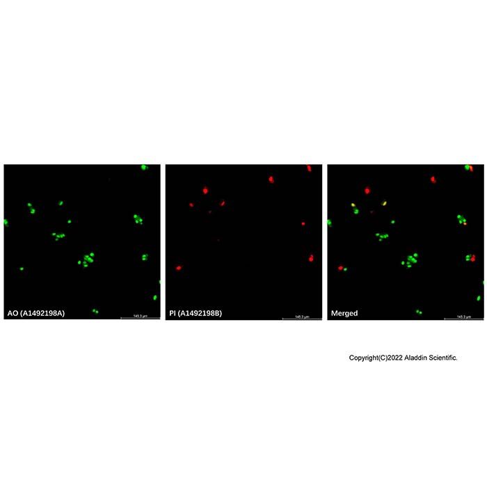
因此,在AO/PI双染后,通过荧光显微镜观察可以清晰区分不同状态的细胞:活细胞显示正常的绿色荧光;早期凋亡细胞显示亮绿色斑点的核形态(仅AO进入);晚期凋亡细胞和坏死细胞则因AO和PI共同作用呈现橙红色或强红色荧光。
注: 每个Test推荐染色的细胞数量为 0.5-1.0 × 10⁶ cells.
使用方法
1. 工作液的配制
(1) 取出试剂,恢复至室温,使用前用双蒸水将AO/PI稀释缓冲液稀释 10 倍。
(2) 配制 AO/PI 染色工作液:本产品中AO染液浓度为10mg/mL,PI染液浓度为1mg/mL,取50μL PI染液 和10μL AO染液加入到 10 mL AO/PI稀释缓冲液中,涡旋混匀,即得到10 mL AO/PI 染色工作液。
注:由于最佳的染色条件可能因细胞类型而异,建议用户根据实际情况单独确定染料的适宜浓度。
2. 染色
a. 对于贴壁细胞:
(a) 将细胞接种在培养皿、多孔细胞培养板或细胞爬片上,按实验设计对细胞进行一定处理。轻轻吸除孔板中的培养液,PBS洗涤约10秒,吸除PBS。
(b) 加入适当体积的AO/PI染色工作液,轻轻晃动使染料均匀覆盖所有细胞。
注:对于在六孔板中培养、汇合度超过80%的贴壁细胞,建议按1mL/well 加入染色工作液,也可根据具体的实验体系进行优化。
(c) 室温避光孵育细胞1-10分钟。
(d) 吸除染色液,用PBS清洗2-3次,然后加入无血清细胞培养液后即可在荧光显微镜下观察,在 Ex/Em = 488/530nm 下观察AO-DNA绿色荧光,在 Ex/Em = 535/617nm 下观察PI-DNA红色荧光,也可以在染色结束后进行流式细胞仪分析检测或使用荧光酶标仪检测细胞的荧光。
b. 对于悬浮细胞:
(a) 细胞按实验设计进行一定处理后,计数。取适当细胞,500×g室温离心5分钟,轻轻吸除培养基后加适量PBS重悬,500×g室温离心5分钟,去除PBS。
(b) 加适量AO/PI染色工作液,使细胞密度约为106个/mL。
(c) 室温避光孵育细胞1-10分钟。
(d) 直接滴加于载玻片上,加盖盖玻片,置于显微镜下观察,在 Ex/Em = 488/530nm 下观察AO-DNA绿色荧光,在 Ex/Em = 535/617nm 下观察PI-DNA红色荧光,也可以在染色结束后进行流式细胞仪分析检测或使用荧光酶标仪检测细胞的荧光。
注:若背景影响严重,可以离心去除染液后使用PBS清洗1-2遍,再置于显微镜下观察。
注意事项
1. AO/PI染色液有一定毒性,请小心操作。
2. 为了您的安全和健康,请穿实验服并戴一次性手套。
3. 荧光染料存在淬灭的问题,建议染色后尽量当天完成检测。
The AO/PI Double Staining Kit is a detection system based on fluorescent nucleic acid dyes, designed to quickly and reliably distinguish live, apoptotic, and necrotic cells in biological samples. Its core principle utilizes the differences in cell membrane permeability and the specific fluorescent reactions upon binding to nucleic acids of the two dyes, Acridine Orange (AO) and Propidium Iodide (PI).
AO can penetrate the cell membranes of all cell types (live, apoptotic, and necrotic). After entering the cell, AO binds to nucleic acids, but its fluorescence color depends on the target: when it binds to double-stranded DNA (dsDNA) in the nucleus, it emits green fluorescence (Ex 488 nm, Em 530 nm); whereas when it binds to single-stranded DNA (ssDNA) or RNA, it emits red fluorescence (Em >640 nm). In contrast, PI cannot penetrate cells with intact cell membranes. It only enters late apoptotic and necrotic cells, whose cell membranes have been compromised, and binds to DNA to produce red fluorescence (Ex 535 nm, Em 617 nm).
Therefore, after AO/PI double staining, observation under a fluorescence microscope allows clear differentiation of cells in different states: live cells display normal green fluorescence; early apoptotic cells show bright green speckled nuclear morphology (only AO enters); late apoptotic and necrotic cells appear orange-red or strong red fluorescence due to the combined effect of AO and PI.
Procedure
1. Preparation of Working Solution
(1) Take out the reagents and allow them to reach room temperature. Dilute the AO/PI Dilution Buffer 10-fold with double-distilled water before use.
(2) Prepare the AO/PI Staining Working Solution: The concentration of AO stock solution in this product is 10 mg/mL, and PI stock solution is 1 mg/mL . Add 50 μL of PI stock solution and 10 μL of AO stock solution to 10 mL of diluted AO/PI Dilution Buffer. Vortex to mix well, resulting in 10 mL of AO/PI Staining Working Solution.
Note: Since optimal staining conditions may vary with cell type, it is recommended that users determine the appropriate dye concentration individually based on practical conditions.
2. Staining
a. For Adherent Cells:
(a) Seed cells in culture dishes, multi-well plates, or on coverslips, and treat the cells as required by the experimental design. Gently aspirate the medium from the wells and wash with PBS for about 10 seconds, then remove the PBS.
(b) Add an appropriate volume of AO/PI Staining Working Solution, and gently swirl to ensure the dye evenly covers all cells.
Note: For adherent cells cultured in 6-well plates with a confluency over 80%, it is recommended to add 1 mL of staining working solution per well. This can be optimized according to the specific experimental setup.
(c) Incubate the cells at room temperature, protected from light, for 1-10 minutes.
(d) Aspirate the staining solution, wash 2-3 times with PBS, then add serum-free culture medium. Observe immediately under a fluorescence microscope: observe AO-DNA green fluorescence at Ex/Em = 488/530 nm, and PI-DNA red fluorescence at Ex/Em = 535/617 nm. Alternatively, analysis by flow cytometry or detection with a fluorescence microplate reader can be performed after staining.
b. For Suspension Cells:
(a) After treating the cells as required by the experimental design, count the cells. Take an appropriate number of cells, centrifuge at 500 × g for 5 minutes at room temperature, gently aspirate the medium, resuspend in an appropriate amount of PBS, and centrifuge again at 500 × g for 5 minutes at room temperature to remove the PBS.
(b) Add an appropriate amount of AO/PI Staining Working Solution to achieve a cell density of approximately 10⁶ cells/mL.
(c) Incubate the cells at room temperature, protected from light, for 1-10 minutes.
(d) Place a drop directly on a glass slide, cover with a coverslip, and observe under a microscope: observe AO-DNA green fluorescence at Ex/Em = 488/530 nm, and PI-DNA red fluorescence at Ex/Em = 535/617 nm. Alternatively, analysis by flow cytometry or detection with a fluorescence microplate reader can be performed after staining.
Note: If background interference is significant, centrifuge to remove the staining solution, wash 1-2 times with PBS, and then observe under the microscope.
Precautions
1. AO/PI staining solutions are somewhat toxic; handle with care .
2. For your safety and health, please wear a lab coat and disposable gloves.
3. Fluorescent dyes are subject to quenching; it is recommended to complete detection on the same day after staining whenever possible.

 J2627429


 危险品化学品经营许可证(带存储)