ATP是细胞能量代谢的核心分子,其含量直接反映生物体及各器官的能量状态。作为关键的能源物质,ATP参与并调控着细胞的各种生理与病理活动,其水平波动会显著影响细胞功能。通常情况下,细胞凋亡、坏死或处于毒性状态时,ATP水平会降低;而在高葡萄糖等特定刺激下,某些细胞的ATP水平则可能升高。ATP的下降往往提示线粒体功能受损,在细胞凋亡过程中常伴随线粒体膜电位的降低。
本试剂盒可用于测定动物组织或细胞样品中ATP含量。己糖激酶(Hexokinase,HK)催化葡萄糖和ATP生成葡萄糖-6-磷酸(G6P),葡萄糖-6-磷酸脱氢酶(Glucose-6-Phosphate Dehydrogenase,G6PD)进一步催化葡萄糖-6-磷酸脱氢生成 NADPH。NADPH可与显色剂反应,产物在460nm有特征吸收峰,以此来测定ATP含量。
| A1508987 | Component | 96 T | Storage conditions | | A1508987A | 裂解液 | 100 mL | -20°C | | A1508987B | 酶试剂A | 1 mL | -20°C. Store in the dark. | | A1508987C | 酶试剂B | 5 mL | -20°C. Store in the dark. | | A1508987D | 酶试剂C | 5 mL | -20°C. Store in the dark. | | A1508987E | WST-8 | 250 μL | -20°C. Store in the dark. | | A1508987F | 1-mPMS | 250 μL | -20°C. Store in the dark. | | A1508987G | 反应缓冲液 | 10 mL | -20°C | | A1508987H | 0.5mM标准品 | 0.6 mL | -20°C. Store in the dark. |
|
使用方法
1. 试剂准备
(1) 检测前,将各试剂完全解冻,解冻后将1-mPMS置于常温待用,其余试剂置于冰上待用。
(2) 酶工作液A的配制:
将酶试剂A: 反应缓冲液按体积比=1:4配制得到酶工作液A。建议现配现用,未使用完可暂存于-20°C。
(3) 显色工作液的配制(1mL为例):
WST-8和1-mPMS完全解冻后,分别取50μL于同一EP管中,加入900μL反应缓冲液,充分混匀后即可使用。每个反应孔需要加50μL显色工作液,具体按需配制,需现用现配。
(4) 40 μmol/L标准品溶液的配制:
将0.5mM ATP标准品: 裂解液按体积比=1:11.5配制,按需配制,未用完的40 μmol/L 标准品可-20°C保存一个月。
(5) 不同浓度标准品的稀释:
| 编号 | ① | ② | ③ | ④ | ⑤ | ⑥ | ⑦ | ⑧ | | 标准品浓度(μmol/L) | 0 | 5 | 10 | 15 | 20 | 25 | 30 | 40 | | 40 μmol/L标准品(μL) | 0 | 100 | 200 | 300 | 400 | 500 | 600 | 800 | | 裂解液 | 800 | 700 | 600 | 500 | 400 | 300 | 200 | 0 |
|
2. 样本准备
(1) 对于贴壁细胞:
吸除培养液,按照6孔板每孔加入200μL裂解液的比例(即相当于细胞培养液量2mL的1/10)加入裂解液来裂解细胞。裂解细胞时为了裂解充分,可以使用移液器进行反复吹打或晃动培养板使裂解液充分接触并裂解细胞。通常细胞在接触裂解液后会立即裂解。裂解后4ºC 12000g离心5分钟,取上清至于冰(盒)上,用于后续的测定。
(2) 对于悬浮细胞:
用离心管离心沉淀细胞,弃上清,轻轻弹散细胞,按照6孔板每孔的细胞量加入200μL裂解液的比例加入裂解液,裂解细胞。裂解细胞时为了裂解充分可以弹击离心管管底或旋涡使裂解液充分接触并裂解细胞。通常细胞在接触裂解液后会立即裂解。裂解后4ºC 12000g离心5分钟,取上清至于冰(盒)上,用于后续的测定。
(3) 对于组织样品:
取新鲜的组织样本,用冷PBS冲洗去除杂质。按照组织样本质量(g):裂解液体积(mL)为1:9的比例(如0.1g组织样本,加入0.9mL裂解液),于4°С条件下进行机械匀浆。将所得组织匀浆沸水浴3min,流水冷却至室温(25°C),4°С,10000g离心10min,取上清至于冰上,用于后续的测定。
(4) 样本预实验:
在正式检测前,建议选择2-3个预期差异大的样本稀释成不同浓度进行预实验。
3. ATP浓度的测定
(1) 标准孔:取100μL不同浓度标准品溶液加入相应的板孔中,再向孔中依次加入50μL酶工作液A、50μL酶试剂B、50μL显色工作液。
(2) 测定孔:取100μL样品加入相应的板孔中,再向孔中依次加入50μL酶工作液A、50μL酶试剂B、50μL显色工作液。
(3) 对照孔:取100μL样品加入相应的板孔中,再向孔中依次加入50μL酶工作液A、50μL酶试剂C、50μL显色工作液。
(4) 振板1-2min混匀,37°C孵育15min,酶标仪460nm处,测定各孔OD值。
4. 结果计算
(1) 标准品拟合曲线:Y = a X + b
细胞样品中ATP含量计算公式:ATP含量(μmol/L) =(ΔA-b)÷a×f
组织样品中ATP含量计算公式:ATP含量(μmol/Kg wet weight)=(ΔA-b)÷a×f÷m×V
注解:
Y:标准孔OD值-空白孔OD值(标准品浓度为0时的OD值);
X:标准品的浓度;
a:标曲的斜率;
b:标曲的截距;
∆A:测定孔OD值-对照孔OD值;
f:样本加入检测体系前的稀释倍数;
m:样本质量,如0.1g;
V:组织匀浆时加入试剂一的体积,如0.9mL。
(2) 性能特点
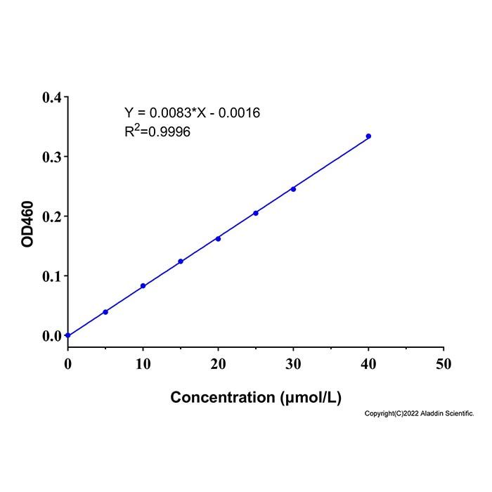
在0-40 μmol/L范围内,本试剂盒有很好的线性关系(示例)。灵敏度为0.5 μmol/L。

图一 标曲绘制示例图
注意事项
1. 试剂盒仅供研究使用,如将其用于临床诊断或任何其他用途,我公司将不对因此产生的问题负责,亦不承担任何法律责任。
2. 实验前请仔细阅读说明书并调整好仪器,严格按照说明书进行实验。
3. 实验中请穿着实验服并戴乳胶手套做好防护工作。
4. 试剂盒检测范围不等同于样本中待测物的浓度范围。如果样品中待测物浓度过高或过低,请对样本做适当的稀释或浓缩。
5. 若所检样本不在说明书所列样本类型之中,建议先做预实验验证其检测有效性。
6. 最终的实验结果与试剂的有效性、实验者的相关操作以及实验环境等因素密切相关。本公司只对试剂盒本身负责,不对因使用试剂盒所造成的样本消耗负责,使用前请充分考虑样本可能的使用量,预留充足的样本。
ATP is a core molecule in cellular energy metabolism, and its content directly reflects the energy status of organisms and their organs. As a key energy substance, ATP participates in and regulates various physiological and pathological activities of cells, and fluctuations in its levels significantly affect cellular functions. Under normal conditions, ATP levels decrease during apoptosis, necrosis, or toxic states; conversely, under specific stimuli such as high glucose, ATP levels may increase in certain cells. A decline in ATP often indicates impaired mitochondrial function, and during apoptosis, it is frequently accompanied by a reduction in mitochondrial membrane potential.
This kit can be used to measure ATP content in animal tissue or cell samples. Hexokinase (HK) catalyzes the reaction between glucose and ATP to produce glucose-6-phosphate (G6P). Glucose-6-phosphate dehydrogenase (G6PD) further catalyzes the dehydrogenation of G6P to generate NADPH. NADPH then reacts with a chromogenic agent, and the resulting product exhibits a characteristic absorption peak at 460 nm, allowing the determination of ATP content.
| A1508987 | Component | 96 T | Storage conditions | | A1508987A | Lysis Buffer | 100 mL | -20°C | | A1508987B | Enzyme Reagent A | 1 mL | -20°C. Store in the dark. | | A1508987C | Enzyme Reagent B | 5 mL | -20°C. Store in the dark. | | A1508987D | Enzyme Reagent C | 5 mL | -20°C. Store in the dark. | | A1508987E | WST-8 | 250 μL | -20°C. Store in the dark. | | A1508987F | 1-mPMS | 250 μL | -20°C. Store in the dark. | | A1508987G | Reaction Buffer | 10 mL | -20°C | | A1508987H | 0.5mM Standard | 0.6 mL | -20°C. Store in the dark. |
|
Instruction for use
1. Reagent Preparation
(1) Before the assay, thaw all reagents completely. After thawing, keep 1-mPMS at room temperature until use, and place the remaining reagents on ice until use.
(2) Preparation of Enzyme Working Solution A:
Mix Enzyme Reagent A and Reaction Buffer at a volume ratio of 1:4 to prepare Enzyme Working Solution A. It is recommended to prepare fresh before use. Any unused portion can be temporarily stored at -20°C.
(3) Preparation of Chromogenic Working Solution (example for 1 mL):
After fully thawing WST-8 and 1-mPMS, transfer 50 μL of each into an EP tube, add 900 μL of Reaction Buffer, and mix thoroughly. Each reaction well requires 50 μL of Chromogenic Working Solution. Prepare the required volume accordingly and use immediately.
(4) Preparation of 40 μmol/L Standard Solution:
Mix 0.5 mM ATP Standard and Lysis Buffer at a volume ratio of 1:11.5. Prepare the required volume as needed. The unused 40 μmol/L Standard Solution can be stored at -20°C for one month.
(5) Dilution of Standard Solutions to Different Concentrations:
| No. | ① | ② | ③ | ④ | ⑤ | ⑥ | ⑦ | ⑧ | | Standard concentration (μmol/L) | 0 | 5 | 10 | 15 | 20 | 25 | 30 | 40 | | 40 μmol/L Standard (μL) | 0 | 100 | 200 | 300 | 400 | 500 | 600 | 800 | | Lysis Buffer | 800 | 700 | 600 | 500 | 400 | 300 | 200 | 0 |
|
2. Sample Preparation
(1) For adherent cells:
Remove the culture medium. Add Lysis Buffer to the cells at a volume of 200 μL per well of a 6‑well plate (i.e., 1/10 of the culture medium volume of 2 mL). To ensure complete lysis, pipette repeatedly or shake the plate so that the Lysis Buffer fully contacts and lyses the cells. Cells typically lyse immediately upon contact with the Lysis Buffer. After lysis, centrifuge at 12,000×g for 5 minutes at 4°C. Transfer the supernatant to ice (or an ice box) for subsequent measurement.
(2) For suspension cells:
Pellet the cells by centrifugation in a tube, discard the supernatant, and gently tap the tube to disperse the cell pellet. Add Lysis Buffer at a volume of 200 μL per well-equivalent of a 6‑well plate. To ensure complete lysis, tap the bottom of the tube or vortex to allow the Lysis Buffer to fully contact and lyse the cells. Cells typically lyse immediately upon contact with the Lysis Buffer. After lysis, centrifuge at 12,000×g for 5 minutes at 4°C. Transfer the supernatant to ice (or an ice box) for subsequent measurement.
(3) For tissue samples:
Take fresh tissue samples and rinse with cold PBS to remove impurities. Add Lysis Buffer at a ratio of tissue mass (g) to Lysis Buffer volume (mL) of 1:9 (e.g., for 0.1 g of tissue sample, add 0.9 mL of Lysis Buffer), and perform mechanical homogenization at 4°C. Place the resulting tissue homogenate in a boiling water bath for 3 minutes, then cool under running water to room temperature (25°C). Centrifuge at 10,000×g for 10 minutes at 4°C, and transfer the supernatant to ice for subsequent determination.
(4) Preliminary sample test:
Before the formal assay, it is recommended to select 2-3 samples with expected large differences, dilute them to different concentrations, and perform a preliminary test.
3. ATP Concentration Determination
(1) Standard wells: Add 100 μL of each standard solution at different concentrations into the corresponding wells of the plate. Then sequentially add 50 μL of Enzyme Working Solution A, 50 μL of Enzyme Reagent B, and 50 μL of Chromogenic Working Solution to each well.
(2) Sample wells: Add 100 μL of sample into the corresponding wells. Then sequentially add 50 μL of Enzyme Working Solution A, 50 μL of Enzyme Reagent B, and 50 μL of Chromogenic Working Solution to each well.
(3) Control wells: Add 100 μL of sample into the corresponding wells. Then sequentially add 50 μL of Enzyme Working Solution A, 50 μL of Enzyme Reagent C, and 50 μL of Chromogenic Working Solution to each well.
(4) Shake the plate for 1-2 minutes to mix, then incubate at 37°C for 15 minutes. Measure the OD of each well at 460 nm using a microplate reader.
4. Result Calculation
(1) Standard curve fitting: Y = aX + b
Formula for ATP content in cell samples:
ATP content (μmol/L) = (ΔA - b) ÷ a × f
Formula for ATP content in tissue samples:
ATP content (μmol/kg wet weight) = (ΔA - b) ÷ a × f ÷ m × V
Explanation:
Y = OD value of standard well - OD value of blank well (OD value when standard concentration is 0);
X = Standard concentration;
a = Slope of the standard curve;
b = Intercept of the standard curve;
ΔA = OD value of sample well - OD value of control well;
f = Dilution factor of the sample before addition to the detection system;
m = Sample mass (e.g., 0.1 g);
V = Volume of Reagent 1 added during tissue homogenization (e.g., 0.9 mL).
(2) Performance characteristics:
This kit exhibits good linearity in the range of 0-40 μmol/L (example shown). The sensitivity is 0.5 μmol/L.
Fig. 1. Example Standard Curve
Precautions
1. This kit is for research use only. If it is used for clinical diagnosis or any other purpose, our company shall not be held responsible for any resulting issues, nor assume any legal liability.
2. Please read the instructions carefully before the experiment and calibrate the instruments accordingly. Strictly follow the instructions during the experiment.
3. Wear a lab coat and latex gloves during the experiment for proper protection.
4. The detection range of the kit is not equivalent to the concentration range of the analyte in the sample. If the analyte concentration in the sample is too high or too low, dilute or concentrate the sample appropriately.
5. If the sample to be tested is not among the sample types listed in the instructions, it is recommended to perform a preliminary experiment to verify the validity of the detection.
6. The final experimental results are closely related to factors such as the validity of the reagents, the operator's technique, and the experimental environment. Our company is only responsible for the kit itself and shall not be liable for sample consumption caused by the use of the kit. Before use, please fully consider the possible amount of sample to be used and reserve sufficient sample.

 ZJ26F0939010


 危险品化学品经营许可证(带存储)