L-氨基酸氧化酶 (L-Amino acid oxidase, L-AAO) 是一种黄素酶,在天然状态下的分子量为110-159kDa。L-AAO广泛分布于药用昆虫、海兔、蛇毒、真菌、细菌和藻类等生物体中。L-AAO能够催化L-氨基酸氧化脱氨,生成α-酮酸和过氧化氢 (H₂O₂),在血小板聚集、水肿形成、出血、抗菌、抗病毒 (如HIV) 作用等方面具有明显的生物学效应;对肿瘤细胞表现出细胞毒性和抗凝作用,为癌症研究提供了新思路。
在L-氨基酸氧化酶 (L-AAO) 的催化下,L-氨基酸(L-Amino acid, L-AA)脱氨形成α-酮酸(Alphaketo acid)、NH₃和 H₂O₂ ,再通过检测 H₂O₂ 与Amplex Red的反应产物试卤灵(Resorufin)的荧光强度或吸光度来最终计算样品中L-氨基酸氧化酶的活力。通过检测试卤灵荧光强度(560 nm 激发、590 nm 发射)或吸光度(OD570),即可实现L-氨基酸氧化酶活性的高灵敏定量检测,信号强度与L-氨基酸氧化酶活性成正比。
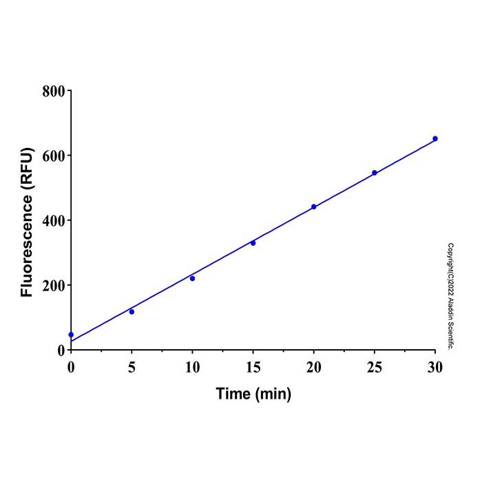
本试剂盒可采用分光光度法和荧光法两种方法进行检测,具有检测灵敏度高、线性范围宽、样品用量少等特点。在样品体积为 50 µL 时,采用分光光度法检测在 2 - 200 μM 浓度范围内有良好的线性关系;采用荧光法检测在0.5 - 50 μM 浓度范围内有良好的线性关系。
A1510318 | 组分 | 物理外观 | 50 T | 100 T | 储存条件 |
A1510318A | 酶混合液 | 液体 | 0.2 mL | 0.3 mL | -20℃.避光保存. |
A1510318B | 阳性对照 | 液体 | 10 μL | 10 μL | -20℃.避光保存. |
A1510318C | 探针 | 液体 | 0.1 mL | 0.2 mL | -20℃.避光保存. |
A1510318D | 底物 | 液体 | 0.3 mL | 0.5 mL | -20℃.避光保存. |
A1510318E | 过氧化氢溶液(3%) | 液体 | 0.5 mL | 1.0 mL | -20℃.避光保存. |
A1510318F | 检测缓冲液 | 液体 | 10.0 mL | 20.0 mL | -20℃.. |
使用方法
1. 试剂盒的准备
1) 融解Amplex Red和Assay Buffer,平衡至室温后混匀备用。其它试剂存放于冰浴备用,使用完毕后宜立即按照试剂盒要求的条件保存。
2) H₂O₂ 标准品的校准
由于 H₂O₂ 不是非常稳定,使用前需自行测定 H₂O₂ 的实际浓度以进行校准。把浓度约为1M的 H₂O₂ 用水稀释100倍,使 H₂O₂ 的浓度约为10mM,测定A240。A240的测定可采用如下的任一方法。
(a) 普通紫外分光光度计法。使用含比色皿架的紫外分光光度计、NanoDrop 2000C、QuickDrop等仪器,配套石英比色皿。确定比色皿光程一般为1cm。用比色皿检测的 H₂O₂ 浓度最接近实际浓度。
(b) 微量紫外分光光度计法。如NanoDrop 2000、NanoDrop One、QuickDrop、含超微量检测板µDrop Plate的 Varioskan等仪器。确定光程:对于NanoDrop 2000、NanoDrop One等,需要取消‘‘自动化光程’’,此时光程一般为0.1cm;Varioskan的超微量检测板µDrop Plate的光程一般为0.05cm。具体的微量紫外分光光度计的光程请参考仪器参数。
(c) 96孔紫外酶标仪法 (须能检测240nm波长)。根据96孔板的参数确定光程,一般200微升样品的光程为0.552cm (样品体积除以96孔单孔孔内横截面面积)。一般建议使用专用的96孔紫外检测板,设置含等量双蒸水的孔作为空白对照 (一般200 μl水在该类96孔板的A240在3.8左右),计算时须减去该空白对照。 在使用非紫外检测专用板的情况下,由于96孔酶标仪在240nm的检测上限有限,建议将 H₂O₂ 稀释至约10mM左右后再进 行浓度测定。
注:以上所有方法都需要设置等量双蒸水或超纯水作为空白对照,并在计算时减去该空白对照。
d) 浓度计算公式:c=A/(ε×b)。
c为样品浓度(单位为mol/L或M);
A为吸光值;
ε为波长依赖的摩尔消光系数(单位为 L·mol⁻¹·cm⁻¹ 或 M⁻¹·cm⁻¹ ),过氧化氢的摩尔消光系数为 43.6 M⁻¹·cm⁻¹ ;b=光程(单位为cm)。
因此: H₂O₂ 浓度(M)=A240/(43.6×b);即: H₂O₂ 浓度(mM)=22.94×A240/b 从而计算出本试剂盒提供的 H₂O₂ 的实际浓度,并根据实际测定出来的浓度进行后续的标准曲线的设置。
示例:将本试剂盒提供的约为1M的 H₂O₂ 用双蒸水稀释100倍后,用96孔酶标仪及一般的96孔板进行检测,每孔200微升,每组3个平行。双蒸水对照组的平均A240为3.750,过氧化氢样品组的平均A240为3.974,差值为0.224,200微升样品的光程为 0.552 cm。代入公式, H₂O₂ 浓度(mM)=22.94×0.224/0.552=9.31,则实际本试剂盒提供的 H₂O₂ 浓度为0.931M。
2.标准曲线的测定
1) H₂O₂ 标准曲线设置吸光度或荧光检测,可选取其中的一种,对于样品量较少或浓度较低的情况,优先推荐采用荧光检测。
a) 吸光度检测。根据校准确定的 H₂O₂ 标准品浓度,取适量的 H₂O₂ Standard,用 Assay Buffer 混 匀,配制成浓度为200 μM的 H₂O₂ 标准溶液。分别取200μM的 H₂O₂ 标准溶液0、0.5、1.25、2.5、5、12.5、25、50 μL加入96孔板的标准品孔中,并相应地用Assay Buffer补足至50 μL,此时,标准曲线的浓度分别为0、2、5、10、20、50、100、200 μM。
b) 荧光检测。根据校准确定的 H₂O₂ 标准品浓度,取适量的 H₂O₂ Standard,Assay Buffer 混 匀,配制成浓度为50 μM的 H₂O₂ 标准溶液。分别取50μM的 H₂O₂ 标准溶液0、0.5、1、2、5、10、20、50 μL加入96孔板 的标准品孔中,并相应地用Assay Buffer补足至50μL,此时,标准曲线的 浓度分别为0、0.5、1、2、5、10、20、50 μM。
2) 阳性对照的设置
根据选择检测方法不同,通过稀释不同梯度的阳性对照,使其落在检测范围之内。
a) 吸光度检测
例如取Positive Control 5 μl,加入Assay Buffer(不同的缓冲体系稀释可能检测的酶活有所不同,推荐用该试剂盒缓冲液稀释)为了减少误差范围推荐梯度稀释,即5 μL+ 95 μL稀释20倍,然后再取5 μL+995 μL 稀释200倍混匀, 再取该混合液20 μL+ 60 μL作为阳性对照1×可进行吸光度发检测。
b) 荧光检测
可以将吸光度法配制的1× 阳性对照母液进行2 - 4倍稀释,如取20μL,1× 的阳性对照加入60μl Assay Buffer,作为荧光检测阳性对照,可根据试验进行调整,保证合理落在曲线范围之内。
注:阳性对照高浓度条件下稳定性良好,稀释后的低浓度可放在四度储存,如要长期保存,推荐用带有高浓度的甘油稀释后进行保存,不同浓度下的甘油会影响酶活检测数据。作为阳性对照组使用检测1-2次即可。
3. 样本制备
推荐使用新鲜样本,若不立即实验,-80℃可保存 1 个月,使用时冰上解冻。
1) 动物组织用冷 PBS 清洗组织,尽可能去除血液。吸干水分,称取 0.1 g,加入 1 mL 预冷的裂解液,冰浴匀浆。10000 g,4℃离心 5 min,取上清置冰上待测。
2) 植物组织称取约 0.1 g 样本,加入 1 mL 预冷的裂解液捣碎,冰浴超声破碎 5 min(功率 20% 或 200 W,超声 3 s,间隔 7 s,重复 30 次),10000 g,4℃离心 5 min,取上清置冰上待测。
3) 细胞(菌)收集每 1×10⁶ 个细胞或细菌,用冷 PBS 清洗后,加入 0.1~0.2 mL 预冷的裂解液冰浴匀浆或超声破碎 5 min。12000 g,4℃离心 5 min,取上清置冰上待测。
4) 血浆、血清、尿液等生物体液去除蛋白后取上清:可使用 10 kDa 超滤管过滤取滤液;或将样本:30% ZnSO₄溶液 = 20:1 混合涡旋,10000 g 室温离心 5 min,取上清。
4. Amplex Red反应工作液的配制
按照每个反应50µl的体积配制适量的Amplex Red反应工作液。均匀 混合40 µLAssay Buffer、2 µL Amplex Red、3 µL Enzyme Solution、5 µL Substrate,即可配制成50 µL Amplex Red反应工作液。根据待检测样品(包括标准品)的数量,配制适量的Amplex Red反应工作液。配制好的 Amplex Red反应工作液置于4ºC或冰浴避光保存,可以在当天使用,建议尽量现配现用。
5. 样本中酶活性测定
1) 按以下体系加样:
试剂 (µL) | 对照孔 | 测定孔 |
探针 | 2 | 2 |
酶溶液 | 3 | 3 |
底物 | / | 5 |
样品 | 50 | 50 |
检测工作液 | 45 | 40 |
注: H₂O₂ 的存在会对L-氨基酸氧化酶的检测产生干扰。如果样品含有 H₂O₂ ,须同时设置背景对照孔,加入不含Substrate 的Amplex Red反应工作液,即配制Amplex Red反应工作液时5µl Substrate用Assay Buffer替代。
2) 反应体系充分混匀后37℃避光孵育 30 min。吸光度法检测,读取OD570;如是荧光法检测,设置激发波长560nm,发射波长590nm进行荧光强度检测。
6. 结果计算
建立标准曲线,将A或RFU带入标准曲线,即可算出在反应时间内样品中L-氨基酸氧化酶催化产生的 H₂O₂ 的浓度(B),如果样品的背景对照信号比较高,样品的信号值应减去样品背景对照的信号值。 H₂O₂ 标准曲线,吸光度检测在2 - 200μM浓度范围内有良好的线性关系,荧光检测在0.2 - 50μM浓度范围内有良好的线性关系。L-氨基酸氧化酶活力的计算公式如下:
L-AAO (U/L)=B×n/T
注:B为根据标准曲线确定的 H₂O₂ 浓度(μM); n为样品总稀释倍数; T为的反应时间(min)。以标准品浓度为横坐标, A 为纵坐标绘制标准曲线,计算样品过氧化物酶活性。
L-氨基酸氧化酶活性单位的定义为:1个酶活力单位(unit, U)为在37ºC条件下,在1分钟内可以催化L-氨基酸反应生成1.0 μmol的 H₂O₂ 。
注意事项
1) Amplex Red 试剂光敏、易氧化,全程避光,开瓶 / 配制后尽快使用。
2) 过氧化物酶浓度过高会导致信号下降,样品需提前稀释。
3) H₂O₂ 工作液不稳定,必须现配现用。
4) 反应体系避免含 DTT、2 - 巯基乙醇等还原剂,否则干扰检测。
5) 每次实验均需重新配制标准品并做标准曲线。
6) 阳性对照稀释后可放四度保存,避免反复冻融。
L-Amino acid oxidase (L-AAO) is a flavoenzyme with a native molecular weight ranging from 110 to 159 kDa. L-AAO is widely distributed in medicinal insects, sea hares, snake venoms, fungi, bacteria, algae and other organisms. It catalyzes the oxidative deamination of L-amino acids to produce α-keto acids and hydrogen peroxide (H₂O₂). L-AAO exerts prominent biological effects including platelet aggregation, edema induction, hemorrhage, antibacterial activity and anti-viral activity (such as against HIV). It also exhibits cytotoxicity toward tumor cells and anticoagulant effects, offering novel research strategies for cancer studies..
Under the catalysis of L-amino acid oxidase (L-AAO), L-amino acids (L-AAs) undergo deamination to generate α-keto acids, ammonia (NH₃) and hydrogen peroxide (H₂O₂). The activity of L-amino acid oxidase in samples is quantified by measuring the fluorescence intensity or absorbance of resorufin, the reaction product formed between H₂O₂ and Amplex Red. Highly sensitive quantitative detection of L-AAO activity can be achieved by measuring the fluorescence intensity of resorufin (excitation at 560 nm, emission at 590 nm) or its absorbance (OD570), and the signal intensity is directly proportional to L-AAO activity.
The kit supports both absorbance and fluorescence detection, featuring high sensitivity, wide linear range, and low sample consumption. With a sample volume of 50 µL:
- Absorbance assay: good linearity from 2 - 200 µM
- Fluorescence assay: good linearity from 0.5 - 50 µM.
A1510318 | Components | Appearance | 50 T | 100 T | Storage |
A1510318A | Enzyme Solution | Liquid | 0.2 mL | 0.3 mL | -20℃.Store in the dark. |
A1510318B | Positive Control | Liquid | 10 μL | 10 μL | -20℃.Store in the dark. |
A1510318C | Amplex Red | Liquid | 0.1 mL | 0.2 mL | -20℃.Store in the dark. |
A1510318D | Substrate | Liquid | 0.3 mL | 0.5 mL | -20℃.Store in the dark. |
A1510318E | H₂O₂ Standard (3%) | Liquid | 0.5 mL | 1.0 mL | -20℃.Store in the dark. |
A1510318F | Assay Buffer | Liquid | 10.0 mL | 20.0 mL | -20℃. |
Usage Method
1. Kit Preparation
1) Thaw Amplex Red and Assay Buffer, equilibrate them to room temperature and mix well for later use. Keep other reagents on ice until use. Immediately store all reagents under the conditions specified in the kit manual after the experiment is completed..
2) Calibration of Hydrogen Peroxide (H₂O₂) Standard
.H₂O₂ is relatively unstable; its actual concentration must be determined for calibration prior to use. Dilute the ~1 M stock H₂O₂ 100-fold with water to obtain an approximate 10 mM H₂O₂ working solution, then measure its absorbance at 240 nm (A₂₄₀). Any one of the following methods can be adopted for A₂₄₀ measurement:
a) Conventional UV Spectrophotometer Method
Use a UV spectrophotometer equipped with a cuvette holder, NanoDrop 2000C, QuickDrop or equivalent instruments paired with quartz cuvettes. The standard path length of cuvettes is 1 cm. Concentrations measured via cuvettes are closest to the true concentration of H₂O₂.
b) Microvolume UV Spectrophotometer Method
Applicable instruments include NanoDrop 2000, NanoDrop One, QuickDrop, and Varioskan fitted with µDrop Plate microvolume detection plates. Path length setup: for NanoDrop 2000, NanoDrop One and similar models, disable the "automatic path length" function, which sets a fixed path length of 0.1 cm; the µDrop Plate for Varioskan has a standard path length of 0.05 cm. Refer to your instrument’s specifications for the exact path length of your microvolume UV spectrophotometer.
c) 96-Well UV Microplate Reader Method (capable of reading absorbance at 240 nm)
Determine the path length according to your 96-well plate specifications. For a sample volume of 200 µL per well, the path length is typically 0.552 cm (calculated as sample volume divided by the cross-sectional area of a single well in the 96-well plate). UV-transparent 96-well plates are strongly recommended. Wells loaded with an equal volume of double-distilled water serve as blank controls (the A₂₄₀ value of 200 µL pure water in such plates is approximately 3.8), and blank absorbance must be subtracted during calculation.
If non-UV-transparent microplates are used, the detection limit of the microplate reader at 240 nm is constrained. In this case, dilute H₂O₂ to roughly 10 mM before concentration measurement.
Note: For all above methods, prepare a blank control with an equal volume of double-distilled water or ultrapure water, and subtract the blank absorbance value in all calculations.
Concentration Calculation Formula: c=A/(ε×b)
· c = sample concentration (mol/L, or M)
· A = absorbance value
· ε = wavelength-dependent molar extinction coefficient (L·mol⁻¹·cm⁻¹ or M⁻¹·cm⁻¹); the molar extinction coefficient of hydrogen peroxide is 43.6 M⁻¹·cm⁻¹
· b = optical path length (cm)
Derived formulas: concentration (M)
H₂O₂ concentration (M) = A240/(43.6×b)
H₂O₂ concentration (mM) = 22.94×A240/b
Calculate the true concentration of the H₂O₂ stock supplied with this kit using the above formula, then prepare standard curves based on the measured actual concentration.
Calculation Example
The ~1 M H₂O₂ stock provided in the kit is diluted 100-fold with double-distilled water, and measured in a standard 96-well microplate reader with 200 µL sample per well, three technical replicates per group.
The average blank A₂₄₀ of double-distilled water = 3.750
The average sample A₂₄₀ of diluted H₂O₂ = 3.974
Absorbance difference = 0.224
Path length for 200 µL sample = 0.552 cm
Substitute into the formula:
H₂O₂ concentration (mM) =22.94×0.224/0.552=9.31
Therefore, the actual concentration of the undiluted H₂O₂ stock from the kit is 0.931 M.
2. Standard Preparation
1) Either absorbance detection or fluorescence detection can be selected for establishing the H₂O₂ standard curve. Fluorescence detection is highly recommended when sample volume is limited or target concentration is low.
a) Absorbance Detection
Based on the calibrated actual concentration of H₂O₂ standard stock, pipette an appropriate volume of H₂O₂ Standard and dilute it with Assay Buffer to prepare a 200 μM H₂O₂ working standard solution.Add 0, 0.5, 1.25, 2.5, 5, 12.5, 25, 50 μL of the 200 μM H₂O₂ working standard into respective standard wells of a 96-well plate, then top up each well to a total volume of 50 μL with Assay Buffer. The final gradient concentrations of the standard curve are 0, 2, 5, 10, 20, 50, 100, 200 μM.
b) Fluorescence Detection
Based on the calibrated actual concentration of H₂O₂ standard stock, pipette an appropriate volume of H₂O₂ Standard and dilute it with Assay Buffer to prepare a 50 μM H₂O₂ working standard solution.Add 0, 0.5, 1, 2, 5, 10, 20, 50 μL of the 50 μM H₂O₂ working standard into respective standard wells of a 96-well plate, then top up each well to a total volume of 50 μL with Assay Buffer. The final gradient concentrations of the standard curve are 0, 0.5, 1, 2, 5, 10, 20, 50 μM.
2) Use freshly prepared standards for each experiment. Match the diluent for samples and standards to minimize error.
a) Absorbance Detection
Example: Take 5 μL Positive Control and dilute with Assay Buffer (different buffer systems may lead to divergent detected enzyme activities; dilution with the kit-provided Assay Buffer is recommended). Serial dilution is suggested to minimize experimental error:
- First dilution: Mix 5 μL Positive Control with 95 μL Assay Buffer for a 20-fold dilution;
- Second dilution: Take 5 μL of the above 20-fold diluted solution and mix with 995 μL Assay Buffer for a total 200-fold dilution;
- Finally, mix 20 μL of the 200-fold diluted solution with 60 μL Assay Buffer to prepare 1× positive control for absorbance measurement.
b) Fluorescence Detection
Dilute the 1× positive control stock prepared for absorbance assay by 2-4 fold. For instance, mix 20 μL 1× positive control with 60 μL Assay Buffer to obtain the working positive control for fluorescence detection. Adjust dilution folds as needed to ensure signals fit the standard curve range.
Notes:The undiluted high-concentration positive control exhibits good stability. Diluted low-concentration positive control can be stored at 4 °C. For long-term preservation, dilute the stock with high-concentration glycerol; glycerol at varying concentrations may interfere with enzyme activity test results. The diluted positive control is only for 1-2 replicate assays as a reference control.
3. Sample Preparation
Fresh samples are recommended. If not assayed immediately, store samples at -80℃ for up to 1 month; thaw on ice before use.
1) Animal Tissue
Rinse tissue with cold PBS to remove blood; blot dry. Weigh 0.1 g, add 1 mL of cold lysis buffer , and homogenize on ice. Centrifuge at 10,000 g, 4℃ for 5 min. Collect supernatant on ice for assay.
2) Plant Tissu
Weigh ~0.1 g sample, add 1 mL of cold lysis buffer r, and homogenize. Sonicate on ice for 5 min (20% or 200 W; 3 sec on, 7 sec off; 30 cycles). Centrifuge at 10,000 g, 4℃ for 5 min. Collect supernatant on ice for assay.
3) Cells / Bacteria
Collect 5×10⁶ cells or bacteria; wash with cold PBS. Add 0.1 mL of cold lysis buffer , then homogenize or sonicate on ice for 5 min. Centrifuge at 12,000 g, 4℃ for 5 min. Collect supernatant on ice for assay.
4) Plasma, Serum, Urine, and Other Biological Fluids
Deproteinize before use:Filter through a 10 kDa ultrafiltration tube and collect filtrate; or
Mix sample : 30% ZnSO₄ solution = 20:1, vortex, centrifuge at 10,000 g, room temperature for 5 min, collect supernatant.
4. Detection Working Solution Preparation
Prepare an appropriate volume of Amplex Red working solution based on a reaction volume of 50 µL per test. Mix 40 µL Assay Buffer, 2 µL Amplex Red, 3 µL Enzyme Solution and 5 µL Substrate thoroughly to prepare 50 µL Amplex Red working solution. Prepare sufficient working solution according to the total number of samples to be tested (including standards).
The prepared Amplex Red working solution should be stored at 4 °C or on ice under light protection and used within the same day. Fresh preparation prior to use is highly recommended.
5. Measurement of Sample Enzyme Activity
1) Set up reactions as follows:
Reagent(μL) | Control Well | Sample Well |
Amplex Red | 2 | 2 |
Enzyme Solution | 3 | 3 |
Substrate | / | 5 |
Sample | 50 | 50 |
Assay Buffer | 45 | 40 |
Note :
a)H₂O₂ interferes with the detection of L-amino acid oxidase. If the sample contains endogenous H₂O₂, background control wells must be set up in parallel. For background controls, prepare Amplex Red working solution without Substrate, replace the 5 µL Substrate with an equal volume of Assay Buffer during working solution preparation
b) To ensure sample readings fall within the linear range of the standard curve, serial dilution gradients are recommended for preliminary estimation of approximate enzyme activity in samples. If the measured value exceeds the standard curve range, dilute the sample with AST Assay Buffer. Record the total dilution factor of the sample as n (e.g., if the sample is diluted 10-fold in advance and 50 µL of this diluted sample is added to the reaction system, then n = 10).
2) Thoroughly mix the reaction system and incubate at 37 °C protected from light for 30 min.
- Absorbance mode: read at 570 nm
- Fluorescence mode: excitation 560 nm, emission 590 nm
6. Data Analysis
Construct a standard curve, then substitute the absorbance (A) or relative fluorescence unit (RFU) value into the curve to calculate the concentration of H₂O₂ (denoted as B) generated by L-amino acid oxidase catalysis in the sample within the reaction duration. If the background control signal of the sample is high, subtract the background signal value from the raw sample signal. For the H₂O₂ standard curve, good linearity is obtained from 2 - 200 μM via absorbance measurement, and from 0.2 - 50 μM via fluorescence measurement.
The formula for calculating L-amino acid oxidase activity is as follows:
- L-Amino acid oxidase activity (U/L) = =B×n/T
- B = H₂O₂ concentration (μM) calculated from the standard curve
- n = total dilution factor of the sample
- T = reaction incubation time (min)
- Plot the standard curve with standard concentration as the X-axis and absorbance (A) as the Y-axis to calculate L-AAO activity in samples.
Definition of L-Amino Acid Oxidase Activity Unit
- One unit (U) of L-Amino acid oxidase activity is defined as the amount of enzyme that catalyzes the oxidation of L-amino acid to produce 1.0 μmol H₂O₂ per minute at 37 °C.
Precautions
1. Amplex Red is light‑sensitive and easily oxidized; protect from light throughout and use promptly after opening/preparation.
2. Excessively high peroxidase concentration reduces signal; dilute samples before assay if needed.
3. H₂O₂ working solution is unstable; must be prepared fresh.
4. Avoid reductants such as DTT or 2‑mercaptoethanol in the reaction, which interfere with detection.
5. Fresh standards and a new standard curve must be used in every experiment..
6. The diluted positive control can be stored at 4 °C; repeated freeze-thaw cycles should be avoided.

 ZJ26F0737528


 危险品化学品经营许可证(带存储)