过氧化氢酶是一种以铁卟啉为辅基的关键抗氧化酶,广泛存在于动物、植物、微生物和培养细胞中。其主要功能是催化过氧化氢分解为水和氧气。H₂O₂是细胞有氧代谢产生的活性氧类物质,能直接或间接氧化细胞内核酸、蛋白质、脂类等生物大分子,导致细胞损伤、加速衰老和解体。过氧化氢酶通过清除体内的H₂O₂,使细胞免遭其毒害,是生物防御体系的关键酶之一。
本试剂盒的原理为:过氧化氢酶催化底物H₂O₂分解为水和氧气,消耗体系中定量的H₂O₂。剩余H₂O₂在HRP作用下与Amplex Red反应生成红色荧光物质试卤灵,荧光强度与剩余H₂O₂浓度成正比。因此,酶活性越高,消耗底物越多,荧光信号越弱。用未加酶的对照孔荧光值减去样本荧光值所得差值(即荧光减少值)直接反映被分解的H₂O₂量,该减少值与酶活性呈正比。根据荧光减少值与标准品活性的线性关系建立标准曲线,从而实现样本酶活性的定量。
产品组分
A1522296 | 组件 | 物理外观 | 100 T | 储存 | 单次测试使用量 |
A1522296A | 裂解液 | 液体 | 30 mL | -20℃ | 按需 |
A1522296B | 检测缓冲液 | 液体 | 30 mL | -20℃ | 按需 |
A1522296C | Amplex Red | 液体 | 200 μL | -20℃.避光 | 2 μL |
A1522296D | 辣根过氧化物酶 | 液体 | 200 μL | -20℃.避光 | 2 μL |
A1522296E | 过氧化氢酶标准品(200 U/mL) | 液体 | 200 μL | -20℃.避光 | 2 μL |
A1522296F | 过氧化氢(1M) | 液体 | 1 mL | -20℃.避光 | 2 μL |
使用方法
1. 样品准备
(1)血液样本。处理好的血清血浆样本直接检测或者用检测缓冲液稀释后进行检测。
(2)细胞样本。收集细胞,PBS洗涤一次,按照每100万细胞加入100-200 μL裂解液的比例加入裂解液,冰浴裂解5-10分钟或超声匀浆。4ºC约12,000×g离心5分钟,取上清用于后续检测。
(3)组织样本。按照每10 mg组织加入100 μL 裂解液的比例,冰浴等低温条件下进行匀浆。4ºC约12,000×g离心3-5分钟,取上清用于后续检测。
以上所有操作均需在4ºC或冰上操作。制备好的细胞或组织样品如果不能立即检测,可以-20ºC或-80ºC冻存。
2、过氧化氢酶标准曲线的设置
取6支EP管,第一支EP管中加入2 µL过氧化氢酶标准品(200 U/mL)和398 µL的检测缓冲液或裂解液(对于不需要裂解液处理的样本使用检测缓冲液,对于需要裂解液处理的样本使用裂解液),将过氧化氢酶稀释为1000 mU/mL。其他每管中加入200 μL检测缓冲液或裂解液。第一只EP管中吸取200 μL第2支EP管,按此步骤往后依次稀释,最后一支EP管为200 μL检测缓冲液或裂解液。此时,各个EP管中过氧化氢酶的浓度依次为1000 、500、250、125、62.5、0 mU/mL。
3、过氧化氢工作液的配制
由于过氧化氢不是非常稳定,使用前需自行测定过氧化氢的实际浓度以进行校准。把浓度约为1 M的过氧化氢用水稀释100倍,使过氧化氢的浓度约为10 mM,测定A240。A240的测定可采用如下的任一方法:普通紫外分光光度计法、微量紫外分光光度计法、96孔紫外酶标仪法,其中用比色皿检测的过氧化氢浓度最接近实际浓度。以光程为1 cm的比色皿校准为例,用下列公式计算计算过氧化氢的浓度:过氧化氢浓度(mM)=22.94 ×A240。得到结果后再乘以稀释倍数100即为过氧化氢校准后的浓度。
以校准后的过氧化氢浓度为准,将过氧化氢稀释为200 µM的过氧化氢工作液。检测样本时,标准孔和样品孔各加入25 µL过氧化氢工作液,按需配置,现配现用,配制完成后尽快使用。
4. 显色液的配制
按照每个反应50 µL的体积配制适量的Amplex Red反应工作液。均匀混合46 µL检测缓冲液、2 µL Amplex Red和2 µL辣根过氧化物酶,即可配制成50 µL Amplex Red反应工作液。根据待检测样品(包括标准品)的数量,配制适量的反应工作液,建议尽量现配现用。具体配制方法参考下表:
样本数 | 1 | 10 | 20 | 50 |
检测缓冲液 | 46 µL | 460 µL | 920 µL | 2300 µL |
Amplex Red | 2 µL | 20 µL | 40 µL | 100 µL |
辣根过氧化物酶 | 2 µL | 20 µL | 40 µL | 100 µL |
总体积 | 50 µL | 500 µL | 1000 µL | 2500 µL |
5. 样品测定
(1)取25 μL标准品和样品加入96孔黑色板中,同时设置仅含裂解液或检测缓冲液的孔为空白对照。再向标准品孔和样品孔中加入25 μL的底物过氧化氢工作液,可以设置一个不含过氧化氢工作液的孔作为背景对照孔,即25 μL的过氧化氢工作液用25 μL的检测缓冲液替代。室温避光反应20 min。
(2)向标准品孔和样品孔中加入显色液50 µL,混匀,室温避光反应20分钟。
(3)使用荧光检测,设置激发波长为530 nm,发射波长为590 nm进行荧光强度检测。
6. 结果计算
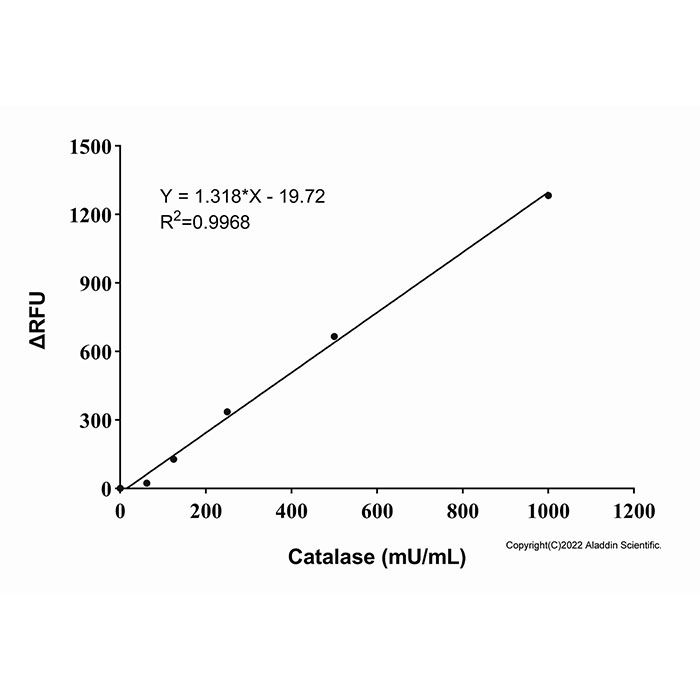
标准曲线中不含过氧化氢酶的空白对照孔的荧光值为A1,标准孔的荧光值为A2,则各个标准孔的荧光减少值为A1-A2。以过氧化氢酶的酶活为横坐标,荧光减少值为纵坐标绘制标准曲线。
标准曲线中不含过氧化氢酶的空白对照孔的荧光值减去样品荧光值为样品荧光减少值。将样品荧光减少值带入标准曲线中,即可得到样品中过氧化氢酶的酶活。
7. 结果展示
标准曲线

注意事项
1. Amplex Red在空气中不太稳定,开启后应尽快使用,且在使用过程中需注意适当避光。
2. Amplex Red须要完全解冻并平衡至室温后再使用,否则会影响检测结果。
3 .为减少稀释液产生的荧光背景带来的误差,样品和标准品的稀释液应该根据样品的种类来定。当样品为裂解液制备的细胞或组织的裂解样品时,应使用裂解液稀释,当样品为血液等其它样品时,宜使用检测缓冲液稀释。
4. 荧光检测时建议使用96孔黑板。
5. 为了您的安全和健康,请穿实验服并戴一次性手套操作。
Catalase is a key antioxidant enzyme with iron porphyrin as its prosthetic group, widely present in animals, plants, microorganisms, and cultured cells. Its main function is to catalyze the decomposition of hydrogen peroxide into water and oxygen. H₂O₂ is a reactive oxygen species produced by cellular aerobic metabolism, which can directly or indirectly oxidize nucleic acids, proteins, lipids, and other biological macromolecules, leading to cell damage, accelerated aging, and disintegration. Catalase protects cells from the toxicity of H₂O₂ by scavenging it, and is one of the key enzymes in the biological defense system.
The principle of this kit is as follows: Catalase catalyzes the decomposition of the substrate hydrogen peroxide (H₂O₂) into water and oxygen, consuming a defined amount of H₂O₂ in the system. The remaining H₂O₂ reacts with Amplex Red in the presence of HRP to generate the red fluorescent compound resorufin, and the fluorescence intensity is proportional to the remaining H₂O₂ concentration. Therefore, higher enzyme activity results in more substrate consumption and weaker fluorescence signal. Subtract the sample fluorescence value from that of the control well without enzyme; the resulting difference (i.e., the fluorescence decrease value) directly reflects the amount of H₂O₂ decomposed, and this decrease is proportional to the enzyme activity. A standard curve is established based on the linear relationship between the fluorescence decrease and the activity of the standard, thereby allowing quantification of enzyme activity in the sample.
Components
A1522296 | Components | Appearance | 100 T | Storage | Quantity Per Test |
A1522296A | Lysis Buffer | Liquid | 30 mL | -20°C | As needed |
A1522296B | Assay Buffer | Liquid | 30 mL | -20°C | As needed |
A1522296C | Amplex Red | Liquid | 200 μL | -20°C. Store in the dark | 2 μL |
A1522296D | Horseradish Peroxidase | Liquid | 200 μL | -20°C. Store in the dark | 2 μL |
A1522296E | Catalase standard (200 U/mL) | Liquid | 200 μL | -20°C. Store in the dark | 2 μL |
A1522296F | Hydrogen peroxide (1 M) | Liquid | 1 mL | -20°C. Store in the dark | 2 μL |
Usage Protocol
1. Sample Preparation
1) Blood samples. For properly prepared serum or plasma samples, they can be directly assayed or diluted with assay buffer before measurement.
2) Cell samples. Collect cells, wash once with PBS. Add lysis buffer at a ratio of 100–200 μL per 1 × 10⁶ cells, and lyse on ice for 5–10 minutes or by sonication homogenization. Centrifuge at approximately 12,000 × g for 5 minutes at 4 °C, and collect the supernatant for subsequent analysis.
3) Tissue samples. Add lysis buffer at a ratio of 100 μL per 10 mg of tissue, and homogenize under low‑temperature conditions such as on ice. Centrifuge at approximately 12,000 × g for 3–5 minutes at 4 °C, and collect the supernatant for subsequent analysis.
All of the above operations should be performed at 4 °C or on ice. If the prepared cell or tissue samples cannot be analyzed immediately, they may be stored at –20 °C or –80 °C.
2. Preparation of Catalase Standard Curve
Take six EP tubes. Add 2 µL of catalase standard (200 U/mL) and 398 µL of assay buffer or lysis buffer (use assay buffer for samples that do not require lysis treatment; use lysis buffer for samples that require lysis treatment) to the first EP tube to dilute the catalase to 1000 mU/mL. Add 200 μL of assay buffer or lysis buffer to each of the remaining tubes. Transfer 200 μL from the first tube to the second tube, and continue this serial dilution through the subsequent tubes. The last tube contains 200 μL of assay buffer or lysis buffer. The resulting catalase concentrations in each tube are 1000, 500, 250, 125, 62.5, and 0 mU/mL, respectively.
3. Preparation of Hydrogen Peroxide Working Solution
Since hydrogen peroxide is not very stable, its actual concentration must be determined by the user before use for calibration. Dilute the approximately 1 M hydrogen peroxide solution 100‑fold with water to obtain a concentration of approximately 10 mM, and measure A₂₄₀. The A₂₄₀ measurement can be performed by any of the following methods: ordinary UV spectrophotometer, micro‑volume UV spectrophotometer, or 96‑well UV microplate reader. Among these, the hydrogen peroxide concentration measured using a cuvette is closest to the actual concentration. Taking calibration using a cuvette with a 1 cm pathlength as an example, calculate the hydrogen peroxide concentration using the following formula: Hydrogen peroxide concentration (mM) = 22.94 × A₂₄₀. Multiply the result by the dilution factor of 100 to obtain the calibrated concentration of hydrogen peroxide.
Based on the calibrated hydrogen peroxide concentration, dilute the hydrogen peroxide to prepare a 200 μM hydrogen peroxide working solution. For sample detection, add 25 μL of the hydrogen peroxide working solution to each standard well and sample well. Prepare the amount needed, and use it immediately after preparation; it should be used as soon as possible.
4. Preparation of Chromogenic Solution
Prepare an appropriate amount of Amplex Red Reaction Working Solution based on a volume of 50 μL per reaction. Mix 46 μL of Assay Buffer, 2 μL of Amplex Red, and 2 μL of Horseradish Peroxidase to prepare 50 μL of Amplex Red Reaction Working Solution. Prepare an appropriate amount according to the number of samples to be tested (including standards). It is recommended to prepare the working solution freshly before use. Refer to the table below for specific preparation methods:
Number of samples | 1 | 10 | 20 | 50 |
Assay Buffer | 46 µL | 460 µL | 920 µL | 2300 µL |
Amplex Red | 2 µL | 20 µL | 40 µL | 100 µL |
Horseradish Peroxidase | 2 µL | 20 µL | 40 µL | 100 µL |
Total volume | 50 µL | 500 µL | 1000 µL | 2500 µL |
5. Sample Determination
1) Pipette 25 μL of standards and samples into a black 96-well plate; wells containing only lysis buffer or assay buffer shall be set as blank controls. Subsequently, add 25 μL of hydrogen peroxide substrate working solution to standard wells and sample wells. Background control wells may be prepared by replacing 25 μL hydrogen peroxide substrate working solution with 25 μL assay buffer. Incubate the plate for 20 min at room temperature in the dark.
2) Add 50 µL chromogenic substrate to standard wells and sample wells, mix thoroughly, and incubate for another 20 min at room temperature with light shielding.
3) Perform fluorescence measurement with excitation wavelength set at 530 nm and emission wavelength at 590 nm to record fluorescence intensity.
6. Result Calculation
The fluorescence value of the blank control well (without catalase) in the standard curve is A1, and the fluorescence value of the standard wells is A2; then the fluorescence decrease value for each standard well is A1 – A2. Plot a standard curve with catalase activity as the abscissa and the fluorescence decrease value as the ordinate.
The fluorescence decrease value of the sample is obtained by subtracting the sample fluorescence value from the fluorescence value of the blank control well (without catalase) in the standard curve. Substitute the fluorescence decrease value of the sample well into the standard curve to obtain the catalase activity in the sample.
7. Results Presentation
Standard curve
Precautions
1. Amplex Red is relatively unstable in the air. Use it as soon as possible after opening, and take care to protect it from light during use.
2. Amplex Red must be completely thawed and equilibrated to room temperature before use; otherwise, the detection results may be affected.
3. To reduce errors caused by fluorescence background from the diluent, the diluent for samples and standards should be selected according to the sample type. When the sample is a cell or tissue lysate prepared with lysis buffer, lysis buffer should be used for dilution; for other samples such as blood, assay buffer should be used for dilution.
4. For fluorescence detection, a black 96‑well plate is recommended.
5. For your safety and health, please wear a lab coat and disposable gloves during operation.

 ZJ26F0737533


 危险品化学品经营许可证(带存储)