
计算溶液所需的质量、体积或浓度。
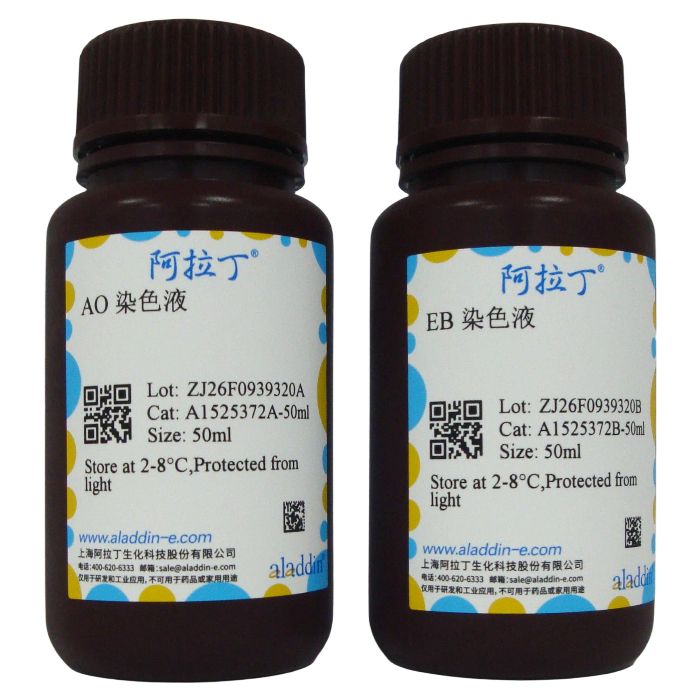
吖啶橙AO/EB双荧光染色试剂盒
有货
BioReagent,生物染色剂,用于显微镜,适用于荧光分析 级 ,适用于对基线干扰要求严格的色谱和分析工作流程。
2-8°C储存,避光。低温运输 。请查阅批次 COA 获取详细规格。
SDS、COA、产品数据表及规格说明书均可下载。可通过批号查询获取批次 COA。
在色谱分析、有机合成和交叉偶联反应领域已被 0 篇同行评审文献引用。
吖啶橙(Acridine Orange), 属于三环杂芳香染料,可以标记DNA、RNA,属于异染性荧光染料,AO常用于细胞内DNA和RNA进行检测。AO与核酸采用插入性结合的方式,AO嵌入核酸双链的碱基对之间,这种结合方式主要为AO与DNA的结合,其荧光发射峰为530nm,激发后呈绿色荧光,其荧光发射峰为640nm,激发后呈红色荧光,少量结合会呈桔黄色或桔红色荧光。因此,AO嵌合到双链DNA分子中显绿色,与DNA单链或RNA结合时发橙红色荧光。Ethidium Bromide嵌合到双链DNA或RNA的碱基对中,无碱基特异性,发红色荧光。
吖啶橙能透过胞膜完整的细胞,嵌入细胞核DNA,与双链DNA结合后发出绿色荧光,溴化乙锭(EB)仅能透过胞膜受损的细胞,嵌入核DNA,发橘红色荧光。凋亡的细胞呈现为染色增强,荧光更为明亮,均匀的圆状或固缩状、团块状结构。非凋亡细胞核呈现荧光深浅不一的结构样特征。二者很容易判别。在荧光显微镜下观察,可见四种细胞形态:活细胞(VN),核染色质着绿色并呈正常结构;早期凋亡细胞(VA),核染色质着绿色呈固缩状或圆珠状;晚期凋亡细胞(NVA),核染色质为橘红色并呈固缩状或圆珠状;非凋亡的死亡细胞(NVN),核染色质着橘红色并呈正常结构。本产品AO、EB溶液浓度分别为100μg/ml,所含稳定剂不影响实验效果。
产品组分及储存条件:
|
使用方法:(仅供参考)
使用前先根据用量,将AO染色液和EB染色液按1:1混合成工作液,现用现配。
1. 对于贴壁细胞或爬片,去掉培养基,用PBS洗两遍去除残余培养基和未贴壁细胞,加入新的PBS;如果需要观察全部细胞特性,保留未贴壁细胞,可以直接在培养基中加入工作液。按照每毫升培养基或PBS中加入20μl工作液即可。室温放置2-5min后于荧光显微镜下观察。
2. 对于悬浮细胞,可直接加入工作液或离心收集细胞后在PBS中加入工作液。按照每毫升培养基或PBS中加入20μl工作液即可。室温放置2-5min后于荧光显微镜下观察。
注意事项:
1. 用能通过活细胞膜与DNA结合后发蓝色荧光的 Hoechist 33342 和只能通过死细胞与DNA结合后发红色荧光的 propidium iodide 对细胞进行双染的方法也比较常用。
2. 如有低温离心机进行离心效果更佳。
3. 操作过程中应注意减少染色溶液暴露于强光下的时间。
4. 含酚红培养基对观察有轻微影响。建议使用无酚红培养基。
5. 染色液工作浓度和染色时间可根据具体实验情况适当调整,以期达到佳效果。
6. AO 和 EB 有一定毒性,请小心操作。所有产品仅限用于研发,必须由专业技术人员操作同时佩戴口罩/手套/实验服并遵守生物实验室安全操作规程!
染色结果:
活细胞:绿色荧光
死细胞:橙色荧光


图解:
(A) 阴性对照细胞(正常细胞):环形细胞核均匀分布在细胞中央。
(B) 实验组 (早期凋亡细胞):AO染色后的细胞核呈黄绿色荧光,以月牙形或颗粒的形态聚集位于细胞的一边。
(C) 实验组 (晚期凋亡细胞):EB染色后的细胞核呈橙红荧光,集中聚集且偏重定位。
(D) 坏死细胞:坏死细胞体积增加,显示平整的橙红荧光和不清晰的轮廓。细胞膜破裂、胞质渗漏、细胞裂解崩解。
Acridine Orange (AO) is a tricyclic heteroaromatic dye capable of labeling DNA and RNA, and it is a metachromatic fluorescent dye. AO is commonly used for the detection of intracellular DNA and RNA. AO binds to nucleic acids via intercalation between base pairs of double-stranded nucleic acids. This binding mode predominates when AO interacts with DNA, producing green fluorescence with an emission peak at 530 nm. When bound to single-stranded nucleic acids, it yields red fluorescence with an emission peak at 640 nm; weak binding results in orange or orange-red fluorescence. Accordingly, AO displays green fluorescence upon intercalation into double-stranded DNA, whereas it emits orange-red fluorescence when bound to single-stranded DNA or RNA. Ethidium Bromide intercalates into the base pairs of double-stranded DNA or RNA without base specificity and emits red fluorescence.
Acridine Orange can cross intact cell membranes and intercalate into nuclear DNA, emitting green fluorescence upon binding to double-stranded DNA. Ethidium Bromide (EB) can only penetrate cells with damaged cell membranes, intercalating into nuclear DNA and producing orange-red fluorescence. Apoptotic cells show intensified, brighter fluorescence with uniformly rounded, condensed or clumped structures, while non-apoptotic cell nuclei display uneven fluorescence patterns, making them easy to distinguish. Four distinct cell morphologies can be observed under a fluorescence microscope: Viable Normal cells (VN): viable cells with normally structured nuclei stained green. Viable Apoptotic cells (VA): early apoptotic cells with condensed or beaded green-stained nuclei. Non-viable Apoptotic cells (NVA): late apoptotic cells with condensed or beaded orange-red stained nuclei. Non-viable Normal cells (NVN): non-apoptotic dead cells with normally structured orange-red stained nuclei. The concentrations of AO and EB in this product are each 100 μg/mL, and the included stabilizer does not affect experimental results.
Product Components and Storage Conditions:
|
Instructions for Use: (For reference only)
Prepare the working solution by mixing AO stain and EB stain at a 1:1 ratio immediately before use, based on the required volume.
1. For adherent cells or cell coverslips: Remove culture medium and rinse twice with PBS to eliminate residual medium and floating cells, then add fresh PBS. If the full cell population (including floating cells) needs to be assessed, add the working solution directly to the culture medium. Add 20 μL of working solution per mL of medium or PBS. Incubate at room temperature for 2–5 minutes and observe under a fluorescence microscope.
2. For suspension cells: Add the working solution directly to the cell suspension, or harvest cells by centrifugation and resuspend in PBS prior to adding the working solution. Add 20 μL of working solution per mL of medium or PBS. Incubate at room temperature for 2–5 minutes and observe under a fluorescence microscope.
Precautions:
1. A common alternative double-staining method uses Hoechst 33342 (which crosses intact live cell membranes and binds DNA to emit blue fluorescence) and propidium iodide (PI, which only penetrates dead cells and binds DNA to emit red fluorescence).
2. Use of a refrigerated centrifuge can improve results for suspension cell processing.
3. Minimize exposure of the staining solution to strong light during handling.
4. Phenol red-containing culture medium may slightly interfere with fluorescence observation. Phenol red-free medium is recommended.
5. The working concentration and incubation time can be appropriately adjusted according to specific experimental conditions to achieve optimal staining results.
6. AO and EB are toxic; handle with extreme caution. This product is for research use only, to be used exclusively by trained laboratory personnel. Always wear a mask, gloves and a lab coat, and follow standard biosafety laboratory protocols.
Staining Results:
Viable cells: Green fluorescence
Dead cells: Orange fluorescence
Fig.
(A) Negative control cells (normal cells): Ring-shaped nuclei are evenly distributed in the center of the cells.
(B) Experimental group (early apoptotic cells): Nuclei stained with AO show yellowish-green fluorescence, accumulating on one side of the cell in a crescent or granular form.
(C) Experimental group (late apoptotic cells): Nuclei stained with EB exhibit orange-red fluorescence, densely aggregated and heavily localized.
(D) Necrotic cells: Necrotic cells are enlarged, displaying diffuse orange-red fluorescence with indistinct outlines. The cell membrane ruptures, cytoplasm leaks out, and the cells disintegrate.
| A1525372 | 物理 | 组件 | 2×50ml | 储存 |
| A1525372A | 液体 | AO 染色液 | 50 ml | 2-8℃. Store in the dark. |
| A1525372B | 液体 | EB 染色液 | 50 ml | 2-8℃. Store in the dark. |
通过匹配包装上的批号来查找并下载产品的 COA,每批产品都进行了严格的验证,您可放心使用!
| 批号(Lot Number) | 证书类型 | 货号 |
|---|---|---|
 ZJ26F0939320 |
分析证书 | A1525372 |
我们的级别选择指南涵盖了目录中所有的变体的纯度、稳定剂状态和应用适用性。
查看 BioReagent 级别指南 → 查看 Biological Stain 级别指南 → 查看 for Fluorescence analysis 级别指南 → 查看 for Microscopy 级别指南 →

 危险品化学品经营许可证(带存储)