Fluorescent secondary antibodies recognize IgG heavy chains and all classes of immunoglobulin light chains from mouse. Ready-to-use secondary antibody concentrations have been optimized and tested by immunofluorescent staining with IF/ICC. No calculations and dilutions are required. Just add an assay dependent volume to stain your cells, and you're ready for the next step in your experiment.
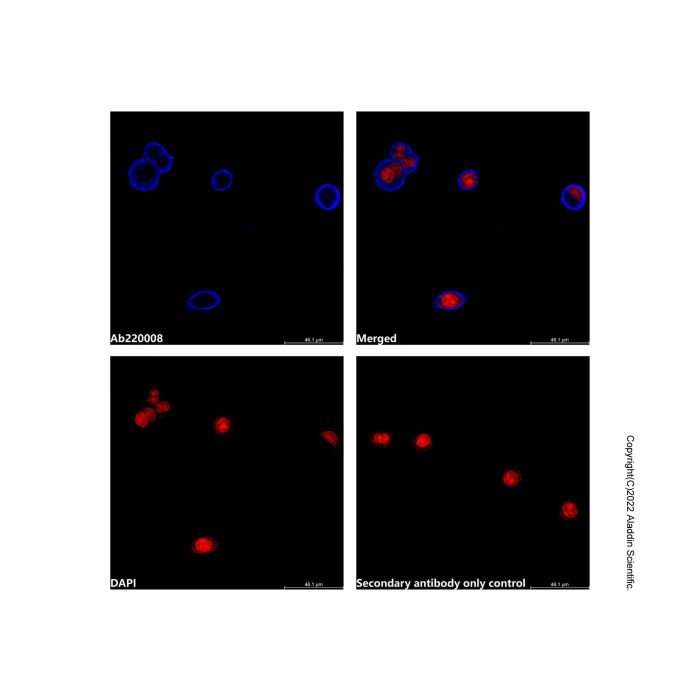
IF analysis of EpCAM (blue) in HT-29 cells. The cells were fixed with 4% Paraformaldehyde Fix Solution (P395744) for 15 minutes and blocked with 5% BSA for 1 hour at room temperature. Cells were stained with 1 μg/mL Catumaxomab (anti-CD3&EpCAM) (Ab175489) in blocking buffer overnight at 4°C. Then incubated with Goat Anti-Mouse IgG H&L (AF405) (Ready to use) (Ab220008) at 2mL/well for 1 hour at room temperature in the dark (blue). A 6-well cell culture plate was used for this experiment. PBS instead of the primary antibody was used as the secondary antibody only control. Cells were counterstained with PI (red). Images were taken on the confocal laser scanning microscope.

 ZJ25F1230410


 危险品化学品经营许可证(带存储)