活死细菌染色试剂盒(SYTO 9/PI)是一款方便且操作简单的试剂盒,利用SYTO 9绿色核酸染料和碘化丙啶(PI)红色荧光核酸染料来进行细菌活力的检测,适用于大量的细菌种属,包括蜡样芽胞杆菌、枯草杆菌、产气荚膜梭菌、大肠杆菌、肺炎克雷伯菌、黄体微球菌、草分枝杆菌、铜绿假单胞菌、沙门氏菌、沙雷氏菌、宋内志贺菌、金黄色葡萄球菌和酿脓链球菌等。
活死细菌染色试剂盒(SYTO 9/PI)的工作原理在于:SYTO 9和PI的光谱特征以及穿透健康细菌细胞的能力不同。用适当比例的SYTO 9与PI混合物染色时,细胞膜完好的细菌发出明亮的绿色荧光,而细胞膜受损的细菌则表现出绿色荧光明显减弱,同时发出较强的红色荧光。 两种染料的最大激发和发射波长分别是480/500nm(SYTO 9)和 490/635nm(PI)。SYTO 9与PI染料在大部分流式细胞仪和荧光显微镜中都是由488nm激光器实现有效激发的,各自的核酸复合物荧光信号可分别在绿色通道和红色通道中检测到,并且几乎没有背景荧光。 本试剂盒兼容于荧光显微镜、荧光酶标仪、流式细胞仪或其它荧光检测仪器。
使用方法
以下实验方案以大肠杆菌检测为例,仅作为指导实验人员设计自己的细菌染色过程的参考。研究人员应用以下染色步骤对革兰氏阳性、阴性菌都是有效的。
1.准备菌悬液
以下步骤描述了制备活细菌(未处理)和死细菌(异丙醇处理)菌悬液的步骤,也可以用来设定流式细胞仪参数,如有必要,还可对染料配比进行优化。培养细菌应使用低磷酸盐生长培养基,SYTO 9 与 PI 可能会结合培养介质中的核酸和其他成分,从而产生一些无关的染色变化。步骤 1.5 的清洗步骤通常可以有效的去除菌悬液中干扰染色的成分,在清洗步骤中不推荐使用磷酸盐,因其可能会降低染色效率。
1.1 取2份1mL细菌悬液于微量离心管中,以 10000×g 离心 1-3 分钟。
1.2 弃上清,一份菌沉淀用 1mL 0.85% NaCl 溶液或其他适当的缓冲溶液(活菌),另一份菌沉淀用1mL 70%异丙醇重悬(死菌)。
1.3 室温下处理30-60min,每隔 15 分钟混匀一次。
1.4 两种菌悬液均以10000×g 离心 1-3 分钟,收集菌沉淀。
1.5 用1mL 0.85% NaCl或其他合适的缓冲溶液清洗,并再次 10000×g 离心 1-3 分钟,收集菌沉淀。
1.6 用1mL 0.85% NaCl 或其他合适的缓冲溶液重悬样品。
1.7 选择步骤,如果未经处理的细菌需要保持最佳生长条件,只需要按照步骤 1.2 准备死菌。对于活菌,可以在使用前直接从液体培养基中分出经过 1 次清洗步骤即可。
2.细菌染色
以下步骤描述了按照步骤 1.1-1.6 制备细菌样品后的染色方法,样品数量增加时,染色步骤需要相应按比例增加。
2.1 取988μL 0.85%NaCl 或其他合适的缓冲溶液到流式细胞分析检测管。
2.2 向步骤2.1中的流式细胞检测管中加入1μL SYTO 9染色液(1000×),1μL PI染色液(1000×)。
2.3 向染料溶液中加入步骤 1.6 中的10μL菌悬液。如果菌悬液非常浑浊或OD600检测值大于1,可以对其进行稀释后再加样,以便得到更准确的检测结果。
2.4 室温下避光孵育15min。
2.5 用荧光显微镜观察,也可以用荧光酶标仪检测或流式细胞仪分析。
3. 荧光观测和结果分析
染色后的细菌可在配备488nm激光器的流式细胞仪或荧光显微镜中进行检测。活死细菌染色试剂盒(SYTO 9/PI)的SYTO 9荧光信号通过绿色通道接收,可选用FITC的滤光片检测;PI的荧光信号通过红色通道接收,可选用德克萨斯红(Texas Red)的滤光片检测。
注意事项
1. 将SYTO 9和PI室温融化,并经过短暂离心后再开启。
2. 两款探针溶液本身就是溶于DMSO中,所以后续无需再单独添加,第一次使用可将SYTO 9和PI根据单次用量分装保存,密封后置于-20℃避光保存,避免反复冻融。
3. 染料有毒,须戴手套操作,做好安全防护。
结果展示

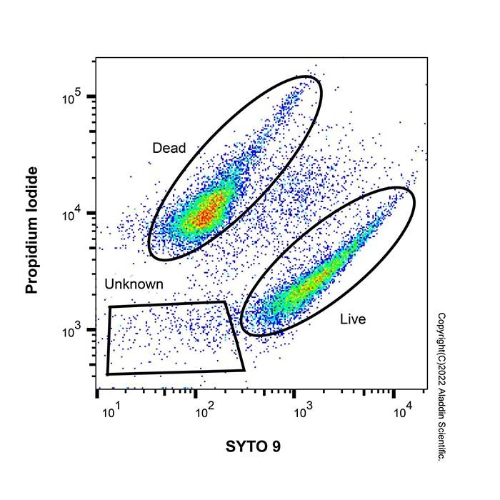
图1. 大肠杆菌的活菌(未处理)、死菌(70% 异丙醇处理)悬液按照一定比例混合后,采用活死细菌染色试剂盒(SYTO 9/PI)(B1511173)染色的流式检测结果。

图2. 活死细菌染色试剂盒(SYTO 9/PI)(B1511173)对大肠杆菌染色的荧光共聚焦显微镜检测效果图。SYTO 9和PI双染叠加(Merged)后可以非常清晰地观察到活细菌和死细菌的荧光染色差别。死细菌组即Dead Bacteria Ratio为100%组,红绿荧光染色数目基本一致,两种荧光几乎完全重合,叠加后呈黄色;活细菌与死细菌混合组即Dead Bacteria Ratio为50%组,两种荧光叠加后,活细菌呈绿色,死细菌呈黄色。
The Live/Dead Bacterial Staining Kit (SYTO 9/PI) is a convenient and easy-to-use kit for assessing bacterial viability using SYTO 9 green fluorescent nucleic acid dye and propidium iodide (PI) red fluorescent nucleic acid dye. It is suitable for a wide range of bacterial species, including Bacillus cereus, Bacillus subtilis, Clostridium perfringens, Escherichia coli, Klebsiella pneumoniae, Micrococcus luteus, Mycobacterium phlei, Pseudomonas aeruginosa, Salmonella, Serratia, Shigella sonnei, Staphylococcus aureus, and Streptococcus pyogenes, among others.
The Live/Dead Bacterial Staining Kit (SYTO 9/PI) operates based on the distinct spectral characteristics of SYTO 9 and PI and their differing abilities to penetrate healthy bacterial cells. When bacteria are stained with an appropriate mixture of SYTO 9 and PI, those with intact cell membranes emit bright green fluorescence, whereas bacteria with damaged cell membranes show markedly reduced green fluorescence and simultaneously emit intense red fluorescence. The maximum excitation and emission wavelengths of the two dyes are 480/500 nm (SYTO 9) and 490/635 nm (PI), respectively. Both SYTO 9 and PI are efficiently excited by a 488 nm laser in most flow cytometers and fluorescence microscopes. The fluorescence signals of their respective nucleic acid complexes can be detected in the green and red channels, with virtually no background fluorescence. This kit is compatible with fluorescence microscopes, fluorescence microplate readers, flow cytometers, or other fluorescence detection instruments.
Protocol
The following protocol uses Escherichia coli as an example and is intended solely as a reference for researchers to design their own bacterial staining procedures. The staining steps described have been shown to be effective for both Gram‑positive and Gram‑negative bacteria.
1. Preparation of Bacterial Suspensions
The following steps describe the preparation of live (untreated) and dead (isopropanol‑treated) bacterial suspensions. These can also be used to set parameters on a flow cytometer and, if necessary, to optimize the dye ratio. Bacteria should be cultured in low‑phosphate growth media, as SYTO 9 and PI may bind to nucleic acids and other components in the culture medium, causing unwanted staining variations. The washing step in 1.5 is generally effective at removing components that interfere with staining; the use of phosphates in the washing step is not recommended because it may reduce staining efficiency.
1.1 Transfer 2×1 mL aliquots of bacterial suspension into microcentrifuge tubes and centrifuge at 10,000×g for 1–3 minutes.
1.2 Discard the supernatant. Resuspend one pellet (live bacteria) in 1 mL of 0.85% NaCl solution or another suitable buffer. Resuspend the other pellet (dead bacteria) in 1 mL of 70% isopropanol.
1.3 Incubate at room temperature for 30–60 minutes, mixing every 15 minutes.
1.4 Centrifuge both suspensions at 10,000×g for 1–3 minutes to pellet the bacteria.
1.5 Wash each pellet with 1 mL of 0.85% NaCl or another suitable buffer, then centrifuge again at 10,000×g for 1–3 minutes to collect the pellets.
1.6 Resuspend each pellet in 1 mL of 0.85% NaCl or another suitable buffer.
1.7 (Optional step) If untreated bacteria need to be maintained under optimal growth conditions, prepare only the dead bacteria as described in step 1.2. For live bacteria, an aliquot can be taken directly from the liquid culture and subjected to a single wash step before use.
2. Staining of Bacteria
The following protocol describes the staining procedure after preparing bacterial samples according to steps 1.1–1.6. For larger numbers of samples, the staining steps should be scaled up proportionally.
2.1 Add 988 μL of 0.85% NaCl or another suitable buffer to a flow cytometry tube.
2.2 Add 1 μL of SYTO 9 staining solution (1000×) and 1 μL of PI staining solution (1000×) to the tube from step 2.1.
2.3 Add 10 μL of the bacterial suspension from step 1.6 to the dye solution. If the bacterial suspension is very turbid or has an OD600 value >1, it can be diluted before addition to obtain more accurate results.
2.4 Incubate for 15 minutes at room temperature, protected from light.
2.5 Observe under a fluorescence microscope, or measure using a fluorescence microplate reader or flow cytometer.
3. Fluorescence Observation and Data Analysis
Stained bacteria can be detected using a flow cytometer or fluorescence microscope equipped with a 488 nm laser. The SYTO 9 fluorescence signal from the Live/Dead Bacterial Staining Kit (SYTO 9/PI) is collected in the green channel, typically using a FITC filter set. The PI fluorescence signal is collected in the red channel, typically using a Texas Red filter set.
Precautions:
1. Thaw SYTO 9 and PI at room temperature and briefly centrifuge them before opening.
2. Both probe solutions are already dissolved in DMSO, so no additional DMSO is required. Upon first use, SYTO 9 and PI may be aliquoted into single‑use portions, stored sealed and protected from light at –20 °C, and repeated freeze‑thaw cycles should be avoided.
3. The dyes are toxic. Gloves must be worn during handling, and appropriate safety precautions should be taken.
Results Presentation
Figure 1. Flow cytometry results of live (untreated) and dead (treated with 70% isopropanol) Escherichia coli suspensions mixed at a certain ratio and stained with the Live/Dead Bacterial Staining Kit (SYTO 9/PI) (B1511173).
Figure 2. Confocal fluorescence microscopy images of Escherichia coli stained with the Live/Dead Bacterial Staining Kit (SYTO 9/PI) (B1511173). After merging the SYTO 9 and PI stains, the difference in fluorescence staining between live and dead bacteria can be clearly observed. In the dead bacteria group (Dead Bacteria Ratio = 100%), the numbers of red and green fluorescent bacteria are essentially the same, and the two fluorescent signals almost completely overlap, appearing yellow after merging. In the mixed live and dead bacteria group (Dead Bacteria Ratio = 50%), after merging the two fluorescent signals, live bacteria appear green, while dead bacteria appear yellow.

 ZJ26F1039739


 危险品化学品经营许可证(带存储)