
计算溶液所需的质量、体积或浓度。
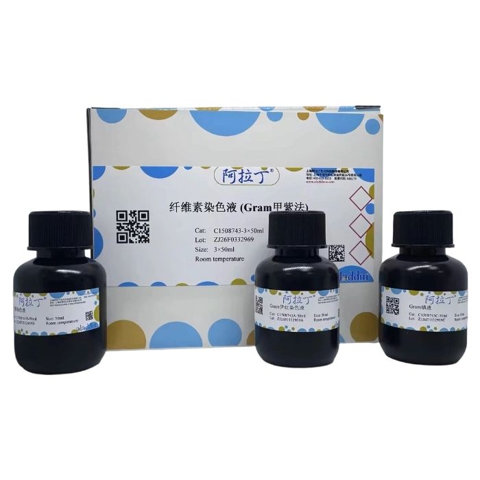
纤维素染色液 (Gram甲紫法)
有货
BioReagent,生物染色剂,用于显微镜 级 ,适用于对基线干扰要求严格的色谱和分析工作流程。
避光,室温。常规运输 。请查阅批次 COA 获取详细规格。
SDS、COA、产品数据表及规格说明书均可下载。可通过批号查询获取批次 COA。
在色谱分析、有机合成和交叉偶联反应领域已被 0 篇同行评审文献引用。
病理的内源性沉着物是色素沉着物的一部分,组织细胞经过一定的病理变化,形成不同形状特点的沉着物质,这种聚合形成的特殊蛋白,经染色后能够显示纤维素蛋白。纤维素是存在于血液内的纤维蛋白分子聚合形成的特殊蛋白质,又称为纤维蛋白,这种蛋白以弯曲细丝纤维素的形式存在于组织内,大多数呈网状结构,有时会呈粗大的纤维素网,陈旧的可凝集成无定型的块状。
纤维素染色液 (Gram甲紫法)主要由Gram伊红染色液、甲紫染色液、Gram碘液组成,是一种简便、廉价的纤维素染色液,染色后纤维素呈蓝色。该试剂仅用于科研领域,不适用于临床诊断或其他用途。
| C1508743 | Component | 3×50 mL | Storage |
| C1508743A | Gram伊红染色液 | 50 mL | RT. Store in the dark. |
| C1508743B | 甲紫染色液 | 50 mL | RT. Store in the dark. |
| C1508743C | Gram碘液 | 50 mL | RT. Store in the dark. |
自备材料:
1、系列乙醇、蒸馏水、去离子水、二甲苯或环保脱蜡透明液、中性树胶
2、吸水纸、显微镜
操作步骤(仅供参考):
1、常规石蜡切片,常规二甲苯或脱蜡透明液脱蜡至水。
2、入Gram伊红染色液染色10min,稍水洗。
3、入甲紫染色液染色3-5min,稍水洗。
4、入Gram碘液染色3-5min。
5、倾去Gram碘液,用吸水纸吸干。
6、取苯胺和二甲苯等量混合作为分化液,分化30s左右。
7、更换新的二甲苯或脱蜡透明液清洗组织,中性树胶封固。
8、镜检。
染色结果:
纤维素: 蓝色
背景: 红色
注意事项:
1、Gram伊红染色液染色后,经过水洗时务必使组织上有足够的红色。
2、苯胺和二甲苯等量混合分化时,注意轻轻摇动,以使组织脱色均匀。
3、试剂开封后请尽快使用,以防影响后续实验效果。
4、为了您的安全和健康,请穿实验服并戴一次性手套操作。
Endogenous pathological deposits are a subset of pigment deposits. Tissue cells undergo certain pathological changes to form deposits with distinct morphological characteristics. These polymerized special proteins can exhibit fibrin after staining. Fibrin, is a special protein formed by the polymerization of fibrin molecules present in the blood. It exists in tissues in the form of curved filamentous cellulose, mostly presenting a reticular structure, and sometimes forming a thick cellulose network. Old cellulose deposits can aggregate into amorphous masses.
Fibrin Staining Solution (Gram Crystal Violet Method) is mainly composed of Gram Eosin Staining Solution, Crystal Violet Staining Solution, and Gram Iodine Solution. It is a simple and cost-effective reagent for cellulose staining, which stains cellulose blue. This reagent is for research use only and not suitable for clinical diagnosis or any other purposes.
| C1508743 | Component | 3×50 mL | Storage |
| C1508743A | Gram Eosin Staining Solution | 50 mL | RT. Store in the dark. |
| C1508743B | Crystal Violet Staining Solution | 50 mL | RT. Store in the dark. |
| C1508743C | Gram Iodine Solution | 50 mL | RT. Store in the dark. |
Materials to Be Prepared by the User
1. Graded ethanol solutions, distilled water, deionized water, xylene or environment-friendly dewaxing and clearing solution, neutral mounting medium
2. Absorbent paper, microscope
Operating Procedures (For Reference Only)
1. Prepare routine paraffin sections, then perform routine dewaxing with xylene or dewaxing and clearing solution and hydrate the sections stepwise to water.
2. Immerse the sections in Gram Eosin Staining Solution for 10 minutes, followed by brief rinsing with water.
3. Transfer the sections to Crystal Violet Staining Solution for 3-5 minutes, then rinse briefly with water.
4. Immerse the sections in Gram Iodine Solution for 3-5 minutes.
5. Pour off the Gram Iodine Solution and blot the sections dry with absorbent paper.
6. Prepare a differentiation solution by mixing aniline and xylene in equal volumes, and differentiate the sections for approximately 30 seconds.
7. Rinse the tissues with fresh xylene or dewaxing and clearing solution, then mount the sections with neutral mounting medium.
8. Observe the sections under a microscope.
Staining Results
Cellulose: Blue
Background: Red
Precautions
1. After staining with Gram Eosin Staining Solution, ensure that the tissues retain sufficient red color during water rinsing.
2. When differentiating with the equal-volume mixture of aniline and xylene, shake the container gently to ensure uniform decolorization of the tissues.
3. Use the reagent as soon as possible after opening to avoid compromising subsequent experimental results.
4. For your safety and health, wear a lab coat and disposable gloves during operation.
通过匹配包装上的批号来查找并下载产品的 COA,每批产品都进行了严格的验证,您可放心使用!
| 批号(Lot Number) | 证书类型 | 货号 |
|---|---|---|
 ZJ26F0332969 |
分析证书 | C1508743 |
我们的级别选择指南涵盖了目录中所有的变体的纯度、稳定剂状态和应用适用性。
查看 BioReagent 级别指南 → 查看 Biological Stain 级别指南 → 查看 for Microscopy 级别指南 →

 危险品化学品经营许可证(带存储)