产品介绍
铁是人体必需的微量元素之一,是生物体内最丰富的过渡金属元素,是血红蛋白、肌红蛋白、细胞色素及其他酶系统的主要成分。铁稳态的任何变化都可能导致铁超载疾病。近年来,活细胞中的游离铁受到广泛关注,游离铁最常以其稳定的氧化还原态存在,即亚铁离子(Fe²⁺)和铁离子(Fe³⁺)。对研究者来说,在研究细胞内的还原环境、金属转运蛋白和Fe²⁺的水溶性时,了解Fe²⁺的行为比了解Fe³⁺的行为更为重要。因此,准确、方便和经济地量化生物组织内亚铁离子(Fe²⁺),对于临床评估铁负荷、研究这些疾病的机制以及评估铁死亡诱导通路的活性具有巨大价值。本试剂盒提供一种荧光探针可以和Fe²⁺特异性结合,探针可以很好的进入到细胞内部,适用于活细胞Fe²⁺的检测。当探针与Fe²⁺反应后,可以生成一种不可逆的橙红色荧光产物(激发波长:543 nm,发射波长:580 nm)。
| C1522840 | 组分 | 物理外观 | 50T | 100T | 储存 | 单次测试使用量 |
| C1522840A | 检测缓冲液 (10×) | 液体 | 15 mL | 30 mL | -20℃. 避光 | 按需 |
| C1522840B | FerroOrange探针 (1000×) | 液体 | 50 μL | 100 μL | -20℃. 避光 | 按需 |
使用方法
1. 试剂准备
1.1 各试剂平衡至室温,若单次实验未能用完,建议剩余试剂分装后-20°C避光保存,避免反复冻融。
1.2. 预先计算好检测缓冲液 (10×)的用量,用双蒸水将检测缓冲液 (10×)稀释为1×的检测缓冲工作液待用。
1.3. 预先计算好FerroOrange探针 (1000×)的用量,用检测缓冲工作液将FerroOrange探针 (1000×)稀释为1×的染色工作液待用。
注1:配制好的检测缓冲工作液和染色工作液必须一次使用完毕,不能冻存。染色工作液中FerroOrange的浓度可以根据染色效果在0.5×-2.5×之间适当调整。
注2:按照样品数和孔板类型计算所需染色工作液量,通常96孔板每孔加入100 μL,24孔板每孔加入250 μL,12孔板每孔加入500 μL,6孔板每孔加入1 mL。以 96 孔板为例,一个孔需要 100 μL的染色工作液,如果20 个样品孔,则需要配制2 mL的染色工作液,即在2 mL的检测缓冲工作液中加入2 µL的FerroOrange探针 (1000×)。剩下的检测缓冲工作液可作为洗涤液备用。
2. 细胞检测步骤
2.1 悬浮细胞:
2.1.1 离心收集细胞,用检测缓冲工作液洗涤 2 次。
2.1.2 弃去上清液,每10⁶ 个细胞加入1 mL稀释好的染色工作液。在 37℃,5% CO₂ 培养箱中孵育 30-60 min。无需清洗,孵育后直接进行检测。
2.1.3 用流式细胞仪或荧光显微镜或荧光酶标仪进行检测。探针与细胞内亚铁离子结合呈现橙红色荧光(Ex/Em=543/580 nm)。
2.2 贴壁细胞:
2.2.1 贴壁细胞可以接种至细胞培养板、微孔板或者制作成细胞爬片。在37℃,5% CO₂ 培养箱中过夜培养。
2.2.2 弃去上清液,用检测缓冲工作液洗涤 2 次。
2.2.3 加入足量的染色工作液,保证没过单层细胞。在 37℃,5% CO₂ 培养箱中孵育30-60 min。
2.2.4 无需清洗,孵育后直接在荧光显微镜下观察细胞。探针与细胞内亚铁离子结合呈现橙红色荧光(Ex/Em=543/580 nm)。
注1:生成的荧光物质容易淬灭,孵育完成后最好在2 h之内进行测定,防止荧光减弱。
注2:如需要对细胞核染色,建议使用阿拉丁的Hoechst 33342 染色液 (O1501802)。
注3:流式细胞仪可以用 PE 的参数设置检测。
注意事项
1. 探针溶液避免反复冻融,使用前需要充分融解混匀,变成液体状态后离心,直至液体离心至管底部再开盖。
2. 检测缓冲工作液和染色工作液必须现配现用,配制好的工作液请在当天使用。
3. 本试剂盒适用于活细胞检测,对死细胞无法发挥出探针的正常性能。
4. 更换培养基会导致探针泄漏到细胞外,因此染色后不要更换培养基。
5. 设置对照组:为了检查铁检测的实验条件,建议准备1 个加入FAS (硫酸亚铁铵)(阳性对照)或铁螯合剂 BPY (2,2'-Bipyridine) (阴性对照)的样品,并观察荧光强度的变化。
6. 荧光染料均存在淬灭问题,保存和使用本试剂盒的过程中请尽量注意避光,以减缓荧光淬灭。建议染色后尽量当天完成检测。
7. 为了您的安全和健康,操作时请穿着实验服并佩戴一次性手套。
结果展示

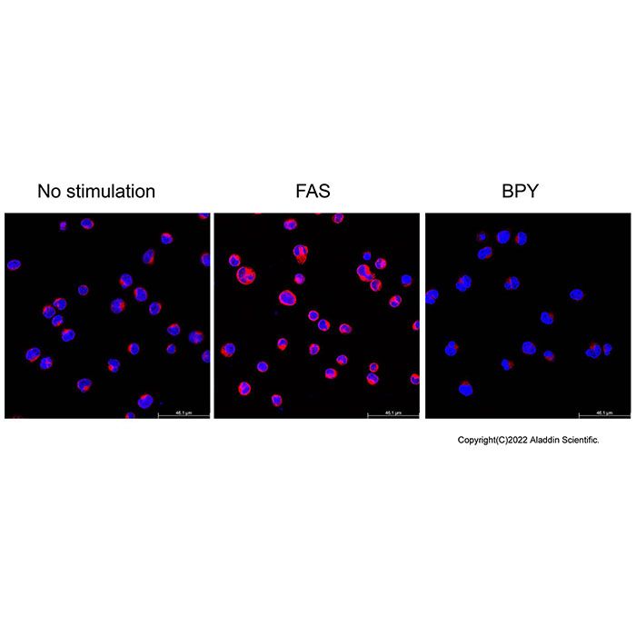
图一:用本试剂盒对Jurkat细胞染色,用荧光共聚焦显微镜检测的染色效果图。左侧图片为正常细胞内亚铁离子(Fe²⁺)呈现较弱的红色荧光,这表明细胞内存在内源性的亚铁离子。中间图片为经100μM硫酸亚铁铵(Ferrous ammonium sulfate, FAS)处理30分钟后,细胞内亚铁离子含量增加,经FerroOrange染色后红色荧光强度显著增强。右侧图片为加入100μM亚铁离子螯合剂2,2'-联吡啶(2,2'-Bipyridine, BPY)后,BPY结合亚铁离子并阻止其与FerroOrange反应,因此红色荧光信号明显降低。实际检测效果会因实验条件、检测仪器等的不同而存在差异,图中效果仅供参考。

图二:用本试剂盒对Jurkat细胞染色,用流式细胞仪检测的染色效果图。黑色:未处理细胞未孵育FerroOrange探针对照;蓝色:未处理细胞孵育FerroOrange探针;红色:细胞先用100μM硫酸亚铁铵(Ferrous ammonium sulfate, FAS)处理30分钟后,再孵育FerroOrange探针;绿色:细胞先用100μM亚铁离子螯合剂2,2'-联吡啶(2,2'-Bipyridine, BPY)处理30分钟后,再孵育FerroOrange探针。
Product Description
Iron is one of the essential trace elements for the human body and is the most abundant transition metal in living organisms. It is a major component of hemoglobin, myoglobin, cytochromes, and other enzyme systems. Any alteration in iron homeostasis may lead to iron overload diseases. In recent years, labile iron in living cells has received extensive attention. Labile iron most commonly exists in its stable redox states, namely ferrous (Fe²⁺) and ferric (Fe³⁺) ions. For researchers, understanding the behavior of Fe²⁺ is more important than that of Fe³⁺ when studying the reducing intracellular environment, metallotransporters, and the water solubility of Fe²⁺. Therefore, accurate, convenient, and economical quantification of ferrous ions (Fe²⁺) in biological tissues is of great value for clinical assessment of iron load, for investigating the mechanisms of these diseases, and for evaluating the activity of ferroptosis‐inducing pathways. This kit provides a fluorescent probe that specifically binds to Fe²⁺. The probe can readily penetrate cell membranes and is suitable for detecting Fe²⁺ in living cells. Upon reaction with Fe²⁺, the probe generates an irreversible orange‑red fluorescent product (excitation wavelength: 543 nm, emission wavelength: 580 nm).
| C1522840 | Component | Appearance | 50T | 100T | Storage | Quantity Per Test |
| C1522840A | Assay Buffer (10×) | Liquid | 15 mL | 30 mL | -20℃. Store in the dark | As needed |
| C1522840B | FerroOrange Probe (1000×) | Liquid | 50 μL | 100 μL | -20℃. Store in the dark | As needed |
Protocol
1. Reagent Preparation
1.1 Equilibrate all reagents to room temperature. If the entire volume is not used in a single experiment, it is recommended to aliquot the remaining reagents and store at –20 °C protected from light, avoiding repeated freeze‑thaw cycles.
1.2 Pre‑calculate the required volume of Assay Buffer (10×), and dilute it with double‑distilled water to prepare 1× Assay Buffer working solution for later use.
1.3 Pre‑calculate the required volume of FerroOrange Probe (1000×), and dilute it with the Assay Buffer working solution to prepare 1× Staining Working Solution for later use.
Note 1: The prepared Assay Buffer working solution and Staining Working Solution must be used immediately in a single experiment and cannot be frozen for storage. The concentration of FerroOrange in the Staining Working Solution can be adjusted between 0.5× and 2.5× as needed to optimize staining.
Note 2: Calculate the required volume of Staining Working Solution according to the number of samples and the plate format. Typically, 100 μL per well is used for a 96‑well plate, 250 μL per well for a 24‑well plate, 500 μL per well for a 12‑well plate, and 1 mL per well for a 6‑well plate. Taking a 96‑well plate as an example: one well requires 100 μL of Staining Working Solution. For 20 sample wells, prepare 2 mL of Staining Working Solution, i.e., add 2 μL of FerroOrange Probe (1000×) to 2 mL of Assay Buffer working solution. The remaining Assay Buffer working solution can be used as a wash buffer.
2. Cell Staining Protocol
2.1 Suspension cells:
2.1.1 Collect cells by centrifugation and wash twice with Assay Buffer working solution.
2.1.2 Discard the supernatant, and resuspend the cells in 1 mL of the diluted Staining Working Solution per 1 × 10⁶ cells. Incubate at 37 °C in a 5 % CO₂ incubator for 30–60 min. No washing is required; proceed directly to detection after incubation.
2.1.3 Detect using a flow cytometer, fluorescence microscope, or fluorescence microplate reader. The probe binds to intracellular ferrous ions, producing orange‑red fluorescence (Ex/Em = 543/580 nm).
2.2 Adherent cells:
2.2.1 Seed adherent cells into cell culture plates, microplates, or onto coverslips, and culture overnight at 37 °C in a 5 % CO₂ incubator.
2.2.2 Discard the supernatant and wash twice with Assay Buffer working solution.
2.2.3 Add sufficient Staining Working Solution to completely cover the monolayer cells. Incubate at 37 °C in a 5 % CO₂ incubator for 30–60 min.
2.2.4 No washing is required. Observe the cells directly under a fluorescence microscope after incubation. The probe binds to intracellular ferrous ions, producing orange‑red fluorescence (Ex/Em = 543/580 nm).
Note 1: The fluorescent product is prone to quenching. It is recommended to measure the fluorescence within 2 hours after incubation to prevent signal loss.
Note 2: If nuclear counterstaining is desired, Aladdin's Hoechst 33342 Staining Solution (O1501802) is recommended.
Note 3: Detection can be performed on a flow cytometer using PE settings.
Precautions:
1. Avoid repeated freeze‑thaw cycles of the probe solution. Before use, ensure the probe is completely thawed and mixed well. After it has become a liquid, centrifuge briefly to collect the solution at the bottom of the tube before opening the cap.
2. The Assay Buffer working solution and Staining Working Solution must be prepared fresh immediately before use and should be used on the same day.
3. This kit is suitable for live‑cell detection and cannot function properly in dead cells.
4. Since probe leakage may occur upon medium exchange, do not replace the culture medium after staining.
5. For control experiments, it is recommended to include one sample treated with FAS (ammonium ferrous sulfate) as a positive control or one sample treated with the iron chelator BPY (2,2'‑bipyridine) as a negative control to monitor changes in fluorescence intensity.
6. Fluorescent dyes are prone to quenching. Please protect the reagents and stained samples from light during storage and use to minimize fluorescence quenching. It is recommended to perform the detection on the same day after staining.
7. For your safety and health, please wear a lab coat and disposable gloves during operation.
Results Presentation
Figure 1: Staining of Jurkat cells using this kit, detected by confocal fluorescence microscopy. The left panel shows normal cells exhibiting weak red fluorescence for ferrous ions (Fe²⁺), indicating the presence of endogenous Fe²⁺. The middle panel shows cells treated with 100 μM ferrous ammonium sulfate (FAS) for 30 minutes, which increases intracellular ferrous ion content; after FerroOrange staining, the red fluorescence intensity is significantly enhanced. The right panel shows cells treated with 100 μM of the ferrous ion chelator 2,2′‑bipyridine (BPY). BPY binds to ferrous ions and prevents them from reacting with FerroOrange, thus resulting in a marked reduction of the red fluorescence signal. Actual staining results may vary depending on experimental conditions and detection instruments; the results shown in the figure are for reference only.
Figure 2: Staining of Jurkat cells using this kit, detected by flow cytometry. Black: Untreated cells without FerroOrange probe incubation (control); Blue: Untreated cells incubated with FerroOrange probe; Red: Cells pre‑treated with 100 μM ferrous ammonium sulfate (FAS) for 30 minutes, then incubated with FerroOrange probe; Green: Cells pre‑treated with 100 μM ferrous ion chelator 2,2′‑bipyridine (BPY) for 30 minutes, then incubated with FerroOrange probe.

 ZJ26F0939187


 危险品化学品经营许可证(带存储)