
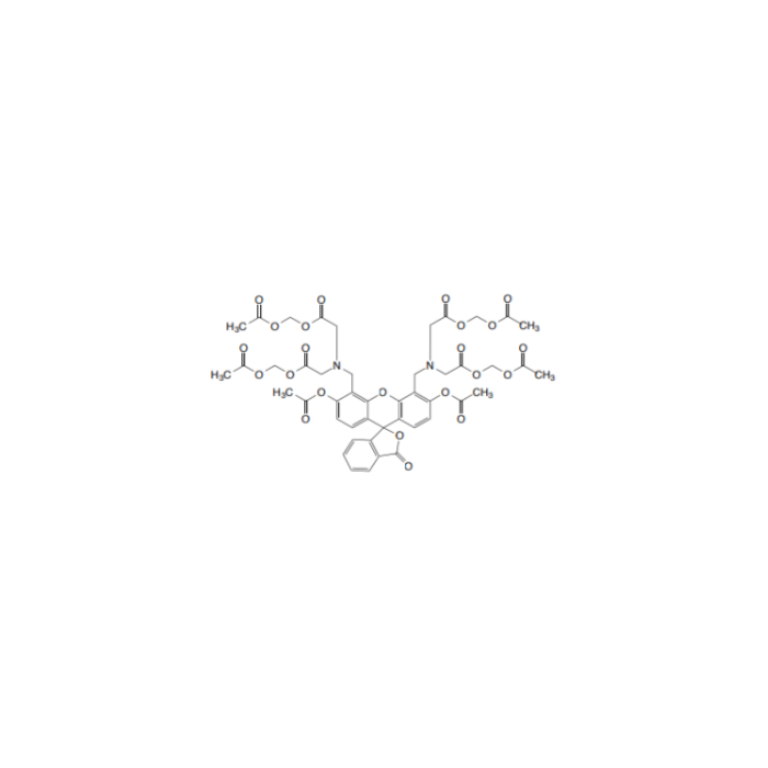
 F2629675
计算溶液所需的质量、体积或浓度。
为什么选择此级别
,适用于对基线干扰要求严格的色谱和分析工作流程。
储存与运输
避光,-20°C储存,干燥。超低温运输 。请查阅批次 COA 获取详细规格。
质量文档
SDS、COA、产品数据表及规格说明书均可下载。可通过批号查询获取批次 COA。
文献证明
在色谱分析、有机合成和交叉偶联反应领域已被 3 篇同行评审文献引用。
概述
产品介绍:
分子式: C46H46N2O23
分子量:994.86
钙⻩绿素-AM (Calcein-AM) 和碘化丙啶(PI) 溶液,分别对活细胞和死细胞染色,可用于同时对活细胞和死细胞进行荧光染色。Calcein-AM的乙酸甲基酯亲脂性很高,使其可透过细胞膜。尽管Calcein-AM本身并不是荧光分子,但通过活细胞内的酯酶作用,Calcein-AM能脱去AM基,产生的Calcein能发出强绿色荧光 (激发: 490 nm,发射: 515 nm)。因此Calcein-AM仅对活细胞染色。另一方面,作为核染色染料的PI不能穿过活细胞的细胞膜。它穿过死细胞膜的无序区域而到达细胞核,并嵌入细胞的DNA双螺旋从而产生红色荧光 (激发: 535 nm,发射: 617 nm)。由于Calcein和PI-DNA都可被490 nm激发,因此可用荧光显微镜同时观察活细胞和死细胞。用545 nm激发,仅可观察到死细胞。由于不同细胞系的最佳染色条件不同,我们建议个别确定Calcein-AM和PI的合适浓度。
I.试剂
Calcein-AM 4mM 50uL in DMSO; Unit Size #500 assays·PI (2mM) 150uL in water
II.操作说明
用荧光显微镜观察细胞形态,以HeLa细胞染色为例,请注意不同的细胞种类、不同浓度,有不同的观察条件。根据细胞条件,摸索不同条件下的细胞贴壁情况和试剂浓度的配制等最佳条件。
1.染色溶液的配制
1) 将Calcein-AM储备液和PI储备液平衡到室温。
2)分别加 2.5 μl Calcein-AM原液和 12.5μl PI原液至5 mL PBS(pH=7.4)中配制成染色工作液。染色工作液中 Calcein-AM的浓度为2 μmol/L,PI的浓度约为 5 μmol/L。
2.细胞染色
1)染色HeLa细胞等贴壁细胞时,先用Trypsin-EDTA等消化细胞,制备成细胞悬液。
2)将细胞悬液离心3分钟(1,000 rpm)。
3)去除上清液,加入PBS缓冲液,细胞数量调整至105-106个/ml。再用移液器充分混匀。
4)由于培养基中的血清等含有酯酶,Calcein-AM遇水会分解,会导致空白上升,所以需要离心数次,用PBS洗涤数次直到完全洗净。
5)将200 μl细胞悬液移至小试管中,加入100 μl染色工作液,在37°C下孵育15分钟。6)在盖玻片上滴加适量的染色的细胞溶液。7)在荧光显微镜下,先用490±10 nm波⻓激发,观察⻩绿色的活细胞,还可以同时观察到红色的死细胞,然后用545 nm波⻓激发,能够看到红色的死细胞。
3.染色试剂的最佳浓度
Calcein-AM和PI最佳浓度根据细胞种类而定,通过以下的操作,可找到不同细胞染色试剂的最佳浓度。
1)通过在0.1%皂苷或0.1-0.5%毛地黄皂苷中孵育10分钟或通过在70%乙醇中孵育30分钟制备死细胞。
2)用0.1-10 μM PI溶液对死细胞染色,以便找到仅对细胞核染色而不对细胞质染色的PI浓度。
3)用0.1-10 μM Calcein-AM溶液对死细胞染色,以便找到不对细胞质染色的Calcein-AM浓度。接着用该浓度的Calcein-AM对活细胞染色以检验活细胞可否被染色。
III.注意事项
1) Calcein-AM的ester部位遇到湿气会分解,使用后请在-20度下密闭冷冻保存,防止水分进入。Calcein-AM储备液用缓冲液或培养基等稀释时尽量现配现用。
2)使用时一定要带手套、眼罩、口罩。万一接触到皮肤的话,迅速使用大量水清洗。
Calcein AM /PI Double Staining Kitis utilized for simultaneous fluorescence staining of viable and dead cells. This kit contains Calcein-AM and Propidium Iodide (PI) solutions, which stains viable and dead cells, respectively(Fig. 1). Calcein-AM, an acetoxymethyl ester of calcein, is highly lipophilic and cell membrane permeable. Though Calcein-AM itself is not a fluorescent molecule, the calcein generated from Calcein-AM by esterase in a viable cell emits a strong green fluorescence (excitationat 490 nm, emission at515 nm). Therefore, Calcein-AM only stains viable cells. On the other hand, PI, a nuclei staining dye, cannot pass through a viable cell membrane. It reaches the nucleus by passing through disordered areas of dead cell membrane, and intercalates with the DNA double helix of the cell to emit red fluorescence (excitation: 535 nm,emmision: 617 nm). Since both calcein and PI-DNA can be excited with 490 nm, simultaneous monitoring of viable and dead cells is possible with a fluorescence microscope. With 545 nm excitation, only dead cells can be observed (Fig. 1). Since optimal staining conditions differ from cell line to cell line, we recommend that a suitable concentration of PI and Calcein-AM be individually determined. Please note that PI is suspected to be highly carcinogenic;careful handling is required.
Required Equipment and Materials:
Microscope with 490 nm excitation filter and 530 nm emission filter;CO2incubator;10 μl and 200 μl adjustable pipettes, PBSSolution A (Calcein-AM);Solution B (PI) Storage Condition: -20oC ;Shipping Condition: blue ice.
Application:
Assay Procedure1)Add 2.5 μl Solution A and 12.5 μl Solution B to 5 ml PBS to prepare assay solution.*2)Wash the cell with PBS several times to remove residual esterase activity.3)Add 100uLof assay solution to200uL105~106CELLSsolution and incubate the mixture at 37oC for 15 min.4)Detect fluorescence using a fluorescence mircoscope with 490 nm excitationfor simultaneous monitoring of viable and dead cells.With 545 nm excitation, only dead cells can be observed.*The following steps may be necessary tooptimizethe suitable concentration of each reagent:1)Prepare dead cells by 10 min incubation in 0.1% saponin or 0.1-0.5% digitonin or by 30 min incubation in 70% ethanol.2)Stain dead cells with 0.1-10 μM PI solution to find a PI concentration that stains the nucleus only, not the cytosol.3)Stain dead cells with 0.1-10 μM Calcein-AM solution to find a Calcein-AM concentration that does not stain the cytosol. Then stainviable cells with that Calcein-AM solution to check whether the viable cell can be stained.
规格
规格或纯度
AM≥90%,PI≥95%
英文名称
Calcein AM /PI Kit
储存与运输
储存条件
避光,-20°C储存,干燥
运输条件
超低温运输
技术文档
高级数据
质检证书(CoA,COO,BSE/TSE 和分析图谱)
此产品的引用文献
溶液计算器
常见问题
本产品应如何储存?
请于?20 °C、避光、干燥条件下保存。请与干燥剂一同密封保存,本品对湿气敏感。
本产品如何运输?
本产品采用保温箱配冰袋运输。收货后请立即拆包,并按上述储存条件保存。
如何获取批次专用COA?
每个订单都会随附分析证书(COA),您也可以从账户订单历史中下载。如需订购前的COA,请联系您的客户代表,或使用PDP上的"申请COA"按钮——我们将提供代表性批次的COA。
试剂盒包含哪些组分?
完整组分列表见PDP和试剂盒说明书。个别组分的替换品可单独订购——请联系您的客户代表。
产品仅提供给经过验证的企业、机构和合格的专业人员用于研发。不适用于人类、动物、诊断或治疗。
Copyright © 2023至今 上海阿拉丁生化科技股份有限公司 保留所有权利。


 危险品化学品经营许可证(带存储)