
计算溶液所需的质量、体积或浓度。
为什么选择此级别
,适用于对基线干扰要求严格的色谱和分析工作流程。
储存与运输
避光,-80℃储存。特低温运输 。请查阅批次 COA 获取详细规格。
质量文档
SDS、COA、产品数据表及规格说明书均可下载。可通过批号查询获取批次 COA。
文献证明
在色谱分析、有机合成和交叉偶联反应领域已被 19 篇同行评审文献引用。
概述
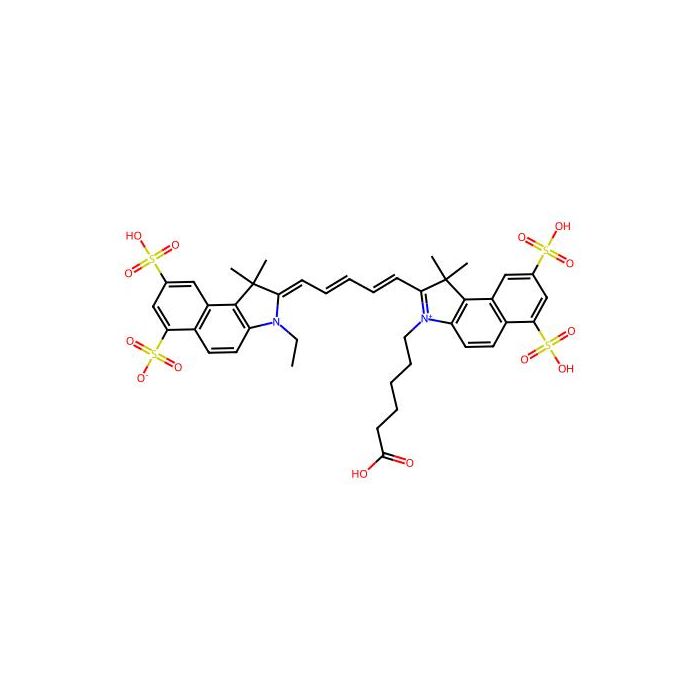
Cy5.5 is a CY dye. CY, short for Cyanine, is a compound consisting of two nitrogen atoms connected by an odd number of methyl units. Cyanine compounds have the characteristics of long wavelength, adjustable absorption and emission, high extinction coefficient, good water solubility and relatively simple synthesis. CY dyes are of en used for the labeling of proteins, antibodies and small molecular compounds. For the labeling of protein antibodies, the combination can be completed through a simple mixing reaction. Below, we introduce the labeling method of protein antibody labeling, which has certain reference significance . Storage: protect from light.
In Vitro
Protocol 1.Protein Preparetion 1) In order to obtain the best labeling effect, please prepare the protein (antibody) concentration as 2 mg/mL. 2) The pH value of protein solution shall be 8.5±0.5. If the pH is lower than 8.0, 1 M sodium bicarbonate shall be used for adjustment. 3) If the protein concentration is lower than 2 mg/mL, the labeling efficiency will be greatly reduced. In order to obtain the best labeling efficiency, it is recommended that the final protein concentration range is 2-10 mg/mL. 4) The protein must be in the buffer without primary amine (such as Tris or glycine) and ammonium ion, otherwise the labeling efficiency will be affected. 2.Dye Preparation (Cy5.5) Add anhydrous DMSO into the vial of Cy5.5 to make a 10 mM stock solution. Mix well by pipetting or vortex. 3.Calculation of dye dosage The amount of Cy5.5 required for reaction depends on the amount of protein to be labeled, and the optimal molar ratio of Cy5.5 to protein is about 10. Example: assuming the required marker protein is 500 μL 2 mg/mL IgG (MW=150,000), use 100 μL DMSO dissolve 1 mg Cy5.5, the required Cy5.5 volume is 5.05 μL, and the detailed calculation process is as follows: 1) mmol (IgG) = mg/mL (IgG) ×mL (IgG) / MW (IgG) =2 mg/mL × 0.5 mL / 150,000 mg/mmol= 6.7×10⁻⁶ mmol 2) mmol (Cy5.5) = mmol (IgG) × 10 = 6.7×10⁻⁶ mmol×10 = 6.7 × 10⁻⁵ mmol 3) μL (Cy5.5) = mmol (Cy5.5) ×MW (Cy5.5) / mg/μL (Cy5.5) = 6.7 ×10⁻⁵ mmol ×753.88 mg/mmol / 0.01 mg/μL = 5.05 μL (Cy5.5) 4.Run conjugation reaction 1) A good volume of freshly prepared 10 mg/mL Cy5.5 is slowly added to 0.5 mL protein sample In solution, gently shake to mix, then centrifuge briefly to collect the sample at the bottom of the reaction tube. Don'tmix well to prevent protein samples from denaturation and inactivation. 2) The reaction tubules were placed in a dark place and incubated gently at room temperature for 60 minutes at intervals.For 10-15 minutes, gently reverse the reaction tubules several times to fully mix the two reactants and raise the bar efficiency. 5.Purify the conjugation The following protocol is an example of dye-protein conjugate purification by using a SepHadex G-25 column. 1) Prepare SepHadex G-25 column according to the manufacture instruction. 2) Load the reaction mixture (From \Run conjugation reaction\) to the top of the SepHadex G-25 column. 3) Add PBS (pH 7.2-7.4) as soon as the sample runs just below the top resin surface. 4) Add more PBS (pH 7.2-7.4) to the desired sample to complete the column purification. Combine the fractions that contain the desired dye-protein conjugate. MCE has not independently confirmed the accuracy of these methods. They are for reference only.
In Vivo
Cy5.5-labeled factor VIIa is developed for imagining cancer. Cy5.5 labeled with these targeting proteins specifically localize to the tumor xenografts for at least 14 days but unconjugated Cy5.5 does not localize to any xenografts or organs. This method of imaging anti-tissue factor in the tumor VECs may be useful in detecting primary tumors and metastases as well as monitoring in vivo therapeutic responses . pH/temperature sensitive magnetic nanogels conjugated with Cy5.5-labled lactoferrin (Cy5.5-Lf-MPNA nanogels) are developed as a promising contrast agent for preoperative MRI and intraoperative fluorescence imaging of glioma. MCE has not independently confirmed the accuracy of these methods. They are for reference only.
规格
规格或纯度
10mM in DMSO
英文名称
Cy5.5
生化机理
Cy5.5 是一种 CY 染料。CY 是 Cyanine 的缩写,是由两个氮原子通过奇数个甲基单元连接而成的化合物。氰基化合物具有波长长、吸收和发射可调、消光系数高的特点。
储存条件
避光,-80℃储存
运输条件
特低温运输
名称和识别符
技术文档
高级数据
质检证书(CoA,COO,BSE/TSE 和分析图谱)
C of A & Other Certificates(BSE/TSE, COO):
输入批号以搜索分析图谱:
此产品的引用文献
溶液计算器
产品仅提供给经过验证的企业、机构和合格的专业人员用于研发。不适用于人类、动物、诊断或治疗。
Copyright © 2023至今 上海阿拉丁生化科技股份有限公司 保留所有权利。


 危险品化学品经营许可证(带存储)