
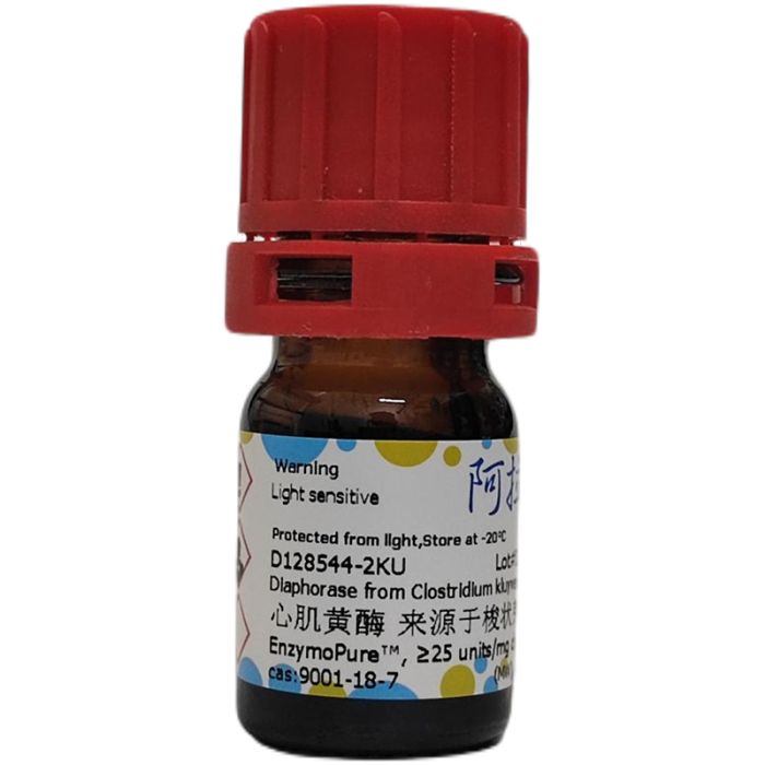
 I2611836
计算溶液所需的质量、体积或浓度。
有货
生物活性
≥25 units/mg dry weight
EnzymoPure™ 级 ,适用于对基线干扰要求严格的色谱和分析工作流程。
避光,-20°C储存。超低温运输 。请查阅批次 COA 获取详细规格。
SDS、COA、产品数据表及规格说明书均可下载。可通过批号查询获取批次 COA。
在色谱分析、有机合成和交叉偶联反应领域已被 1 篇同行评审文献引用。
心肌黄酶 (黄递酶) 催化还原型二 / 三磷酸吡啶核苷酸(氢供体)与氢受体发生反应,氢受体通常为染料的无色还原态。
黄递酶是一类广泛存在的黄素结合酶,可催化各类染料(氢受体)被还原型二、三磷酸吡啶核苷酸(即 NADH、NADPH)还原。首个被纯化的该类酶来源于心肌(Straub,1939)。约二十年后证实,心肌黄递酶即为硫辛酰胺脱氢酶(Massey,1958、1963)。已有文献报道并从多种细菌、植物及哺乳动物器官中纯化得到其他类型黄递酶。
本实验室观测到克氏梭菌细胞部分纯化提取物具备黄递酶活性;该提取物最初被报道含有 NADH、NADPH 氧化酶活性(Ciotti and Kaplan,1957),后续将其应用于脱氢酶与乙醇的偶联比色检测(Teller,1958)。该检测方法基于 2,6‑二氯酚靛酚的褪色反应;后改用四唑染料进行优化,四唑染料被还原后可生成有色产物(Brower and Woodbridge,1970;Nachlas et al.,1960)。
克氏梭菌心肌黄酶 (黄递酶) 酶学性质: 若无特殊说明,以下数据均引自 Kaplan 等人(1969)的文献。
底物特异性:NADH、NADPH 均可作为还原底物;但该酶不催化两种辅酶之间的氢交换。克氏梭菌黄递酶不能还原氧气,也不能还原细胞色素 C。
组成:每分子酶结合一分子黄素单核苷酸(FMN);该酶的氨基酸组成已完成测定。
心肌黄酶 (黄递酶) 酶学性质:
蛋白登录号:Q97E86(丙酮丁醇梭菌)
分子量:24 000
最适 pH:8.5;pH 7.4 与 pH 9.4 条件下酶活下降 50%;以 NADH 或 NADPH 为底物时,最适 pH 保持一致。
激活剂:实验发现,过量黄素单核苷酸(FMN)可轻微促进该酶与二氯酚靛酚的反应。
抑制剂:N‑乙基马来酰亚胺浓度低于 5 mM 即可使黄递酶失活。
稳定性:该酶具备较好稳定性;干粉长期保存需放置冷冻环境;酶溶液尤其需要避光,避免强光照射。
稳定剂:NADH、NADPH、FMN 可保护该酶,抵御尿素、胍引起的蛋白变性。
动力学常数:文献报道,NADH 的 Km 值为 9 × 10⁻⁵ mol/L;NADPH 的 Km 值显著更低。
检测方法
通过比色法测定 2,6‑二氯酚靛酚的还原程度,计算 NADH 的氧化量。本检测方法基于 Edelhoch 等人 (1952) 与 Mahler 等人 (1952) 发表的方法进行修改,优化后反应速率线性更好,初速率与酶浓度呈正比。
1 个酶活单位 (U) 定义:在规定条件下,25 ℃、pH 8.5,每分钟还原 1 μmol 2,6‑二氯酚靛酚所需的酶量。
试剂
1. 0.2 mol/L Tris‑HCl 缓冲液,pH 8.5
2. 0.12 mol/L 氯化锌(溶于 0.005 mol/L 盐酸)
3. 0.006 mol/L NADH:每日现配。
4. 0.0024 mol/L 2,6‑二氯酚靛酚 (DCPIP):每日现配。
5. 酶稀释液:0.2 mol/L Tris‑HCl(pH 8.5),内含 0.3 mol/L 氯化钾、0.025% 黄素单核苷酸 (FMN)、0.025% 牛血清白蛋白。每日现配,避光保存,避免强光照射。
酶样品处理
将冻干粉末用 0.2 mol/L Tris‑HCl pH 8.5 配制成 10 mg/mL。临使用前,用酶稀释液进一步稀释至 0.1‑0.15 mg/mL。
操作步骤 分光光度计设置:波长 600 nm,温度 25 ℃。
向比色皿中按如下移液:
| 试剂 | Test(样品管) | Blank(空白管) |
| 0.2 M Tris‑HCl 缓冲液,pH 8.5 | 2.6 ml | 2.6 ml |
| 0.12 M 氯化锌 | 0.1 ml | 0.1 ml |
| 0.006 M NADH | 0.1 ml | 0.1 ml |
| 0.0024 M 2,6‑二氯酚靛酚 (DCPIP) | 0.1 ml | — |
| 酶稀释液 | — | — |
将比色皿置于分光光度计中孵育,600 nm 下监测空白比色皿吸光度 8‑10 min,从曲线初始线性段计算 ΔA₆₀₀。
向样品比色皿加入 0.1 mL 0.0024 mol/L DCPIP,随即立即加入 0.1 mL 稀释后的酶液。记录 3‑4 min 内 A₆₀₀吸光度下降。取曲线初始线性区间计算 ΔA₆₀₀。
注意:反应线性区间仅维持 1‑2 min。
计算
比活 (U/mg) = (样品每分钟 ΔA₆₀₀ − 空白每分钟 ΔA₆₀₀) ÷[19 × (酶质量 mg ÷ 反应混合液体积 mL)]
Diaphorase catalyzes the reaction of a reduced di- or tri-phosphopyridine nucleotide hydrogen donor with a hydrogen acceptor, usually a dye in the leucoform. The diaphorases are a ubiquitous class of flavin-bound enzymes that catalyze the reduction of various dyes which act as hydrogen acceptors from the reduced form of di- and tri- phosphopyridine nucleotides, i.e., NADH, NADPH. The first such enzyme to be purified was that from heart muscle (Straub 1939). Almost twenty years later heart diaphorase was shown to be identical to lipoyl dehydrogenase (Massey 1958, 1963). Other diaphorases have been described and purified from various bacteria, plants and mammalian organs. Diaphorase activity of a partially purified extract of Clostridium kluyveri cells, originally described as a source of NADH and NADPH oxidase (Ciotti and Kaplan 1957), was observed in this laboratory and its use applied to the coupled, colorimetric determinations of dehydrogenases and ethanol. (Teller 1958). These methods, based on the decolorization of 2,6-dichlorophenolindophenol were improved by the substitution of a tetrazolium dye which becomes chromogenic on reduction. (Brower and Woodbridge 1970; Nachlas et al. 1960).
Characteristics of Diaphorase from Clostridium kluyveri:
Unless otherwise noted, the following data have been obtained from Kaplan et al. (1969).
Specificity: Either NADH or NADPH may be used as reductants. However, no exchange of hydrogen between the coenzymes is catalyzed. Neither oxygen nor cytochrome C is reduced by Cl. kluyveri diaphorase.
Composition: The enzyme contains one molecule of flavin mononucleotide per molecule. The amino acid composition has been determined.
Characteristics of Diaphorase
Protein Accession Number: Q97E86 (Clostridium acetobutylicum)
Molecular Weight: 24,000
Optimal pH: 8.5. At pH 7.4 and 9.4 the activity is reduced 50%. Both NADH and NADPH share this optimum.
Activators: We have found that excess flavin mononucleotide (FMN) appears to stimulate the reaction with dichlorophenol-indophenol slightly. Inhibitors: N-ethylmaleimide inactivates diaphorase at concentrations of under 5 mM.
Stability: Though of reasonable stability, if held for prolonged periods, the dry enzyme should be stored in a freezer. Solutions, especially, should always be kept out of strong light.
Stabilizers: NADH, NADPH and FMN protect the enzyme against denaturation by urea and guanidine.
Constants: Km for NADH was reported to be 9 × 10⁻⁵ and for NADPH it was much lower.
Diaphorase - Assay
The oxidation of NADH is determined by measuring colorimetrically the reduction of 2,6-dichlorophenolindophenol. The assay is based on that described by Edelhoch et al. (1952) and Mahler et al. (1952) with modifications to provide greater linearity of reaction rate and proportionality of initial rate to enzyme concentration. One unit is defined as the amount of enzyme which reduces one micromole of 2,6-dichlorophenolindophenol per minute at 25°C and pH 8.5 under the specified conditions.
Reagents
0.2 M Tris⋅HCl buffer, pH 8.5
0.12 M Zinc chloride in 0.005 M HCl
0.006 M NADH. Prepare fresh daily.
0.0024 M Dichlorophenolindophenol (DCPIP) Prepare fresh daily.
Enzyme diluent: 0.2 M Tris⋅HCl pH 8.5 containing 0.3 M potassium chloride, 0.025% flavin mononucleotide (FMN), and 0.025% bovine serum albumin. Prepare fresh daily and protect from strong light.
Enzyme Dissolve lyophilized powders at 10 mg/ml in 0.2 M Tris⋅HCl pH 8.5. Immediately prior to use, dilute further in enzyme diluent to a concentration of 0.1-0.15 mg/ml.
Procedure
Adjust spectrophotometer to 600 nm and 25°C.
Pipette into cuvettes as follows:
| Test | Blank | |
| 0.2 M Tris⋅HCl, pH 8.5 | 2.6 ml | 2.6 ml |
| 0.12 M ZnCl₂ | 0.1 ml | 0.1 ml |
| 0.006 M NADH | 0.1 ml | 0.1 ml |
| 0.0024 M 2,6‑Dichlorophenolindophenol (DCPIP) | — | 0.1 ml |
| Enzyme diluent | — | — |
Incubate in spectrophotometer at 600 nm and determine A₆₀₀ of blank cuvette for 8‑10 minutes. Determine ΔA₆₀₀ from initial linear portion of curve. To test cuvette add 0.1 ml of 0.0024 M DCPIP followed immediately by 0.1 ml of diluted enzyme. Record decrease in A₆₀₀ for 3‑4 minutes. Calculate ΔA₆₀₀ from the initial linear portion of the curve. The reaction is linear for no more than 1‑2 minutes.
Calculation
Units/mg = [ ΔA₆₀₀/min (Test) − ΔA₆₀₀/min (Blank) ] ÷ [ 19 × (mg enzyme / ml reaction mixture) ]


 危险品化学品经营许可证(带存储)