储存缓冲液:100% Isopropyl Alcohol
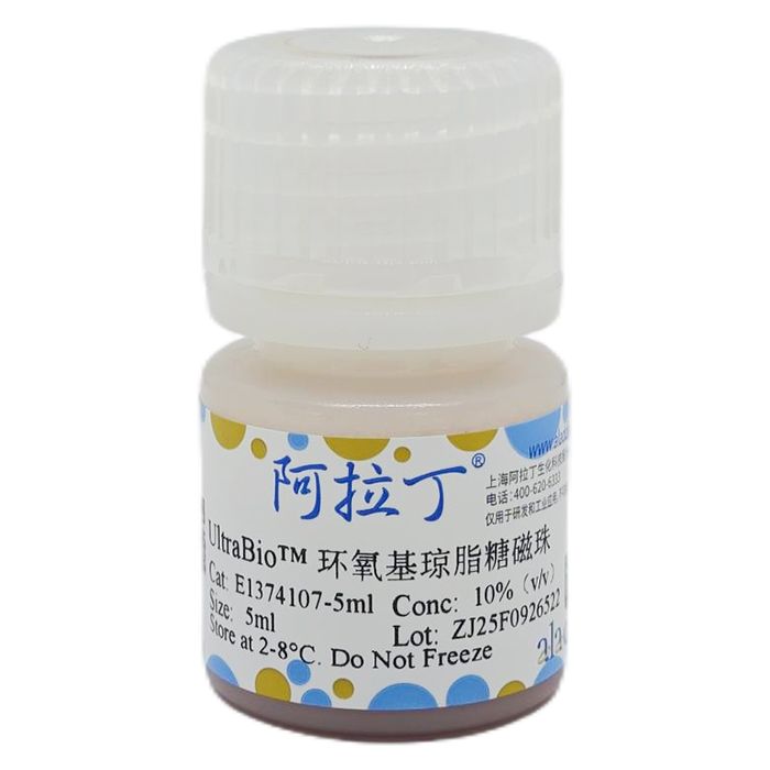
UltraBio™ 环氧基琼脂糖磁珠以交联琼脂糖磁珠为基质,拥有良好的刚性和磁响应性,表面修饰有丰富的环氧基(-Epoxy),能够与含氨基或者巯基的生物配体,如:蛋白质、抗体、寡聚核苷酸和药物分子等通过共价偶联的方法实现结合,是一种分子生物学和医学研究的重要载体工具。可用于各种类型的IP,pull-down,蛋白抗体纯化,免疫检测等领域。
本产品每毫升磁珠悬液含约10%琼脂糖磁珠沉淀。
包装规格:10%(v/v)悬液
参数
| 指标
|
基质
| 琼脂糖
|
配基
| Epoxy
|
| 含量 | Slurry: 10%
|
粒径
| 50 μm (20-80)
|
配基密度
| 40~60 µmol/mL (100%, v/v) |
保存
| 2-8℃ (100% 异丙醇) |
使用说明:
一、偶联准备
1、离心管、磁力架
2、反应缓冲液:
0.2M PB, 1mM EDTA, pH 8.5(适用于含半胱氨酸的蛋白)
0.1M Na₂CO₃-NaHCO₃, pH 10.5(适用于不含半胱氨酸的蛋白)
3、清洗缓冲液:50mM Tris-HCl, 0.15M NaCl, pH 7.2
4、封闭液:0.25M Ethanolamine, 0.1M Tris-HCl, pH 10.5
5、需要偶联的目标蛋白、抗体、寡核苷酸等生物配体。
二、操作流程
1、将磁珠混合均匀,取1mL 磁珠 (10%, V/V) 加入到2mL离心管中,磁性分离去除上清液。
注:“磁性分离”指将离心管置于外加磁场中,至磁珠吸附完全,约需要30s。
2、加入500µL反应缓冲液,混合重悬磁珠,磁性分离去除上清液(重复3次)。
3、加入0.5-1mg抗体或者蛋白质溶液(提前用100-500µL 反应缓冲液溶解),室温旋转混合12-16h(37℃反应12h效果更好),磁性分离去除上清液。
注:可加入极少量的还原剂TCEP,反应缓冲液通氮气除氧,效果更佳。
4、加入1mL封闭缓冲液,室温旋转混合3h(37℃反应效果更好),磁性分离去除上清液。
注:此步骤也可以用 0.2% BSA 分散于结合液中进行封闭处理。
5、(选做)
用0.5 M NaCl, 0.1 M 的乙酸-乙酸钠缓冲液(pH 4.0)、0.5 M NaCl, 0.1M 的硼酸-四硼酸钠缓冲液(pH 8.0)和去离子水充分洗涤(重复交替清洗3次)。
注:此步骤可在最大程度上去除非特异性结合抗体。
6、加入1mL清洗缓冲液,混合重悬磁珠,磁性分离去除上清液(重复3次)
7、将上述磁珠分散于0.5mL PBS, pH 7.4短期保存,或分散于PBS, pH 7.4, 0.1% BSA, 0.02% NaN₃长期保存。
注意事项:
1、偶联过程中不应含有除目标配体外含伯胺基团的物质,如:甘氨酸、BSA、Tris-HCl等,请注意您的样品溶液是否含有上述物质。
2、磁珠使用和保存过程中应避免冻融。
3、UltraBio™ 环氧基琼脂糖磁珠不可干燥。
4、不同抗体和蛋白与UltraBio™ 环氧基琼脂糖磁珠的结合能力不同,客户可自行优化加入不同的抗体或者蛋白量。
UltraBio™ Epoxy Magnetic Agarose Beads utilize cross-linked agarose magnetic beads as the matrix, offering excellent rigidity and magnetic responsiveness. The beads are surface-modified with abundant epoxy groups (-Epoxy), enabling covalent coupling with biological ligands containing amino or thiol groups, such as proteins, antibodies, oligonucleotides, and drug molecules. This product serves as a crucial carrier tool for molecular biology and medical research, applicable in various applications including immunoprecipitation (IP), pull-down assays, protein/antibody purification, and immunoassays.
Parameter
| Specification
|
Matrix
| Agarose
|
Ligand
| Epoxy
|
Content
| Slurry: 10%
|
Particle Size
| 50 μm (20-80)
|
Ligand Density
| 40~60 µmol/mL (100%, v/v) |
Storage
| 2-8°C (in 100% Isopropanol) |
Instructions for Use:
I. Coupling Preparation
Equipment: Microcentrifuge tubes, Magnetic separation rack.
Reaction Buffer:
0.2 M PB, 1 mM EDTA, pH 8.5 (For proteins containing cysteine)
0.1 M Na₂CO₃-NaHCO₃, pH 10.5 (For proteins without cysteine)
Wash Buffer: 50 mM Tris-HCl, 0.15 M NaCl, pH 7.2
Blocking Solution: 0.25 M Ethanolamine, 0.1 M Tris-HCl, pH 10.5
Target Ligand: Biological ligand for coupling (e.g., target protein, antibody, oligonucleotide).
II. Coupling Procedure
Resuspend the magnetic beads thoroughly. Transfer 1 mL of bead slurry (10%, V/V) to a 2 mL microcentrifuge tube. Perform magnetic separation and discard the supernatant.
Add 500 µL of the appropriate Reaction Buffer. Resuspend the beads thoroughly. Perform magnetic separation and discard the supernatant. Repeat this wash step twice more (3 washes total).
Add 0.5-1 mg of the antibody or protein solution (pre-dissolved in 100-500 µL Reaction Buffer). Incubate with end-over-end rotation at room temperature for 12-16 hours (reaction at 37°C for 12 hours is recommended for better results). Perform magnetic separation and discard the supernatant.
Add 1 mL of Blocking Solution. Incubate with end-over-end rotation at room temperature for 3 hours (reaction at 37°C is recommended for better results). Perform magnetic separation and discard the supernatant.
Optional:
Wash the beads thoroughly by alternating between:
0.5 M NaCl in 0.1 M Sodium Acetate-Acetic Acid Buffer, pH 4.0
0.5 M NaCl in 0.1 M Boric Acid-Sodium Tetraborate Buffer, pH 8.0
Deionized Water
Perform magnetic separation and discard the supernatant after each wash. Repeat this alternating wash cycle twice more (3 cycles total).
Note: This step helps minimize non-specifically bound antibodies/proteins.
Add 1 mL of Wash Buffer. Resuspend the beads thoroughly. Perform magnetic separation and discard the supernatant. Repeat this wash step twice more (3 washes total).
Resuspend the coupled beads in 0.5 mL of PBS, pH 7.4 for short-term storage OR in PBS, pH 7.4, containing 0.1% BSA and 0.02% NaN₃ for long-term storage.
Precautions:
Avoid Primary Amines: The coupling reaction should NOT contain substances with primary amine groups other than the target ligand (e.g., glycine, BSA, Tris-HCl). Please verify if your sample solution contains such interfering agents.
Avoid Freeze-Thaw: Beads should be protected from freeze-thaw cycles during use and storage.
Do Not Dry: UltraBio™ Epoxy Magnetic Agarose Beads must not be allowed to dry out.
Ligand Optimization: The binding capacity of UltraBio™ Epoxy Magnetic Agarose Beads varies for different antibodies and proteins. Users may need to optimize the amount of antibody or protein added.

 ZJ26F1039790


 危险品化学品经营许可证(带存储)