本试剂盒是一种基于点击化学(Click Chemistry)增强型TUNEL技术的细胞凋亡检测试剂盒,用于高灵敏度检测凋亡细胞中的DNA片段化。
细胞凋亡时,内源性核酸内切酶被激活,将核小体间的基因组DNA切割成180-200 bp的片段,暴露出大量的3'-OH末端。本试剂盒利用末端脱氧核苷酸转移酶(TdT)催化EdUTP(5-ethynyl-dUTP,一种炔基修饰的dUTP掺入到断裂DNA的3'-OH末端。随后,在一价铜离子催化下,EdUTP中的炔基与Azide 488(叠氮化物标记的AF488荧光探针)发生高特异性点击反应(Click reaction),使凋亡细胞DNA断裂处被绿色荧光标记。
产品组分
E1456515 | 组分 | 20 T | 50 T | 储存条件 |
E1456515A | Recombinant TdT Enzyme | 20 μL | 50 μL | -20℃. |
E1456515B | 5-EdUTP | 40 μL | 100 μL | -20℃. |
E1456515C | 反应缓冲液(5×) | 200 μL | 500 μL | -20℃. |
E1456515D | Azide 488 | 20 μL | 50 μL | -20℃. 避光保存. |
E1456515E | CuSO₄
| 1 mL | 2.5 mL | -20℃. |
E1456515F | Cu(Ⅰ)保护剂 | 200 μL | 500 μL | -20℃. |
E1456515G | Click Additive | 20mg | 50mg | -20℃. |
E1456515H | Hoechst 33342 (1000×) | 20 μL | 50 μL | -20℃. 避光保存. |
使用方法
1. 实验材料(自备)
1) 1×PBS (pH 7.4);
2) 4%多聚甲醛(固定液,PBS配制);
3) 通透液:0.1-0.2% Triton X-100(PBS配制)或20 μg/mL蛋白酶K;
4) DNase I(用于阳性对照处理);
5) 抗荧光淬灭封片剂;
6) 盖玻片、载玻片、湿盒;
7) 蒸馏水或去离子水;
8) 二甲苯、梯度乙醇(石蜡切片用);
2. 细胞爬片/细胞涂片
1) 洗涤:用PBS洗涤细胞一次。
2) 固定:加入适量4%多聚甲醛,室温固定30分钟或4°C固定1-2小时。
3) PBS洗涤3次,每次5分钟。
4) 通透:加入适量0.1% Triton X 100,室温通透15分钟。PBS 洗涤2次。
5) 转步骤6。
3. 石蜡组织切片
1) 石蜡组织切片脱蜡与水化:二甲苯中脱蜡5-10分钟。换用新鲜的二甲苯,再脱蜡5-10分钟。无水乙醇5分钟。90%乙醇2分钟。70%乙醇2分钟,蒸馏水2分钟。
2) 通透:将蛋白酶K (P274341,2 mg/mL)用PBS稀释至终浓度为20 μg/mL,每个样本滴加100 μL覆盖样本,37℃孵育15-30分钟。
注:孵育时间过长会增加组织切片在后续洗涤步骤中从载玻片上脱落的风险,过短则可能造成透性处理不充分,影响标记效率;不同组织的最佳作用温度和时间需自行摸索。
3) 洗涤:PBS洗涤3次,每次5分钟。
注:这一步必须把蛋白酶K洗涤干净,否则会严重干扰后续的标记反应。
4) 转步骤6。
4. 冰冻组织切片
1) 固定:将切片样本浸入4%多聚甲醛中,室温固定30分钟。PBS缓慢漂洗3次,每次5分钟。
2) 通透:将蛋白酶K (2 mg/mL)用PBS稀释至终浓度为20 μg/mL,每个样本滴加100 μL覆盖样本,37℃孵育10分钟。
注:孵育时间过长会增加组织切片在后续洗涤步骤中从载玻片上脱落的风险,过短则可能造成透性处理不充分,影响标记效率;不同组织的最佳作用温度和时间需自行摸索。
3) PBS漂洗切片3次,每次5分钟。用免疫组化笔圈好样本轮廓,以便下游标记。将处理好的切片放在湿盒中保持湿润。
注:若在后续实验操作中发现免疫组化笔画的轮廓圈被破坏,需及时补画。
4) 转步骤6。
5. 阳性对照(选做)
1) 将DNase I (10 U/μL)用DNase I 稀释液至终浓度为100 U/mL。
2) 加入50 μL DNase I 到已处理的样品上,室温孵育10分钟。
3) 弃掉DNase I,PBS洗涤2次。
6. TUNEL反应液(现配现用)
1) 平衡:每个样本滴加100 μL TdT Reaction Buffer,37°C湿盒中平衡10-30分钟。
2) 参考以下比例配制工作液,配制时须在冰上避光操作并充分混合均匀,每个样本(50 μL体系)所需体积计算:
TUNEL反应液组分 | 1个样品 | 5个样品 | 10个样品 |
Recombinant TdT Enzyme | 1 μL | 5 μL | 10 μL |
5-EdUTP | 2 μL | 10 μL | 20 μL |
反应缓冲液(5×) | 10 μL | 50 μL | 100 μL |
ddH₂O | 37 μL | 185 μL | 370 μL |
总体积 | 50 μL | 250 μL | 500 μL |
注:1) TUNEL反应缓冲液和TdT Enzyme需完全溶解混匀后使用。2)步骤以96孔板一个孔或一个切片的检测为例进行说明。48孔板、24孔板的一个孔,可以参考96孔板的用量,12孔板或6孔板的一个孔液体用量可以适当加大。
3) 标记反应:吸除平衡液(注意勿干片),每个样本滴加50 μL标记工作液,盖上盖玻片或封口膜,放入湿盒中37°C避光孵育60分钟。
4) 终止反应:PBS洗涤3次,每次5min。
7. 点击反应(荧光标记)
1) 配制Click Additive Solution:对于20T包装,用0.36 mL超纯水溶解试剂盒中提供的1管Click Additive,混匀至全部溶解,即为Click Additive Solution;对于50T包装,加入0.9 mL超纯水溶解试剂盒中提供的1管Click Additive,混匀至全部溶解,即为Click Additive Solution。配制后可以适当分装后在-20ºC保存。
2) 参考以下比例配制Click反应工作液(现配现用),每个样本(50 μL体系)所需体积计算:
Click反应液组分 | 1个样品 | 5个样品 | 10个样品 |
Azide 488 | 1 μL | 5 μL | 10 μL |
CuSO₄ | 2 μL | 10 μL | 20 μL |
Cu(Ⅰ)保护剂 | 5 μL | 25 μL | 50 μL |
Click Additive Solution | 10 μL | 50 μL | 100 μL |
ddH₂O | 33 μL | 165 μL | 330 μL |
总体积 | 50 μL | 250 μL | 500 μL |
注:50 μL生物素标记液适合涂片、切片或96孔板、48孔板、24孔板或12孔板的一个孔,如果是6孔板中的一个孔生物素标记液宜使用100 μL。孵育时注意防护防止工作液挥发。
3) 每个样本滴加50 μL Click反应工作液,室温避光孵育30 min。
4) PBS洗涤3次,每次5 min。
8. 细胞核复染与封片
1) Hoechst 33342染色:用PBS按1:1000稀释Hoechst 33342,每个样本滴加适量稀释液,室温避光孵育5-10分钟。
2) PBS漂洗3次,每次5分钟。
3) 滴加抗荧光淬灭封片剂,盖玻片封片。
4) 荧光显微镜下立即观察拍照;若无法立即观察,4°C避光保存。
注意事项
1. 推荐使用4%多聚甲醛(PBS配制),避免使用乙醇、甲醇或酸性固定液,否则会导致标记效率显著降低。
2. 蛋白酶K通透时间需优化,过长易导致切片脱落,过短则标记效率低。建议首次实验进行时间梯度摸索。
3. 所有孵育步骤必须保持样本湿润,建议使用湿盒,孵育时可覆盖盖玻片或封口膜。
4. Azide 488和Hoechst 33342荧光染料见光易淬灭,所有荧光相关步骤需避光。
5. TdT对温度敏感,使用前后需严格置于冰上,避免反复冻融。
6. 标记反应后充分洗涤可有效降低背景。
7. 极少数类型的细胞凋亡不发生DNA断裂,不适用TUNEL法检测;个别坏死细胞也可能呈TUNEL阳性,建议结合其他凋亡指标联合判断。
8. 本产品仅限科研使用,不得用于临床诊断或治疗。
结果展示

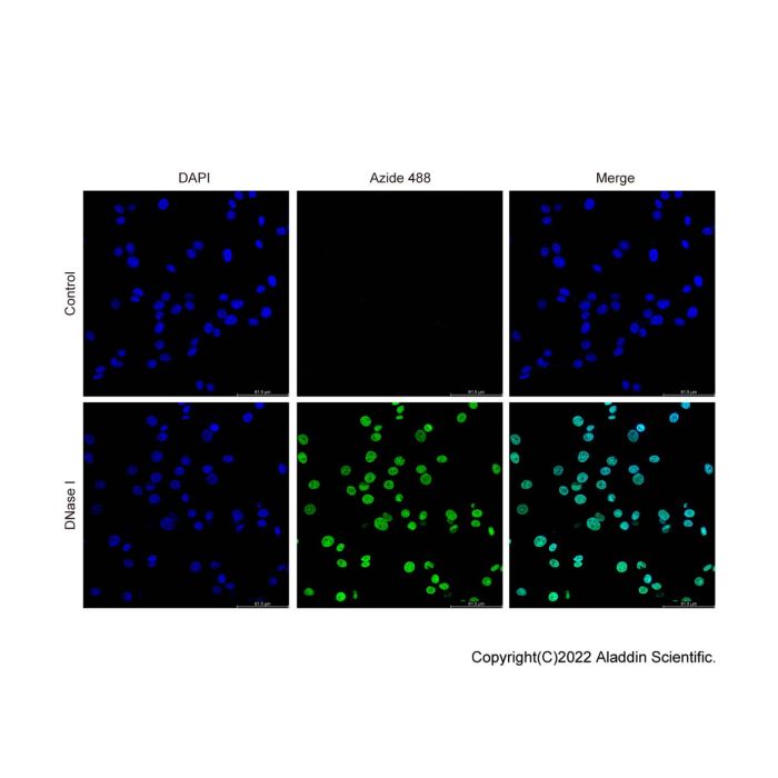
图1. TUNEL细胞凋亡检测试剂盒检测HeLa细胞的效果图。对HeLa细胞进行不同处理后的TUNEL染色结果。上层(对照组):未经处理的正常HeLa细胞;下层(DNase I组):经DNase I (D106200)处理的HeLa细胞,TUNEL阳性细胞呈绿色荧光(Azide 488,伪彩显示),细胞核以Hoechst 33342复染呈蓝色荧光。Merge图为各通道叠加图像。
This kit is an apoptosis detection assay based on click chemistry-enhanced TUNEL technology, designed for high-sensitivity detection of DNA fragmentation in apoptotic cells.
During apoptosis, endogenous endonucleases are activated and cleave genomic DNA at the internucleosomal linker regions into 180-200 bp fragments, generating abundant 3′-OH termini. This kit utilizes terminal deoxynucleotidyl transferase (TdT) to catalyze the incorporation of EdUTP (5-ethynyl-dUTP, an alkyne-modified dUTP) onto the 3′-OH ends of fragmented DNA. Subsequently, in the presence of a monovalent copper ion catalyst, the alkyne group in EdUTP undergoes a highly specific click reaction with Azide 488 (an AF488 fluorescent probe conjugated to an azide moiety), thereby labeling the DNA break sites in apoptotic cells with green fluorescence.
Components
T1456509 | Components | 20 T | 50 T | Storage |
T1456509A | Recombinant TdT Enzyme | 20 μL | 50 μL | -20°C. |
T1456509B | 5-EdUTP | 40 μL | 100 μL | -20°C. |
T1456509C | Reaction Buffer(5×) | 200 μL | 500 μL | -20°C. |
T1456509D | Azide 488 | 20 μL | 50 μL | -20°C. Store in the dark |
T1456509E | CuSO₄ | 1 mL | 2.5 mL | -20°C. |
T1456509F | Copper Protectant | 200μL | 500μL | -20°C. |
T1456509G | Click Additive | 20mg | 50mg | -20°C. |
T1456509H | Hoechst 33342(1000×) | 20 μL | 50 μL | -20°C. Store in the dark |
Protocol
1. Experimental Materials (Self-Provided)
1) 1×PBS (pH 7.4)
2) 4% Paraformaldehyde (fixative solution, prepared in PBS).
3) Permeabilization solution: 0.1-0.2% Triton X-100 (prepared in PBS) or 20 μg/mL Proteinase K.
4) DNase I (for positive control treatment).
5) Anti-fluorescence fading mounting medium.
6) Coverslips, glass slides, and humidified chamber.
7) Distilled water or deionized water.
8) Xylene and graded ethanol (for paraffin-embedded sections).
2. Cell Smears/Cytospin Preparations
1) Wash: Wash the cells once with PBS.
2) Fixation: Add an appropriate volume of 4% paraformaldehyde and fix at room temperature for 30 minutes or at 4°C for 1-2 hours.
3) Wash three times with PBS, 5 minutes each.
4) Permeabilization: Add an appropriate volume of 0.1% Triton X-100 and permeabilize at room temperature for 15 minutes. Wash twice with PBS.
5) Continue to Step 6.
3. Paraffin-Embedded Tissue Sections
1) Deparaffinization and Rehydration: Deparaffinize in xylene for 5-10 minutes. Transfer to fresh xylene and repeat for another 5-10 minutes. Then incubate in absolute ethanol for 5 minutes, 90% ethanol for 2 minutes, 70% ethanol for 2 minutes, and distilled water for 2 minutes.
2) Permeabilization: Dilute Proteinase K (P274341, 2 mg/mL) with PBS to a final concentration of 20 μg/mL. Add 100 μL of this solution to each sample to cover the tissue, and incubate at 37°C for 15-30 minutes.
Note: Prolonged incubation may increase the risk of tissue detachment from slides during subsequent washing steps, whereas insufficient incubation may result in inadequate permeabilization and compromised labeling efficiency. The optimal incubation temperature and duration should be empirically determined for different tissue types.
3) Wash: Wash three times with PBS, 5 minutes each.
Note: It is critical to thoroughly remove Proteinase K at this step, as residual enzyme activity will severely interfere with subsequent labeling reactions.
4) Continue to Step 6.
4. Frozen Tissue Sections
1) Fixation: Immerse the section slides in 4% paraformaldehyde and fix at room temperature for 30 minutes. Gently rinse three times with PBS, 5 minutes each.
2) Permeabilization: Dilute Proteinase K (2 mg/mL) with PBS to a final concentration of 20 μg/mL. Add 100 μL of this solution to each sample to cover the tissue, and incubate at 37°C for 10 minutes.
Note: Prolonged incubation may increase the risk of tissue detachment from slides during subsequent washing steps, whereas insufficient incubation may result in inadequate permeabilization and compromised labeling efficiency. The optimal incubation temperature and duration should be empirically determined for different tissue types.
3) Rinse the sections three times with PBS, 5 minutes each. Use a hydrophobic barrier pen to delineate the sample contours for downstream labeling. Keep the processed sections in a humidified chamber to maintain moisture.
Note: If the hydrophobic barrier ring is found to be damaged during subsequent experimental procedures, it should be promptly reapplied.
4) Continue to Step 6.
5. Positive Control (Optional)
1) Dilute DNase I (10 U/μL) with DNase I dilution buffer to a final concentration of 100 U/mL.
2) Add 50 μL DNase I solution onto the pretreated sample, and incubate at room temperature for 10 minutes.
3) Remove DNase I solution and wash the sample twice with PBS.
6. TUNEL Reaction Labeling Master Mix(Prepare Fresh Immediately Before Use)
1) Equilibration: Dilute the 5× Reaction Buffer with ddH₂O to prepare 1× TdT Reaction Buffer. Add 100 μL of the 1× TdT Reaction Buffer to each sample, and incubate in a humidified chamber at 37°C for 10-30 minutes to allow equilibration.
2) Prepare the labeling working solution according to the proportions provided below. All operations must be performed on ice, protected from light, and thoroughly mixed. Calculate the required volume for each sample (50 μL system) as follows:
Components of the TUNEL Labeling Working Solution | One sample | Five samples | Ten samples |
Recombinant TdT Enzyme | 1 μL | 5 μL | 10 μL |
5-EdUTP | 2 μL | 10 μL | 20 μL |
Reaction Buffer(5×) | 10 μL | 50 μL | 100 μL |
ddH₂O | 37 μL | 185 μL | 370 μL |
Total Volume | 50 μL | 250 μL | 500 μL |
Note: 1) The TUNEL Reaction Buffer and TdT Enzyme must be completely dissolved and mixed thoroughly before use. 2) The following procedure is described for one well of a 96-well plate or one tissue section. For one well of a 48-well or 24-well plate, the volumes used for a 96-well plate may be adopted as a reference. For one well of a 12-well or 6-well plate, the reagent volumes should be increased appropriately to ensure complete coverage of the sample.
3) Labeling Reaction: Aspirate the equilibration buffer (taking care not to allow the slides to dry out). Add 50 μL of the labeling working solution to each sample, cover with a coverslip or sealing film, and incubate in a humidified chamber at 37°C in the dark for 60 minutes.
4) Termination Reaction: Wash 3 times with PBS, 5 minutes each wash.
7. Click Reaction (Fluorescence Labeling)
1) Preparation of Click Additive Solution: For the 20T kit, dissolve the provided 1 vial of Click Additive in 0.36 mL of ultrapure water, and mix thoroughly until completely dissolved to obtain the Click Additive Solution. For the 50T kit, dissolve the provided 1 vial of Click Additive in 0.9 mL of ultrapure water, and mix thoroughly until completely dissolved to obtain the Click Additive Solution. After preparation, the solution may be appropriately aliquoted and stored at -20°C.
2) Prepare the Click Reaction Working Solution according to the proportions below (prepare fresh immediately before use). The required volume is calculated per sample (50 μL system):
Components of the Click Reaction Working Solution | One sample | Five samples | Ten samples |
Azide 488 | 1 μL | 5 μL | 10 μL |
CuSO₄ | 2 μL | 10 μL | 20 μL |
Cu(Ⅰ)保护剂 | 5 μL | 10 μL | 50 μL |
Click Additive Solution | 10 μL | 50 μL | 100 μL |
ddH₂O | 33 μL | 165 μL | 330 μL |
Total Volume | 50 μL | 250 μL | 500 μL |
Note: 50 μL of the biotin labeling solution is suitable for smears, sections, or one well of a 96-well, 48-well, 24-well, or 12-well plate. For one well of a 6-well plate, 100 μL of the biotin labeling solution is recommended. During incubation, take precautions to prevent evaporation of the working solution.
3) Add 50 μL of the Click Reaction Working Solution to each sample and incubate at room temperature in the dark for 30 minutes.
4) Wash 3 times with PBS, 5 minutes each wash.
8. Nuclear Counterstaining and Mounting
1) Hoechst 33342 Staining: Dilute Hoechst 33342 with PBS at a ratio of 1:1000. Add an appropriate amount of the diluted solution to each sample and incubate at room temperature in the dark for 5-10 minutes.
2) Rinse 3 times with PBS, 5 minutes each wash.
3) Add an anti-fade mounting medium and seal with a coverslip.
4) Observe and capture images immediately under a fluorescence microscope. If immediate observation is not possible, store at 4°C in the dark.
Notes
1. It is recommended to use 4% paraformaldehyde (prepared in PBS). Avoid using ethanol, methanol, or acidic fixatives, as they may significantly reduce labeling efficiency.
2. The permeation time with Proteinase K requires optimization. Excessive permeation may cause sections to detach, while insufficient permeation may result in low labeling efficiency. For first-time experiments, a time-gradient optimization is recommended.
3. All incubation steps must be performed with the samples kept moist. The use of a humidified chamber is recommended, and coverslips or sealing film may be applied during incubations.
4. Azide 488 and Hoechst 33342 fluorescent dyes are light-sensitive and prone to photobleaching. All fluorescence-related steps must be carried out in the dark.
5. TdT is temperature-sensitive. Strictly keep it on ice before and after use, and avoid repeated freeze-thaw cycles.
6. Thorough washing after the labeling reaction can effectively reduce background staining.
7. Apoptosis in a very small number of cell types does not involve DNA fragmentation and is therefore not detectable by the TUNEL method. Occasional necrotic cells may also show TUNEL positivity. It is advisable to combine TUNEL results with other apoptosis indicators for confirmatory assessment.
8. This product is for research use only and shall not be used for clinical diagnosis or treatment.
Results Presentation
Figure 1. Detection of HeLa cells using the TUNEL Apoptosis Detection Kit. HeLa cells were subjected to TUNEL staining under different conditions. Top panel (Control): Untreated normal HeLa cells. Bottom panel (DNase I): HeLa cells treated with DNase I (D106200) as a positive control for DNA fragmentation. TUNEL-positive cells are shown in green (Azide 488, pseudocolored), and cell nuclei are counterstained with Hoechst 33342 (blue). Merged images (Merge) are shown in the rightmost column.

 ZJ26F0938998


 危险品化学品经营许可证(带存储)