艾滋病病毒 (HIV) 是单链 RNA 病毒,属于逆转录病毒 (retrovirus) 科,慢病毒 (lentivirus) 亚科,HIV 分为 HIV-1 型和 HIV-2 型,世界各地的 AIDS 主要由 HIV-1 型引起,HIV-2 型在西非洲呈地方性流行。HIV-1 可进一步分为不同的亚型,包括 M 亚型组 (主要亚型组)、O 亚型组和 N 亚型组。HIV 呈 20 面体立体对称球形颗粒,表面有刺突状结构的糖蛋白,直径约为 100nm,分为包膜与核心两部分。病毒的最外层为包膜,其中嵌有外膜糖蛋白 (gp120) 和跨膜糖蛋白 (gp41)。HIV-1 的跨膜蛋白为 gp41,HIV-2 的跨膜蛋白为 gp36。
实验原理:
本试剂盒采用双抗原夹心法酶联免疫吸附试验 (ELISA)。往预先包被有免疫缺陷病毒 I+II 型捕获抗原的微孔中,依次加入样本、阳性对照品、阴性对照品、HRP 标记的检测抗原,中间经过温育和洗涤,用底物 TMB 显色,TMB 在过氧化物酶 (HRP) 的催化下转化成蓝色,并在酸的作用下转化成最终的黄色。颜色的深浅和样本中的免疫缺陷病毒 I+II 型抗体 (HIV I+II Ab) 呈正相关。用酶标仪在 450nm 波长下测定吸光度 (OD 值),判断样本阴阳性。
特异性:可检测样本中的 HIV I+II Ab,且与其类似物无明显交叉反应。
注意事项:
1. 严格按照规定的时间和温度进行温育以保证准确结果。所有试剂都必须在使用前达到室温20-25℃。使用后立即冷藏试剂。
2. 洗板不正确可能导致不准确的结果。在加入底物前确保尽量吸干孔内液体。整个过程中不要让微孔干燥时间过长。
3. 清洁板底残留的液体和手指印,否则影响OD值。
4. 底物显色液应呈无色的颜色,已经变蓝的底物液不能使用。
5. 避免试剂和样本的交叉污染以免造成错误结果。
6. 在储存和温育时避免强光直接照射。
7. 任何反应试剂不能接触漂白溶剂或漂白溶剂所散发的强烈气体。任何漂白成分都会破坏试剂盒中试剂的生物活性。
8. 不能使用过期产品,不同货号和批号组分不得混用。
9. 试剂盒以外来源的重组蛋白可能会出现与本试剂盒抗体不匹配而不被识别的情况。
10. 如果可能传播疾病,所有的样本都应管理好,按照规定的程序处理样本和检测装置。
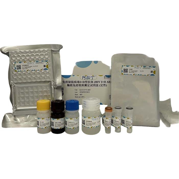
试剂盒组成:
项目号
| 物理外观 | 组件名称
| 48T | 96T | 储存
| | EJ1515775A | — | 预包被酶标板 | 48孔 | 96孔 | 2-8℃. | | EJ1515775B | 固体 | 阳性对照品 | 1支 | 2支 | 2-8℃. | | EJ1515775C | 固体 | 阴性对照品 | 1支 | 2支 | 2-8℃. | | EJ1515775D | 液体 | 通用稀释液 | 1×20 mL | 2×20 mL | 2-8℃. | | EJ1515775E | 液体 | 浓缩HRP-抗原 (100×) | 60 μL | 120 μL | 2-8℃. | | EJ1515775F | 液体 | 洗涤液 (20×) | 1×10 mL | 2×10 mL | 2-8℃. | | EJ1515775G | 液体 | 底物 (TMB) | 5 mL | 10 mL | 2-8℃. | | EJ1515775H | 液体 | 终止液 | 3 mL | 6 mL | 2-8℃. | | EJ1515775I | — | 封板膜 | 4张 | 4张 | 2-8℃. |
备注: EJ1515775B、 EJ1515775C、EJ1515775E、 EJ1515775F需按说明书进行稀释 |
样本处理及要求:
1. 试剂盒检测范围不等同于样本中待测物的浓度范围,建议实验前通过相关文献预估样本中待测物的浓度并通过预实验确定样本的实际浓度。如果样本中待测物浓度过高或过低,请对样本做适当的稀释或浓缩。
2. 若所检样本不在说明书所列样本类型之中,建议做预实验验证其检测有效性。
3. 血清:将收集于血清分离管的全血样本在室温放置 2 小时或 2–8℃过夜,然后 1000×g 离心 20 分钟,取上清即可,或将上清置于 - 20℃或 - 80℃保存,但应避免反复冻融。
4. 血浆:用 EDTA 或肝素作为抗凝剂采集样本,并将样本在采集后的 30 分钟内于 2–8℃ 1000×g 离心 15 分钟,取上清即可检测,或将上清置于 - 20℃或 - 80℃保存,但应避免反复冻融。
5. 组织匀浆:用预冷的 PBS (0.01M, pH=7.4) 冲洗组织,去除残留血液(匀浆中裂解的红细胞会影响检测结果),称重后将组织剪碎。将剪碎的组织与对应体积的 PBS(一般按 1:9 的重量体积比,比如 1g 的组织样本对应 9mL 的 PBS,具体体积可根据实验需要适当调整,并做好记录。推荐在 PBS 中加入蛋白酶抑制剂)加入玻璃匀浆器中,于冰上充分研磨或匀浆机研磨。为了进一步裂解组织细胞,可以对匀浆液进行超声破碎,或反复冻融。最后将匀浆液于 5000×g 离心 5~10 分钟,取上清检测。
6. 细胞培养物上清:请 1000×g 离心 20 分钟,取上清即可检测,或将上清置于 - 20℃或 - 80℃保存,但应避免反复冻融。
7. 细胞裂解液:贴壁细胞用预冷 PBS 轻轻清洗,然后用胰蛋白酶消化,1000×g 离心 5 分钟后收集细胞;悬浮细胞可直接离心收集。收集的细胞用预冷 PBS 清洗 3 次,每 1×10⁶个细胞中加入 150–200μL PBS 重悬(推荐在 PBS 中加入蛋白酶抑制剂;若含量很低可适当减少 PBS 体积)并通过反复冻融或超声使细胞破碎。将提取液于 2–8℃,1500×g 离心 10 分钟,取上清检测。
8. 其它生物样本:1000×g 离心 20 分钟,取上清即可检测。
9. 样本外观:样本应清澈透明,悬浮物应离心去除。
10. 样本保存:样本收集后若在 1 周内进行检测的可保存于 4℃,若不能及时检测,请按一次使用量分装,冻存于 - 20℃(1 个月内检测),或 - 80℃(6 个月内检测),避免反复冻融,样本溶血会影响最后检测结果,因此溶血样本不宜进行此项检测。
实验所需自备试验器材:
1. 酶标仪 (450nm)
2. 高精度移液器及吸头:0.5–10μL、5–50μL、20–200μL、200–1000μL
3. 37℃恒温箱
4. 蒸馏水或去离子水
检测前准备工作:
1. 请提前 10 分钟从冰箱中取出试剂盒,平衡至室温。
2. 阳性和阴性对照品工作液配制:分别加入 1mL 通用稀释液至冻干对照品中,静置 15 分钟待其完全溶解后轻轻混匀。
3. HRP-抗原工作液配制:使用前 15 分钟将 100× 浓缩HRP-抗原于 1000×g 离心 1 分钟,以通用稀释液将 100× 浓缩HRP-抗原稀释成 1× 工作浓度 (例:10μL 浓缩液 + 990μL 通用稀释液),现配现用。
4. 1× 洗涤液配制:取 10mL 20× 洗涤液到 190mL 蒸馏水中(从冰箱中取出的浓缩洗涤液可能有结晶,属于正常现象,可放置室温,待结晶完全溶解后再配制)。
操作步骤:
1. 从室温平衡 10 分钟后的铝箔袋中取出所需板条,剩余板条用自封袋密封放回 4℃。
2. 加样:分别将样本或阳性、阴性对照品按照 100μL 每孔加入相应孔中,空白孔加入 100μL 通用稀释液。盖上封板膜后 37℃温育 60 分钟。(建议:将待测样本用通用稀释液最低稀释 1 倍后再加入酶标板内测试,从而减少基质效应对测试结果的影响。所有的待测样本和阳性、阴性对照品在检测中建议设立复孔)。
3. 洗板:弃去液体,每孔加入 300μL 1× 洗涤液,静置 1 分钟,甩去洗涤液,吸水纸上拍干,如此重复洗板 3 次(也可用洗板机洗板)。
4. 加HRP-抗原:每孔加入 100μL HRP-抗原工作液,盖上封板膜后 37℃温育 30 分钟。
5. 洗板:弃去液体按步骤 3 洗涤方法,洗板 5 次。
6. 加底物:每孔加入 90μL 底物 (TMB),盖上封板膜,37℃避光温育 15 分钟。
7. 加终止液:取出酶标板,每孔直接加入 50μL 终止液,立即在 450nm 波长处测定各孔的 OD 值。
实验结果计算:
计算公式:
S/P值=(待检样本OD值-阴性对照品平均OD值)/(阳性对照品平均OD值-阴性对照品平均OD值)
(注:“P” 表示阳性对照品;“N” 表示阴性对照品;“S” 等表示待检样本)
判定:
1. 试验结果有效的条件是:阳性对照品孔的平均 OD 值 >0.20,阴性对照品孔的平均 OD 值 <0.20。
2. 检测样本 S/P 值 ≥ 0.2 时,判定为阳性样本;检测样本 S/P 值 <0.2 时,判定为阴性样本。
试剂盒性能:
重复性:板内变异系数小于 10%,板间变异系数小于 10%。
Human Immunodeficiency Virus (HIV) is a single-stranded RNA virus belonging to the family Retroviridae and subfamily Lentivirinae. HIV is classified into HIV-1 and HIV-2. Globally, most Acquired Immunodeficiency Syndrome (AIDS) cases are caused by HIV-1, while HIV-2 is endemic in West Africa. HIV-1 is further divided into multiple subgroups, including Group M (the major pandemic group), Group O and Group N.HIV is an icosahedral spherical particle approximately 100 nm in diameter with spike-like glycoproteins on its surface. Structurally, it consists of an envelope and a core. The outermost layer is the viral envelope embedded with surface glycoprotein gp120 and transmembrane glycoprotein gp41. The transmembrane protein of HIV-1 is gp41, and that of HIV-2 is gp36.
Experimental Principle
This kit adopts the double-antigen sandwich enzyme-linked immunosorbent assay (ELISA). Add samples, positive controls, negative controls and HRP-conjugated detection antigen sequentially into the microplates pre-coated with HIV Type I & II capture antigen, followed by incubation and washing steps. The chromogenic substrate TMB is then added for color development. Catalyzed by horseradish peroxidase (HRP), TMB turns blue and finally changes to yellow after the addition of acid.The color intensity is positively correlated with the concentration of anti-HIV Type I & II antibodies (HIV I+II Ab) in the samples. Measure the absorbance (OD value) at 450 nm using a microplate reader to determine whether the samples are positive or negative.Specificity: The kit is capable of detecting HIV I+II Ab in samples with no significant cross-reactivity to its analogs.
Precautions
1. Strictly follow the specified incubation time and temperature to ensure accurate test results. Allow all reagents to equilibrate to room temperature (20–25 °C) before use. Refrigerate the reagents immediately after use.
2. Improper washing will lead to inaccurate results. Ensure that residual liquid in each well is thoroughly removed before adding the substrate. Do not let the micropores dry out for an extended period during the entire assay.
3. Wipe off residual liquid and fingerprints on the bottom of the microplate, as they may interfere with OD value readings.
4. The TMB substrate solution shall be colorless. Discard the substrate solution if it has turned blue.
5. Avoid cross-contamination between reagents and samples to prevent erroneous test results.
6. Protect from direct strong light during storage and incubation.
7. None of the reaction reagents shall come into contact with bleach solutions or strong fumes released by bleach. Any bleaching components will destroy the biological activity of the kit reagents.
8. Do not use expired products. Do not mix components from different catalog numbers/lots.
9. Recombinant proteins from external sources may fail to be recognized due to mismatching with the antibodies supplied in this kit.
10. All samples that may transmit infectious diseases shall be properly managed. Dispose of samples and testing devices in accordance with standard procedures.
Kit Components
| Item No. | Appearance | Components | 48T | 96T | Storage | | EJ1515775A | — | Pre-coated Assay Plate | 48 wells | 96 wells | 2-8℃. | | EJ1515775B | Solid | Positive Control | 1 vial | 2 vials | 2-8℃. | | EJ1515775C | Solid | Negative Control | 1 vial | 2 vials | 2-8℃. | | EJ1515775D | Liquid | Universal Diluent | 1×20 mL | 2×20 mL | 2-8℃. | | EJ1515775E | Liquid | HRP-antigen (100×) | 60 μL | 120 μL | 2-8℃. | | EJ1515775F | Liquid | Wash Buffer (20×) | 1×10 mL | 2×10 mL | 2-8℃. | | EJ1515775G | Liquid | TMB Substrate | 5 mL | 10 mL | 2-8℃. | | EJ1515775H | Liquid | Stop Solution | 3 mL | 6 mL | 2-8℃. | | EJ1515775I | — | Plate Sealer | 4 pieces | 4 pieces | 2-8℃. |
Note: EJ1515775B, EJ1515775C, EJ1515775E, EJ1515775F shall be diluted in accordance with the instructions. |
Sample Handling and Requirements
1. The detection range of the kit does not equal the concentration range of the analyte in samples. It is recommended to estimate the analyte concentration by referring to relevant literature and confirm the actual concentration via pre-experiments before formal testing. Appropriately dilute or concentrate samples if the analyte concentration is too high or too low.
2. If the sample type is not specified in this manual, perform a pre-experiment to verify the detection performance.
3. Serum: Place whole blood samples collected in serum separation tubes at room temperature for 2 hours or store at 2–8 °C overnight. Centrifuge at 1000×g for 20 minutes and collect the supernatant. The supernatant can be stored at −20 °C or −80 °C. Avoid repeated freeze-thaw cycles.
4. Plasma: Collect samples using EDTA or heparin as anticoagulant. Within 30 minutes after collection, centrifuge the samples at 1000×g and 2–8 °C for 15 minutes, then collect the supernatant for testing. The supernatant can be stored at −20 °C or −80 °C. Avoid repeated freeze-thaw cycles.
5. Tissue homogenate: Rinse tissues with pre-cooled PBS (0.01 M, pH=7.4) to remove residual blood (Lysed red blood cells in homogenate will interfere with test results). Weigh and mince the tissues. Combine minced tissues with PBS at a weight-to-volume ratio of 1:9 (e.g., 1 g tissue with 9 mL PBS; adjust volume appropriately and keep records. Addition of protease inhibitors to PBS is recommended). Fully grind the mixture in a glass homogenizer or tissue grinder on ice. For thorough cell lysis, perform ultrasonication or repeated freeze-thaw treatment. Finally, centrifuge the homogenate at 5000×g for 5–10 minutes and collect the supernatant for testing.
6. Cell culture supernatant: Centrifuge at 1000×g for 20 minutes and collect the supernatant for testing. The supernatant can be stored at −20 °C or −80 °C. Avoid repeated freeze-thaw cycles.
7. Cell lysate: Wash adherent cells gently with pre-cooled PBS, digest with trypsin, and collect cells by centrifugation at 1000×g for 5 minutes. Suspension cells can be collected by direct centrifugation. Rinse collected cells 3 times with pre-cooled PBS. Resuspend every 1×10⁶ cells in 150–200 μL PBS (addition of protease inhibitors is recommended; reduce PBS volume if the analyte content is low). Lyse cells by repeated freeze-thaw or ultrasonication. Centrifuge the lysate at 1500×g and 2–8 °C for 10 minutes and collect the supernatant for testing.
8. Other biological samples: Centrifuge at 1000× g for 20 minutes and collect the supernatant for testing.
9. Sample appearance: Samples shall be clear and transparent. Remove suspended substances by centrifugation.
10. Sample storage: Test samples within 1 week can be stored at 4 °C. For delayed testing, aliquot samples for single use and store at −20 °C (test within 1 month) or −80 °C (test within 6 months). Avoid repeated freeze-thaw cycles. Hemolyzed samples are not suitable for this assay as hemolysis will affect test results.
Required Laboratory Equipment
1. Microplate reader (450 nm)
2. High-precision pipettes & tips: 0.5–10 μL, 5–50 μL, 20–200 μL, 200–1000 μL
3. 37°C incubator
4. Distilled or deionized water
Pre-Assay Preparation
1. Take the kit out of the refrigerator 10 minutes in advance and equilibrate it to room temperature.
2. Preparation of positive and negative control working solutions: Add 1 mL of universal diluent to each lyophilized control respectively, let stand for 15 minutes until completely dissolved, then mix gently.
3. Preparation of HRP antigen working solution: Centrifuge the 100× concentrated HRP antigen at 1000× g for 1 minute 15 minutes before use. Dilute the 100× concentrated HRP antigen to 1× working concentration with universal diluent (e.g., 10 μL concentrate + 990 μL universal diluent). Prepare freshly before use.
4. Preparation of 1× wash buffer: Add 10 mL 20× wash buffer into 190 mL distilled water. Crystals may form in concentrated wash buffer after refrigeration, which is normal. Allow to stand at room temperature until crystals are fully dissolved before dilution.
Assay Procedure
1. Take required strips from the aluminum foil bag after 10 minutes of room temperature equilibration. Seal the remaining strips with a zip bag and store at 4 °C.
2. Sample loading: Add 100 μL of sample, positive control or negative control into corresponding wells. Add 100 μL universal diluent to blank wells. Cover with plate sealer and incubate at 37 °C for 60 minutes. (Recommendation: Dilute test samples at least 1-fold with universal diluent to reduce matrix effects. It is recommended to set up duplicate wells for all samples and controls.)
3. Washing: Discard liquid in wells. Add 300 μL 1× wash buffer to each well, let stand for 1 minute, discard buffer and pat dry on absorbent paper. Repeat washing 3 times (an automated microplate washer is also acceptable).
4. Add HRP antigen: Add 100 μL of HRP antigen working solution to each well, cover with plate sealer, and incubate at 37℃ for 30 minutes.
5. Washing: Repeat the washing procedure described in Step 3 for 5 times.
6. Substrate addition: Add 90 μL of TMB substrate to each well, cover with plate sealer, and incubate at 37℃ for 15 minutes in the dark.
7. Stop reaction: Add 50 μL stop solution to each well immediately. Measure the OD value of each well at 450 nm right away.
Calculation of Test Results
Calculation Formula
S/P ratio = (OD value of test sample − Average OD value of negative control) / (Average OD value of positive control − Average OD value of negative control)
(Note: "P" = Positive Control; "N" = Negative Control; "S" = Test Sample)
Result Interpretation
1. Valid assay criteria: The average OD value of positive control wells >0.20; the average OD value of negative control wells <0.20.
2. A sample is judged positive when S/P ratio ≥ 0.2; a sample is judged negative when S/P ratio <0.2.
Kit Performance
Repeatability: Intra-assay coefficient of variation (CV) <10%, Inter-assay coefficient of variation (CV) <10%.

 ZJ26F0535651


 危险品化学品经营许可证(带存储)