
Catalytic Mechanisms of Creatinine Metabolism-Related Enzymes and Their Applications in Renal Function Testing
Catalytic Mechanisms of Creatinine Metabolism-Related Enzymes and Their Applications in Renal Function Testing
Creatinine-related enzymes can convert creatinine into measurable products such as ammonia and hydrogen peroxide through direct deamination or multienzyme cascades, providing the reaction basis for biochemical renal function testing and enzyme sensor construction.
Keywords: creatinine; creatinine deiminase; creatininase; creatinase; sarcosine oxidase; renal function testing; enzyme-coupled reaction; enzyme sensor
1 Creatinine Formation, Metabolism, and Clinical Significance
1.1 Creatinine Formation
Creatinine is a nitrogen-containing metabolite formed through the spontaneous dehydration and cyclization of creatine and phosphocreatine. Most creatine in the body is distributed in skeletal muscle and participates in energy buffering through the creatine–phosphocreatine system. A small proportion of creatine and phosphocreatine continuously undergoes nonenzymatic conversion to creatinine, which then enters the bloodstream. Creatinine production is associated with total muscle mass, muscle metabolic status, and dietary intake.
1.2 Fate and Excretion of Creatinine In Vivo
Creatinine undergoes little further conversion in the body. It is mainly filtered through the glomeruli into the urine and is generally not substantially reabsorbed by the renal tubules, although a certain degree of tubular secretion may occur. When the glomerular filtration rate decreases, creatinine clearance is reduced and creatinine accumulates in the blood. Therefore, serum or plasma creatinine concentrations usually increase as renal function declines.
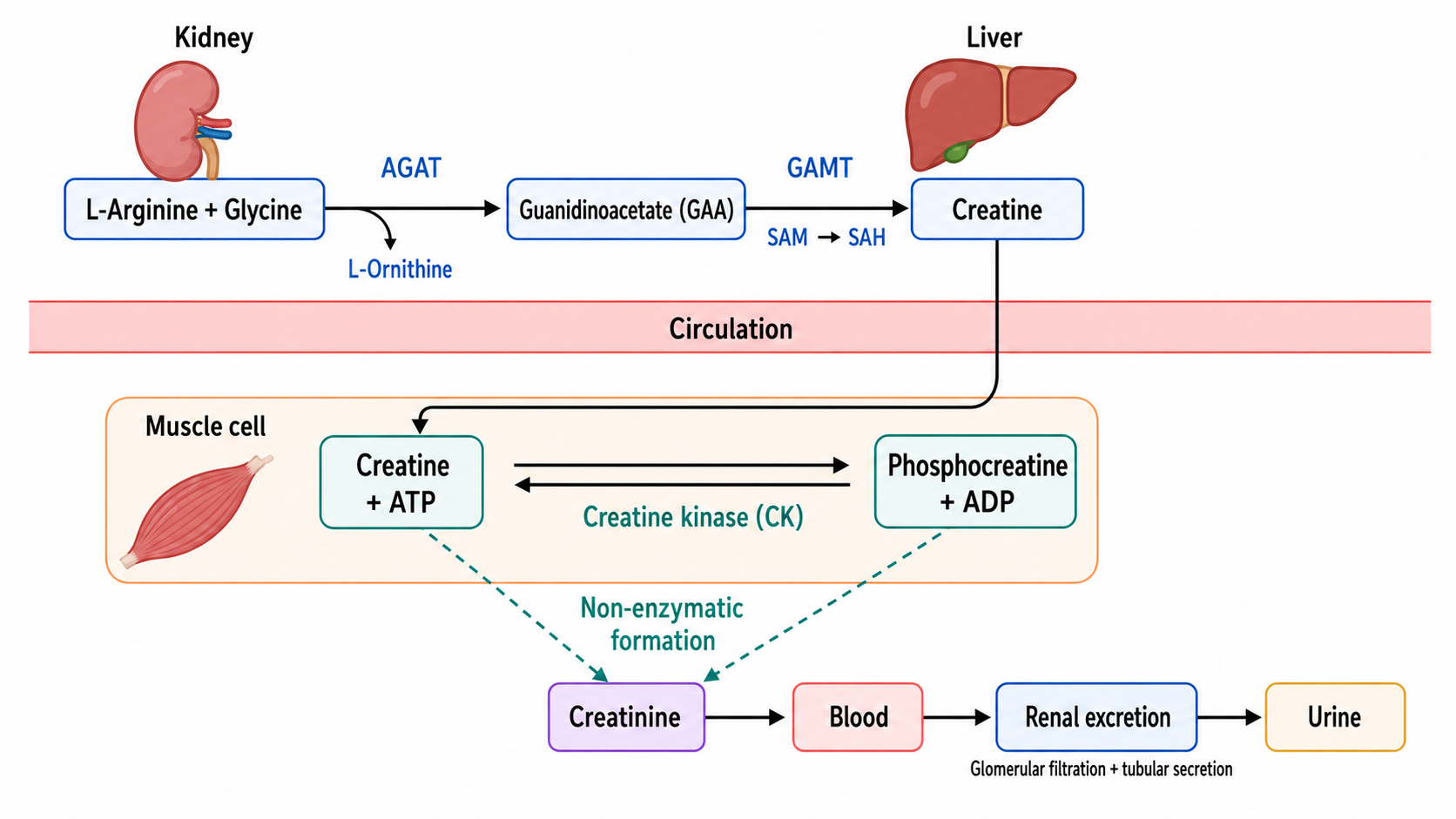
Figure 1. Creatine synthesis, creatine–phosphocreatine cycling, and creatinine formation and renal excretion
1.3 Clinical Significance of Creatinine Testing
Serum creatinine is a commonly used indicator for evaluating renal function and estimating the glomerular filtration rate. Urinary creatinine can be used to calculate creatinine clearance and to normalize the concentrations of urinary protein, urinary albumin, and other urinary analytes. Creatinine concentrations are also affected by age, sex, muscle mass, diet, exercise, muscle injury, and certain medications. A single creatinine result cannot fully represent glomerular filtration function and should be interpreted together with the estimated glomerular filtration rate and other renal function indicators.
1.4 Major Methods for Creatinine Testing
Laboratory methods for creatinine testing mainly include the picrate method and enzymatic methods. The picrate method is simple and relatively inexpensive but is readily affected by ketone bodies, glucose, proteins, bilirubin, and certain medications. Enzymatic methods use creatinine deiminase or a cascade consisting of creatininase, creatinase, and sarcosine oxidase to generate measurable signals and generally provide better analytical specificity.
2 Molecular Characteristics and Catalytic Mechanism of Creatinine Deiminase
2.1 Enzyme Sources and Reaction Type
Creatinine deiminase, also known as creatinine deaminase or creatinine iminohydrolase, is mainly derived from microorganisms capable of utilizing creatinine. This enzyme directly catalyzes the hydrolytic deamination of creatinine without first converting creatinine into creatine:
Creatinine + H₂O → N-methylhydantoin + NH₃
The ammonia generated by the reaction can be detected through colorimetric, potentiometric, pH-based, or enzyme-coupled methods. Compared with multienzyme cascade methods, the creatinine deiminase method involves fewer reaction steps and is suitable for rapid analysis and the construction of immobilized-enzyme sensors.
2.2 Substrate Binding and Catalytic Process
After creatinine enters the substrate-binding pocket of the enzyme, its cyclic nitrogen-containing structure is positioned within the catalytic center. An activated water molecule attacks the reactive carbon atom of the creatinine ring, followed by proton transfer and C–N bond cleavage, producing N-methylhydantoin and releasing ammonia. Some microbial creatinine deiminases are metal-dependent hydrolases in which metal ions participate in water activation, substrate positioning, and stabilization of reaction intermediates. Because enzymes from different sources differ in subunit composition, metal dependence, and catalytic residues, their responses to EDTA, buffer components, and externally added metal ions are not identical.
2.3 Conversion of the Ammonia Signal
The ammonia generated by creatinine deiminase can be coupled to a glutamate dehydrogenase reaction. In the presence of α-ketoglutarate and NADH or NADPH, ammonia is converted into glutamate while the reduced coenzyme is oxidized. Creatinine content can therefore be calculated by monitoring the decrease in absorbance near 340 nm.
Ammonia can also be detected using an ammonium ion-selective electrode, a pH-sensitive element, or an ammonia color reaction. Because pre-existing ammonia in samples, reagents, and the laboratory environment also generates a detection signal, sample blanks, reagent blanks, or pretreatment procedures are required.
3 Substrate Specificity and Enzymatic Properties of Creatinine Deiminase
3.1 Substrate Specificity
Creatinine deiminase primarily recognizes the cyclic nitrogen-containing structure and spatial conformation of creatinine. Creatine is an open-chain compound and generally cannot enter the active site in the same manner. Certain cyclic nitrogen-containing compounds may compete for the substrate-binding site. Therefore, when establishing a detection method, the cross-reactivity of creatine, urea, uric acid, ammonium ions, and structurally related analogues should be evaluated.
3.2 pH, Temperature, and Metal Ions
Creatinine deiminase generally exhibits relatively high activity under neutral to mildly alkaline conditions, although the specific optimum range depends on the enzyme source. The pH affects both the protonation states of catalytic residues and the ratio between ammonia and ammonium ions. Increasing the temperature can accelerate the reaction, but excessively high temperatures may reduce enzyme stability. Metal-dependent creatinine deiminases may be inhibited by chelating agents such as EDTA. Whether external metal ions are required should be determined according to the specific enzyme preparation.
3.3 Reaction Kinetics and Stability
Within a relatively low creatinine concentration range, the reaction rate increases as the substrate concentration increases. As the enzyme gradually approaches saturation, the reaction rate reaches a plateau. For quantitative testing, the sample concentration should remain within the linear range of the method, and the enzyme amount and reaction time should be sufficient to achieve stable substrate conversion.
Storage temperature, the number of freeze–thaw cycles, buffer composition, and immobilization method also affect residual enzyme activity and lot-to-lot consistency. In coupled assays, the downstream signal-generating reaction should not become the rate-limiting step.
4 Cascade Reactions of Creatininase, Creatinase, and Sarcosine Oxidase
4.1 Multienzyme Cascade Route
The multienzyme-coupled method sequentially uses creatininase, creatinase, and sarcosine oxidase to convert creatinine into creatine, sarcosine, and hydrogen peroxide. The hydrogen peroxide is then detected by peroxidase-mediated color development or an electrochemical method. The basic route is:
Creatinine → Creatine → Sarcosine → H₂O₂ → Colorimetric or electrochemical signal
Insufficient activity of any enzyme in the cascade may cause the accumulation of intermediates or a decrease in the final signal. Therefore, downstream enzymes are generally maintained in relative excess so that the overall reaction rate primarily reflects the creatinine content of the sample.
4.2 Individual Enzymatic Reactions
(1)Creatininase Reaction
Creatininase catalyzes the hydrolysis of the creatinine ring to generate creatine:
Creatinine + H₂O → Creatine
This reaction is the first step through which creatinine enters the creatine metabolic pathway in the multienzyme method. Insufficient creatininase activity causes incomplete creatinine conversion and directly limits the strength of the downstream signal.
(2)Creatinase Reaction
Creatine amidinohydrolase, also known as creatinase, further catalyzes the hydrolysis of creatine to generate sarcosine and urea:
Creatine + H₂O → Sarcosine + Urea
Creatine originally present in the sample can also enter this reaction. Therefore, detection systems generally use a prereaction, a two-reagent design, or a sample blank to subtract the signal generated by endogenous creatine.
(3)Sarcosine Oxidase Reaction
Sarcosine oxidase uses molecular oxygen to oxidize sarcosine, producing glycine, formaldehyde, and hydrogen peroxide:
Sarcosine + O₂ + H₂O → Glycine + Formaldehyde + H₂O₂
This step depends on oxygen. Sample viscosity, mixing of the reaction solution, and oxygen diffusion through an immobilized-enzyme membrane can all affect the rate of hydrogen peroxide generation. Endogenous sarcosine in the sample may also generate background signals unrelated to creatinine.
(4)Peroxidase Color Reaction
The generated hydrogen peroxide can oxidize a chromogen in the presence of peroxidase to form a colored product. The color intensity can then be used to calculate the creatinine concentration. Ascorbic acid, glutathione, and certain medications may consume hydrogen peroxide or reduce the colored product, resulting in falsely low results.
5 Comparison Between the Creatinine Deiminase Method and the Multienzyme-Coupled Method
5.1 Major Differences Between the Two Detection Routes
Comparison Item | Creatinine Deiminase Method | Multienzyme-Coupled Method |
Initial reaction | Direct hydrolytic deamination of creatinine | Creatinine is first hydrolyzed to creatine |
Core enzyme or enzymes | Creatinine deiminase | Creatininase, creatinase, and sarcosine oxidase |
Major products | N-Methylhydantoin and ammonia | Creatine, sarcosine, and hydrogen peroxide |
Detection signal | Ammonia, ammonium ions, pH, or changes in coenzyme absorbance | Hydrogen peroxide-based colorimetric, fluorescence, or electrochemical signal |
Reaction steps | Fewer | More |
Major advantages | Short reaction route, facilitating rapid testing and enzyme immobilization | Mature automated systems and multiple signal-conversion options |
Major interferences | Endogenous ammonia, ammonium salts, and environmental ammonia | Endogenous creatine, sarcosine, reducing substances, and peroxides |
Key quality-control considerations | Ammonia blank and stability of the ammonia detection system | Enzyme activity ratios, oxygen supply, and color-development efficiency |
5.2 Method Selection and Result Interpretation
The creatinine deiminase method reduces problems associated with multienzyme activity ratios and incomplete conversion of intermediates. However, without effective ammonia blank correction, it may produce a systematic positive bias. The multienzyme-coupled method is suitable for automated biochemical analyzers but requires correction for endogenous creatine and sarcosine and appropriate matching of the enzymatic activities and color-development reactions at each step.
The performance of the two methods cannot be judged solely by the number of enzymes involved. Method selection should comprehensively consider the sample type, creatinine concentration range, instrument platform, reaction speed, and major interfering substances. Results should be verified using calibrators, quality-control materials, and spike-recovery experiments.
6 Reaction Conditions and Interfering Factors in Enzymatic Creatinine Testing
6.1 Control of Reaction Conditions
(1)pH, Temperature, and Reaction Time
The optimal pH values of creatininase, creatinase, sarcosine oxidase, and peroxidase are not completely identical. The reaction system therefore requires a compromise condition that maintains effective activity of all enzymes. Increasing the temperature can accelerate the reaction but may also reduce enzyme stability and increase blank color development. An endpoint method must allow the reaction to reach a stable state, whereas a kinetic method requires a time window in which the reaction rate maintains a stable relationship with the creatinine concentration.
(2)Enzyme Activity Ratios
Creatininase determines the efficiency with which creatinine enters the cascade pathway. Creatinase and sarcosine oxidase should rapidly convert the generated products to prevent the accumulation of creatine or sarcosine. Downstream enzymes are generally maintained in relative excess to ensure that the final signal is primarily controlled by the creatinine concentration.
(3)Oxygen Supply and Color-Development System
Sarcosine oxidase depends on molecular oxygen. Increased sample viscosity, inadequate mixing, or an excessively thick immobilized-enzyme layer may limit oxygen diffusion and cause falsely low results. The concentrations of peroxidase and the chromogenic substrate should be sufficient to respond to the generated hydrogen peroxide while avoiding increased blank values caused by chromogen auto-oxidation.
6.2 Major Interfering Factors
(1)Endogenous Creatine and Sarcosine
Creatine in a sample can directly enter the creatinase reaction, whereas sarcosine can directly enter the sarcosine oxidase reaction. Both generate background signals unrelated to creatinine. In a two-reagent method, the signal generated by endogenous creatine and sarcosine can first be measured, after which creatininase is added to initiate the complete cascade and the difference is calculated.
(2)Reducing Substances
Ascorbic acid, glutathione, and certain medications may consume hydrogen peroxide or interfere with color development, producing falsely low results. The addition of ascorbate oxidase can reduce interference from ascorbic acid, although the effects of other reducing substances on the reaction system should still be evaluated.
(3)Bilirubin, Hemoglobin, and Lipemia
Bilirubin and hemoglobin may absorb light near the detection wavelength, whereas lipemia increases light scattering. Sample blanks, dual-wavelength measurements, and auxiliary reactions can reduce these effects but cannot replace appropriate quality assessment of icteric, hemolyzed, and lipemic samples.
6.3 Sample Types and Method Validation
Serum, plasma, and urine differ in creatinine concentration ranges and matrix composition. Their linear ranges, dilution conditions, and quality-control criteria should therefore be established separately. Method validation should include precision, linearity, recovery, detection capability, interference resistance, and reagent stability. Enzymatic methods using different enzyme sources, reaction routes, and calibration systems may also exhibit method-dependent biases.
7 Applications of Creatinine Deiminase in Biochemical Testing and Enzyme Sensors
7.1 Biochemical Testing
(1)Coenzyme Absorbance Method
Creatinine deiminase can be coupled with glutamate dehydrogenase to quantify creatinine by monitoring the decrease in NADH or NADPH absorbance. This method does not depend on peroxidase-mediated color development and may reduce certain interferences related to hydrogen peroxide and chromogens. However, endogenous ammonia and background coenzyme absorbance in the sample must be controlled.
(2)Ammonia Colorimetric Method
Ammonia generated by creatinine deiminase can be detected through a color reaction. This method is suitable for microplate, flow-analysis, and dry-chemistry systems. Although the reaction route is relatively short, ammonia-containing reagents, environmental ammonia, and reagent-container sealing can affect blank stability.
7.2 Enzyme Sensors
(1)Ammonium Ion Potentiometric Sensor
Creatinine deiminase can be immobilized near an ammonium ion-sensitive membrane, allowing creatinine to be measured by detecting the potential change caused by generated ammonium ions. Ammonium ions originally present in the sample produce a background signal. Therefore, a reference channel, blank reaction, or pretreatment procedure is required.
(2)pH-Based Enzyme Sensor
The creatinine deimination reaction can alter the local acid–base equilibrium. A pH-sensitive electrode, ion-sensitive field-effect transistor, or optical pH probe can convert this change into a detection signal. Excessively high buffer capacity weakens the response, whereas excessively low buffer capacity makes the system more susceptible to the intrinsic pH of the sample.
(3)Multienzyme Electrochemical Sensor
Creatininase, creatinase, and sarcosine oxidase can be co-immobilized on an electrode surface, and creatinine can be determined by measuring the electrical current generated by hydrogen peroxide. Enzyme-layer thickness, oxygen diffusion, hydrogen peroxide mass transfer, and electrode-surface fouling all affect response speed and the linear range.
7.3 Evaluation of Immobilized-Enzyme Performance
Immobilization can improve enzyme reusability and storage stability but may also alter enzyme conformation and substrate diffusion. In addition to sensitivity and linear range, sensor evaluation should examine selectivity, response time, drift, number of reuse cycles, lot-to-lot variation, and storage stability. Enzyme-free controls, substrate-free controls, and spike experiments using actual samples should be performed to confirm the source of the signal.
8 Products
8.1 Core Enzymes for Creatinine Testing and Cascade-Reaction-Related Products
Catalog | Product Name | Grade & Purity | Research Stage | Main Application |
Creatinine Deiminase (CNI) | Bioactive, recombinant, ActiBioPure™, high-performance, EnzymoPure™, ≥90% (SDS-PAGE), ≥10 U/mg enzyme powder; ≥100 U/mg protein | Direct creatinine deimination | Converts creatinine into N-methylhydantoin and ammonia for deiminase-based testing and enzyme sensor construction | |
Creatinine deiminase (CNI) | Bioactive, recombinant, ActiBioPure™, high-performance, EnzymoPure™, ≥20 U/mg enzyme powder; ≥100 U/mg protein | Direct creatinine deimination | Used for creatinine deimination reactions, enzyme activity comparison, and development of ammonia-signal detection systems | |
Creatininase (CAH) | Bioactive, recombinant, ActiBioPure™, high-performance, EnzymoPure™, ≥90% (SDS-PAGE), ≥50 U/mg enzyme powder; ≥100 U/mg protein | First step of the multienzyme cascade | Catalyzes the hydrolysis of creatinine to creatine for construction of enzymatic creatinine detection systems | |
Creatinine amidohydrolase (CAH) | BioReagent, EnzymoPure™, ≥90% (SDS-PAGE), ≥400 U/mg | First step of the multienzyme cascade | Used for high-activity creatinine conversion and optimization of cascade-reaction conditions | |
Creatininase (CAH) from Microorganism | Bioactive, ActiBioPure™, native, high-performance, EnzymoPure™, ≥450 U/mg enzyme powder | Native creatininase research | Used to compare the activity, stability, and detection performance of native and recombinant creatininase | |
Creatine amidinohydrolase (CR) | Recombinant, BioReagent, ActiBioPure™, high-performance, EnzymoPure™, ≥90% (SDS-PAGE), ≥5.8 U/mg enzyme powder | Second step of the multienzyme cascade | Catalyzes the conversion of creatine into sarcosine and urea for multienzyme-coupled creatinine testing | |
Sarcosine oxidase (SOX) | Bioactive, recombinant, ActiBioPure™, high-performance, EnzymoPure™, ≥90% (SDS-PAGE), ≥15 U/mg enzyme powder; expressed in E. coli | Third step of the multienzyme cascade | Oxidizes sarcosine and generates hydrogen peroxide for colorimetric and electrochemical detection | |
Recombinant Sarcosine Oxidase (SOX) | Bioactive, recombinant, ActiBioPure™, high-performance, EnzymoPure™, ≥25 U/mg enzyme powder; ≥40 U/mg protein | Third step of the multienzyme cascade | Used for sarcosine oxidation, hydrogen peroxide generation, and optimization of cascade systems |
8.2 Creatinine, Creatine, and Sarcosine Standards and Substrate-Related Products
Catalog | Product Name | Grade & Purity | Research Stage | Main Application |
Creatinine | ≥99% | Creatinine standard | Used to prepare standard curves and evaluate linearity, recovery, and enzymatic reaction efficiency | |
Creatinine | 10 mM in Water | Creatinine standard solution | Used for rapid establishment of standard curves and validation of detection systems | |
Creatinine-(methyl-d₃) | ≥98 atom% D, ≥97% | Isotope internal standard | Used for mass-spectrometric testing, methodological comparison, and correction of creatinine quantification | |
Creatinine-(methyl-¹³C) | ≥99 atom% ¹³C | Isotope internal standard | Used for confirmation of creatinine analytical methods and mass-spectrometric quantification | |
Creatine, anhydrous | Moligand™, ≥98% | Creatinase substrate | Used to evaluate creatinase activity and interference from endogenous creatine | |
Creatine | Moligand™, 10 mM in Water | Creatine standard solution | Used for measurement of creatine background and validation of cascade reactions | |
Creatine monohydrate | ≥98% | Creatinase substrate | Used for creatine hydrolysis reactions and evaluation of analytical interference | |
Creatine monohydrate | 10 mM in Water | Creatine standard solution | Used for creatinase reactions, spike recovery, and correction of endogenous creatine | |
Creatine-(methyl-d₃) monohydrate | ≥98 atom% D | Isotopic creatine standard | Used for creatine quantification and analysis of creatine background interference | |
Creatine-(methyl-¹³C) monohydrate | ≥99 atom% ¹³C | Isotopic creatine standard | Used for creatine metabolism studies and analytical method validation | |
Phosphocreatine | ≥92% | Creatine–phosphocreatine metabolism | Used to investigate the origin of creatinine formation, energy metabolism, and substrate conversion | |
Phosphocreatine disodium salt hydrate | ≥97% | Phosphocreatine research | Used to investigate the creatine–phosphocreatine system and creatinine formation | |
Creatine Phosphate Sodium Salt | 10 mM in Water | Phosphocreatine standard solution | Used for creatine metabolism and phosphocreatine conversion experiments | |
Creatine Phosphate Sodium Salt | Moligand™, ≥98% | Phosphocreatine metabolism | Used for research on creatine-mediated energy buffering and creatinine formation | |
Sarcosine | UltraBio™, Ultra pure | Sarcosine oxidase substrate | Used to evaluate SOX activity, hydrogen peroxide generation, and cascade efficiency | |
Sarcosine | Moligand™, ≥98% | Sarcosine standard | Used for substrate-specificity studies and investigation of endogenous sarcosine interference | |
Sarcosine | Moligand™, 10 mM in Water | Sarcosine standard solution | Used for SOX reactions, spike recovery, and signal-conversion evaluation | |
Sarcosine | for Synthesis, Moligand™ | Sarcosine substrate research | Used for basic studies related to sarcosine oxidation | |
Sarcosine hydrochloride | ≥99% | Evaluation of sarcosine salt responses | Used to compare the responses of different sarcosine salt forms in SOX reaction systems |
8.3 Products for Creatinine and Creatine Content Testing
Catalog | Product Name | Grade & Purity | Research Stage | Main Application |
Creatinine (Cr) Content Assay Kit (SOX, Micro Method) | BioReagent | Micro-scale creatinine testing | Used to determine creatinine content in research samples and validate multienzyme-coupled methods | |
Creatinine (Cr) Content Assay Kit (SOX, Colorimetric Method) | BioReagent | Colorimetric creatinine testing | Used for creatinine analysis in samples and research on enzymatic detection methods | |
Creatinine (Cr) Content Assay Kit (PA Rate, Micro Method) | BioReagent | Picrate rate-method testing | Used for creatinine determination and comparison between picrate and enzymatic methods | |
Creatine Content Assay Kit (Micro Method) | BioReagent | Creatine background testing | Used to detect endogenous creatine in samples and evaluate its interference with enzymatic creatinine testing | |
Creatine Content Assay Kit (Enzymatic, Micro Method) | BioReagent | Enzymatic creatine testing | Used for creatine content determination and evaluation of creatinase reactions | |
Creatine Content Assay Kit (Enzymatic, Colorimetric Method) | BioReagent | Colorimetric creatine testing | Used for creatine background correction and interference analysis in multienzyme-coupled creatinine testing |
The reliability of creatinine testing methods depends on enzyme substrate specificity, cascade-reaction efficiency, and the signal-conversion method. Stable and interpretable renal function test results can be obtained only by selecting an appropriate detection route according to the sample type and major interfering substances and by simultaneously controlling blanks, enzyme activity ratios, and the calibration system.
For more related articles, please see below:
[2] Creatinine Content Detection: Main Methods, Reaction Principles, and Result Control
[3] Experimental determination of serum creatinine (Cr) by the deproteinization endpoint method


 危险品化学品经营许可证(带存储)