
Differences Between Animal-Derived Collagen and Recombinant Collagen in Skincare: Sequence and Structure, Safety, Skin Effects, and Evidence Evaluation
Differences Between Animal-Derived Collagen and Recombinant Collagen in Skincare: Sequence and Structure, Safety, Skin Effects, and Evidence Evaluation
Abstract
Animal-derived collagen and recombinant collagen differ in amino acid sequence, post-translational modifications, triple-helical structure, molecular-weight distribution, impurity profiles, and manufacturing quality control. These differences may affect the film-forming properties, stability, immunogenicity, pathogen-related risks, and sites of action in the skin of the raw materials.
Intact collagen is a hydrophilic macromolecule. On intact skin, it primarily improves the condition of the skin surface by forming a film, binding water, and reducing water evaporation. Hydrolyzed collagen and certain recombinant collagen fragments have lower molecular weights, better water solubility, and greater potential for deposition in the stratum corneum. However, their actual delivery depth remains dependent on molecular conformation, formulation vehicles, and the condition of the skin barrier.
Existing studies have separately shown that certain animal-derived collagen products and recombinant collagen materials may improve skin hydration, erythema, or the condition of damaged tissue. Because the raw materials, formulations, methods of use, and study populations vary among studies, human studies directly comparing the skincare effects of the two types of collagen under standardized conditions are currently lacking. When evaluating a collagen-containing skincare product, particular attention should be paid to the precise category, structural form, molecular weight, quality control of the raw material, and human efficacy data for the finished product.
Keywords: animal-derived collagen; recombinant human collagen; recombinant humanized collagen; triple helix; immunogenicity; skin repair; transepidermal water loss
1. Definitions of Raw Materials and Scope of Comparison
1.1 Animal-Derived Collagen
Animal-derived collagen is commonly extracted from tissues such as bovine hide, bovine Achilles tendon, porcine skin, porcine tendon, fish skin, or fish scales. After extraction, collagen may also undergo acid treatment, enzymatic treatment, heat treatment, or hydrolysis. Therefore, even among raw materials of animal origin, molecular structure and primary applications may differ.
Common Forms of Animal-Derived Collagen
Raw Material Form | Main Structural Characteristics | Primary Role in Skincare Formulations |
Native collagen | Largely retains the triple helix and some fibrous structures | Film formation, water binding, and maintenance of moisture on the skin surface |
Atelocollagen | Portions of the non-helical N- and C-terminal telopeptides are removed | Reduces the risk of telopeptide-related immune responses and is used to prepare collagen solutions, gels, or materials |
Gelatin | Collagen is thermally denatured, causing dissociation of the triple helix | Film formation, thickening, and improvement of formulation texture |
Hydrolyzed collagen | Collagen polypeptide chains are cleaved into peptides of different lengths | Improves water solubility and formulation compatibility |
Collagen membranes, sponges, or fibers | Collagen forms continuous or porous material structures | Covers the skin, absorbs water, and maintains a moist environment |
1.2 Recombinant Collagen
Recombinant collagen is produced using recombinant deoxyribonucleic acid technology. The manufacturing process generally includes target-sequence design, expression-vector construction, expression in host cells, protein purification, and structural characterization. Common expression systems include bacteria, yeast, and mammalian cells.
According to the nomenclature interpretation for medical devices issued by the National Institutes for Food and Drug Control of China, recombinant collagen biomaterials may be classified as follows:
① Recombinant human collagen: Has the full-length amino acid sequence encoded by the gene of a specific type of human collagen and forms a triple-helical structure.
② Recombinant humanized collagen: May be classified as Type A or Type B according to whether it contains non-human collagen amino acid sequences. Type A does not contain non-human collagen amino acid sequences and includes the full-length amino acid sequence encoded by a gene for a specific type of human collagen without a triple-helical structure, partial amino acid sequence fragments, or combinations of functional fragments that do not contain non-human collagen amino acid sequences such as linker amino acids or tag amino acids. Type B refers to materials in which non-human collagen amino acid sequences, such as linker amino acids or tag amino acids, are added to combinations of functional fragments of human collagen.
③ Recombinant collagen-like protein: Consists of artificially designed or modified sequences and has relatively low sequence homology with human collagen.[1]
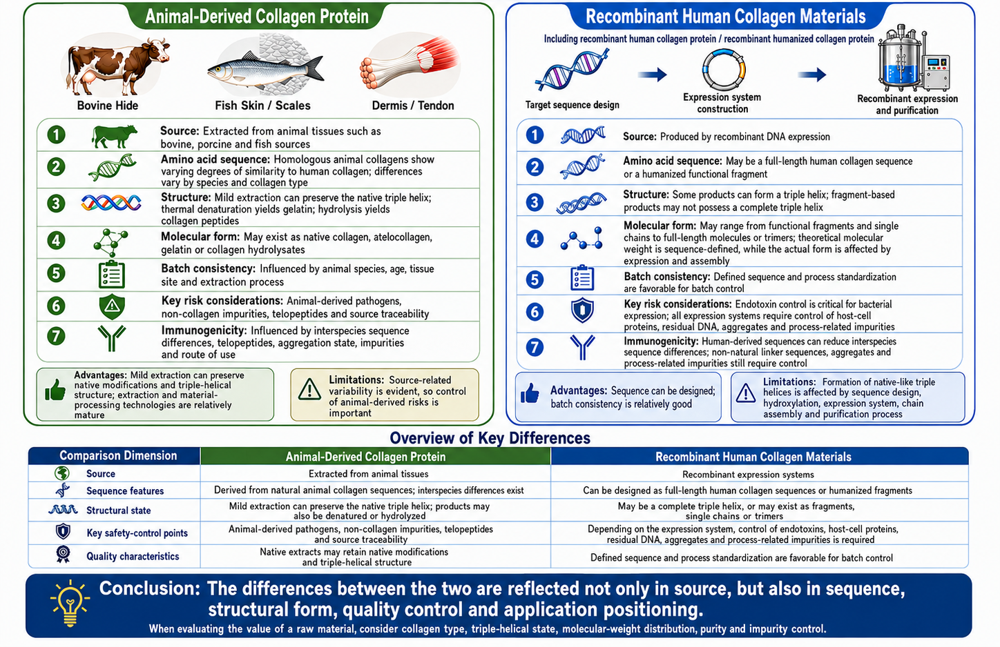
The differences between animal-derived collagen and recombinant human collagen materials begin with the source of the raw material and extend to amino acid sequence, molecular structure, batch-to-batch consistency, safety controls, and factors affecting immunogenicity. The figure below summarizes the principal characteristics of the two types of materials and the key points for their evaluation.
2. Differences in Amino Acid Sequence and Triple-Helical Structure
2.1 Molecular Structural Basis of Collagen
The principal fibrillar collagens in the dermis are types I and III. Type I collagen generally consists of two α1(I) chains and one α2(I) chain. The three polypeptide chains intertwine to form a triple helix, which subsequently assembles into collagen fibrils and collagen fibers.
The collagen helical region contains numerous glycine-X-Y, or Gly-X-Y, repeating sequences. Glycine is small enough to be positioned within the interior of the triple helix, while proline and hydroxyproline frequently occur at the X and Y positions. Proline helps restrict the conformation of the peptide chains, while hydroxyproline contributes to the stability of the triple helix.[2]
The material properties of collagen are jointly determined by multiple levels of structure:
① The amino acid arrangement determines the primary structure;
② The composition and arrangement of the three collagen chains determine the triple-helical structure;
③ Further assembly of triple-helical molecules determines the fibrillar and fibrous structures;
④ Hydroxylation, glycosylation, and crosslinking affect thermal stability, mechanical properties, and degradation behavior.
Collagen raw materials with similar amino acid compositions may nevertheless exhibit different solubility, film-forming properties, and biological performance because of differences in triple-helix content, degree of crosslinking, and aggregation state.
2.2 Sequence and Structural Differences Between Animal and Human Collagen
Type I collagen from humans, cattle, pigs, and other mammals has a high degree of structural similarity. Animal collagen can therefore form triple-helical and fibrous structures similar to those of human type I collagen. Nevertheless, differences among species remain in local amino acid composition, telopeptide regions, degree of hydroxylation, glycosylation patterns, and natural crosslinking status.
Comparative studies of human-derived and animal-derived type I collagen materials have shown that collagen source and processing methods affect gelation rate, fiber morphology, swelling properties, mechanical characteristics, and cellular responses.[3]
Collagen in animal tissues has already undergone proline hydroxylation, three-chain assembly, and fiber formation in vivo. Mild extraction conditions are conducive to preserving these native structures. Strong acids, strong alkalis, prolonged heating, or extensive hydrolysis may cause dissociation of the triple helix, cleavage of peptide chains, or alteration of the fibrous structure.
Animal origin indicates the tissue from which the raw material was obtained, but does not mean that the extracted finished material still retains an intact triple helix. Structural assessments should be based on test results for the finished material.
2.3 Structural Characterization Parameters for Recombinant Collagen
Recombinant technology can control the amino acid sequence of the target protein, but triple-helix formation, degree of hydroxylation, and aggregation state are also affected by the expression system and purification process.
Conventional Escherichia coli lacks the complete prolyl hydroxylation system required for human collagen synthesis. Co-expression of prolyl hydroxylase, engineering of host cells, or use of an appropriate eukaryotic expression system can increase the degree of hydroxylation and triple-helix stability of recombinant collagen. Relevant studies generally evaluate hydroxylation sites, circular dichroism, and biological functions in combination.[4]
Common structural and quality characterization parameters for recombinant collagen include:
① Collagen type and amino acid sequence coverage.
These parameters are used to confirm whether the raw material is derived from type I, type III, or another collagen type, and to determine whether it consists of a full-length sequence, a single α-chain, a local fragment, or a repeated combination of sequences.
② Non-native amino acid sequences.
These parameters are used to confirm whether the product contains linker peptides, purification tags, or artificially designed sequences. Such sequences may affect protein folding, purification behavior, degradation, and immunogenicity.
③ Molecular weight and molecular-weight distribution.
These parameters are used to confirm whether the target protein has the designed molecular weight and to identify truncated forms, degradation fragments, and components with abnormal molecular weights.
④ Chain composition, trimer formation, and aggregate levels.
Intact triple-helical collagen requires the correct assembly of three collagen chains. The proportions of single chains, trimers, and high-molecular-weight aggregates may affect material stability and biological behavior.
⑤ Degree and sites of proline hydroxylation.
When a raw material is claimed to possess an intact triple helix or a native-like collagen structure, hydroxyproline content and hydroxylation sites provide important reference information. For shorter recombinant humanized collagen fragments, hydroxyproline content should be interpreted in relation to the designed sequence and intended structure of the fragment and cannot be used alone to determine product quality.
⑥ Triple-helical conformation.
Circular dichroism can detect spectral characteristics associated with a collagen-like triple helix, but the results generally need to be interpreted together with chain-assembly, thermal-stability, or other structural test results.
⑦ Thermal stability.
The thermal denaturation temperature reflects the stability of the triple helix under increasing temperature and is affected by hydroxylation, amino acid composition, crosslinking, and solution conditions.
⑧ Purity and process-related impurities.
These include host-cell proteins, residual deoxyribonucleic acid, endotoxins, culture-medium residues, and purification-medium residues. Product-related variants of recombinant proteins, including oxidation, deamidation, degradation, and aggregation, should also be considered.[5]
The same evaluation criteria do not apply uniformly to all recombinant collagens. For intact triple-helical collagen, particular emphasis should be placed on confirming three-chain assembly, hydroxylation, and thermal stability. For recombinant humanized collagen fragments, particular emphasis should be placed on confirming sequence identity, molecular weight, purity, aggregation state, and designed function. The testing parameters should correspond to the structural claims made for the raw material.
3. Molecular Weight and Sites of Action in the Skin
3.1 Intact Collagen Acts Primarily on the Skin Surface
Intact type I collagen consists of three long polypeptide chains and is a hydrophilic macromolecule. The stratum corneum is composed of corneocytes and intercellular lipids and provides a strong barrier against hydrophilic macromolecules. Transdermal delivery of proteins and peptides is jointly affected by molecular weight, spatial conformation, charge, hydrophilicity, stability, and formulation vehicles.[6]
On intact skin, native animal collagen and intact recombinant collagen generally act primarily on the skin surface through the following mechanisms:
① Formation of a hydrophilic protein film;
② Adsorption and binding of water;
③ Reduction of water evaporation from the skin surface;
④ Reduction of external friction;
⑤ Improvement of dryness, tightness, and roughness.
These effects do not require collagen to enter the dermis. Intact collagen can provide moisturization by forming a film on the skin surface, but this does not demonstrate that it has entered the dermis or supplemented human collagen fibers.
3.2 Transdermal Characteristics of Hydrolyzed Collagen and Recombinant Collagen Fragments
After enzymatic or chemical hydrolysis, the peptide-chain length and molecular weight of collagen decrease, while water solubility generally increases. Recombinant technology can also be used to directly produce shorter functional collagen fragments.
A lower molecular weight facilitates dispersion of the raw material in the aqueous phase and may increase its deposition in the stratum corneum or hair follicles. An ex vivo mouse-skin study found that certain peptides in deer-sinew collagen hydrolysates were able to pass through skin samples. The study also evaluated the effects of the permeated components on cellular oxidative stress and type I collagen secretion.[7]
Results obtained from ex vivo skin can be used to assess penetration trends, but they cannot directly represent the effective concentration achieved in the human facial dermis after use. Actual transdermal behavior is also affected by the following factors:
① Skin source and integrity;
② Molecular weight, charge, and conformation of the peptides;
③ Amount of product applied and contact time;
④ Hair-follicle and sebaceous-gland pathways;
⑤ Penetration-enhancing ingredients or delivery vehicles;
⑥ The concentration of peptides actually detected in the skin;
⑦ Whether the peptides remain active after entering the skin.
When hydrolysis of collagen leads to a decrease in molecular weight, the original triple-helical and fibrous structures are generally reduced or disrupted. Native collagen is more suitable for film formation and material construction, whereas collagen peptides and recombinant fragments are more suitable for water-based formulations and research on functional fragments.
4. Differences in Safety and Immunogenicity
4.1 Safety Risks of Animal-Derived Collagen
The safety risks of animal-derived collagen arise primarily from animal tissues and the extraction and manufacturing processes. Key control parameters include:
① Animal species, age, and health status;
② Tissue site and collection conditions;
③ Bacterial, viral, and parasitic contamination;
④ Risk of transmissible spongiform encephalopathy associated with bovine-derived materials;
⑤ Residual non-collagen proteins, nucleic acids, and endotoxins;
⑥ Effectiveness of pathogen inactivation or removal processes.
Guidance issued by the U.S. Food and Drug Administration for animal-derived materials used in medical devices states that animal tissues may present a risk of infectious pathogens if improperly collected, stored, or manufactured. Risk management should cover traceability of animal sources, tissue collection, manufacturing processes, and validation of pathogen clearance.[8]
4.2 Manufacturing Risks of Recombinant Collagen
Producing collagen through non-animal-tissue routes, such as bacterial or yeast expression, can reduce pathogen risks associated with animal tissues and variability between raw-material batches. Different expression systems introduce their corresponding process-related impurities.
Common Expression Systems for Recombinant Collagen and Key Quality-Control Considerations
Expression System | Main Quality-Control Parameters |
Bacterial expression | Endotoxins, host-cell proteins, residual DNA, misfolded proteins, and aggregates |
Yeast expression | Host-cell proteins, residual DNA, culture-medium residues, and differences in glycosylation |
Mammalian-cell expression | Cell-bank quality, adventitious viruses, host-cell proteins, residual DNA, and viral-clearance capability |
The ICH Q5A(R2) guideline issued by the International Council for Harmonisation of Technical Requirements for Pharmaceuticals for Human Use requires evaluation of cell substrates, viral testing, and viral clearance for biotechnology products derived from human or animal cell lines.[9]
These requirements primarily apply to biotechnology-derived medicinal products, but they illustrate the types of risks involved in recombinant protein manufacturing. Recombinant collagen reduces risks associated with animal tissues, while still requiring controls for risks arising from host cells, culture processes, purification processes, and protein aggregation.
4.3 Immunogenicity of Animal-Derived and Recombinant Collagen
Immunogenicity refers to the ability of a substance to induce a specific immune response. The immunogenicity of collagen is affected by the following factors:
① Differences between its amino acid sequence and that of human collagen;
② Non-helical N- and C-terminal telopeptides;
③ Protein denaturation, oxidation, and aggregation;
④ Non-collagen proteins and host-cell impurities;
⑤ Endotoxins and other process-related residues;
⑥ Exposure dose and method of use;
⑦ Whether the skin barrier is intact.
Early studies of intradermally implanted bovine collagen found that antibodies against bovine collagen implants could be detected in some participants who developed clinical adverse reactions.[10]
This study involved intradermal implantation, in which the protein came into direct contact with living tissues and immune cells. These exposure conditions differ from those of ordinary topical products applied to intact skin.
Some antigenic epitopes in animal collagen are located in the telopeptide regions. Pepsin treatment can remove portions of the telopeptides to produce atelocollagen and reduce the risk of telopeptide-related immune responses.[11]
Recombinant human collagen uses human collagen sequences and can therefore reduce interspecies sequence differences between humans and animals. However, non-native linker peptides, purification tags, misfolding, protein aggregation, host-cell proteins, and endotoxins may still affect immune responses.
Factors Affecting the Immunogenicity of the Two Types of Collagen
Comparison Parameter | Animal-Derived Collagen | Recombinant Human Collagen or Recombinant Humanized Collagen |
Sequence-related factors | Local sequence differences exist between human and animal collagen | Human collagen sequences can reduce interspecies sequence differences |
Telopeptide-related factors | Depend on whether atelocollagen processing has been performed | Depend on sequence design and whether non-native linker sequences are present |
Sources of impurities | Animal-tissue proteins, nucleic acids, endotoxins, and extraction residues | Host-cell proteins, residual DNA, endotoxins, and culture- and purification-process residues |
Aggregation and denaturation | Affected by extraction, sterilization, and storage conditions | Affected by expression, refolding, purification, and storage conditions |
Topical application to intact skin | The stratum corneum restricts the entry of macromolecules into living tissues | The stratum corneum likewise restricts the entry of macromolecules into living tissues |
Recombinant human collagen has an advantage in reducing interspecies sequence differences. The actual immunogenicity of a raw material must nevertheless be evaluated in relation to protein purity, aggregation state, the finished formulation, and the site of application.
5. Skin Mechanisms of Action and Research Evidence for Repair
5.1 Surface Film Formation and Moisturization
Collagen contains numerous groups capable of interacting with water and can form a continuous or semi-continuous film on the skin surface. This film can reduce water evaporation and friction and improve dryness, tightness, and roughness.
A study of a native bovine-collagen sheet mask evaluated skin hydration, roughness, periocular wrinkles, ultraviolet-induced erythema, and tolerability. The results showed improvements in certain hydration, erythema, and appearance-related parameters.[12]
The study evaluated a specific finished collagen mask product. The structure of the sheet material, application time, hydration state, and other formulation factors all contributed to the results. The observed effects therefore cannot be attributed entirely to the animal origin of the material or to collagen as a single ingredient.
5.2 Restoration of the Skin Barrier
Restoration of the skin barrier involves corneocyte maturation, organization of intercellular lipids, and improvement of local inflammatory conditions. The surface film formed by collagen can reduce water loss and external friction, thereby providing a relatively stable surface environment for restoration of the damaged stratum corneum.
Certain collagen peptides and recombinant functional fragments have shown the potential to affect keratinocyte migration, fibroblast adhesion, inflammatory mediators, or extracellular-matrix expression in cellular or animal studies. Such effects require the raw material to reach the relevant living cells while maintaining sufficient concentration and activity.
In cell-culture experiments, collagen or collagen fragments can come into direct contact with cells. Ordinary topical products must first be delivered through the stratum corneum. Cell-based experiments are therefore suitable for explaining potential mechanisms, but cannot independently demonstrate that a finished skincare product will produce the same effects.
5.3 Repair of Damaged Skin and Wounds
In open wounds, implantable materials, and tissue engineering, collagen can form extracellular-matrix-like structures that provide an environment for cell adhesion, migration, and new-tissue formation. Collagen dressings can also absorb some wound exudate and maintain a moist environment.
One study of recombinant humanized type III collagen used an ultraviolet-induced photoaging animal skin model and evaluated the effects of the material through implantation. Improvements were observed in skin condition, tissue structure, and collagen deposition.[13]
Another study evaluated recombinant type I collagen and recombinant humanized collagen fragments in mouse wound and ultraviolet-induced photoaging models and observed improvements in wound closure and repair of photodamage.[14]
These studies support the research value of recombinant collagen in damaged tissues and biomaterial applications. Healthy intact skin, barrier-damaged skin, and open wounds differ in permeability and conditions of cellular exposure. Results from animal implantation and wound studies therefore cannot directly represent the effects of daily-use creams, serums, or facial masks.
6. Skincare Efficacy Parameters and Interpretation of Evidence
6.1 Transepidermal Water Loss
Transepidermal water loss (TEWL) refers to the rate at which water diffuses through and evaporates from the skin. It is a commonly used indicator for evaluating skin-barrier function. Higher TEWL is generally associated with impaired skin-barrier function.[15]
A reduction in TEWL after use of a collagen product may result from two types of effects:
① Formation of a surface film by the product, resulting in a short-term reduction in water evaporation;
② Gradual restoration of the stratum corneum structure, resulting in a more sustained improvement in barrier function.
When evaluating long-term repair effects, assessments should consider not only immediate TEWL but also the effects of continuous use, the duration of effects after discontinuation, stratum corneum hydration, and the clinical condition of the skin. The testing process should also control environmental temperature, relative humidity, the test site, and the acclimatization time of participants.
6.2 Stratum Corneum Hydration
Stratum corneum hydration reflects the hydration status of the superficial layer of the skin. Both animal-derived collagen and recombinant collagen can increase local hydration through their hydrophilicity and film-forming properties.
Glycerol, hyaluronic acid, oils, occlusive agents, and other film-forming agents in a complete formulation may also affect the measurement results. An improvement in hydration following use of a single product can demonstrate that the product has a moisturizing effect, but cannot directly establish collagen source as the determining factor.
6.3 Redness and Erythema
Redness may be associated with ultraviolet exposure, dryness, cleansing-induced damage, laser treatment, inflammatory responses, or vasodilation. Different types of redness arise through different mechanisms.
Collagen films may improve certain forms of dryness-related or irritation-related redness by increasing hydration, reducing friction, and decreasing external irritation. Improvements in ultraviolet-induced erythema parameters were observed in a study of a native bovine-collagen mask, but the results apply to the specific product and test conditions used in that study.[12]
Evidence regarding improvements in erythema, inflammation, and tissue condition with recombinant collagen is derived largely from cellular studies, animal injury models, implantation applications, or professional care following disruption of the skin barrier. These findings cannot be directly translated into the effects of daily topical products in healthy populations.
Overall, existing evidence includes studies in which animal-derived collagen products and recombinant collagen materials improved hydration, TEWL, erythema, or the condition of damaged tissue. However, the studies differ considerably in raw-material form, concentration, base formulation, method of use, and study population. Human trials directly comparing the two types of collagen under standardized conditions are currently lacking. The efficacy of a specific product should be assessed in relation to collagen structure, molecular weight, the formulation system, and human evaluation results.
7. Advantages, Limitations, and Selection Criteria for the Two Types of Collagen
7.1 Advantages and Disadvantages of Animal-Derived and Recombinant Collagen
Raw Material Type | Main Advantages | Main Limitations | More Suitable Applications |
Native animal-derived collagen | Hydroxylation and native assembly have already occurred in vivo; mild extraction can preserve triple-helical and fibrous structures; film-forming and material-processing technologies are relatively mature; available in relatively large supply | Affected by animal species, age, and tissue source; animal-derived pathogens, telopeptides, and xenogeneic proteins require control; processing conditions may disrupt the triple helix | Collagen membranes, moist coverings, film-forming moisturizers, and products emphasizing native structure |
Hydrolyzed animal-derived collagen | Good water solubility; readily incorporated into aqueous formulations; mature cost and supply systems | Triple-helical and fibrous structures have been disrupted or reduced; peptide composition is relatively complex; molecular-weight distribution may vary among batches | Moisturizing serums, emulsions, mask essences, and water-soluble formulations |
Recombinant human collagen | Defined sequence; good batch-to-batch consistency; reduced risks associated with animal tissues; specific collagen types can be designed | Full-length expression, hydroxylation, and triple-helix assembly are technically demanding; host-cell impurities, endotoxins, and aggregates require control; intact macromolecules remain restricted by the stratum corneum | Materials emphasizing sequence traceability, non-animal-tissue origin, and intact collagen structure |
Recombinant humanized collagen fragments | Sequence and molecular weight can be adjusted; facilitates functional-fragment screening and formulation adaptation; good manufacturing consistency | May lack intact triple-helical structure and the ability to assemble into fibers; function depends on the specific sequence; different raw materials cannot be assumed to have the same effects based on their names alone | Functional peptides, formulations combining multiple active ingredients, and research on delivery systems |
7.2 Information to Verify When Evaluating Products
When evaluating collagen-containing skincare products, the following information should be reviewed:
① Raw-material category and structural form.
Confirm whether the raw material is native collagen, atelocollagen, gelatin, hydrolyzed collagen, recombinant human collagen, or recombinant humanized collagen, and determine its collagen type, sequence coverage, and whether it possesses an intact triple-helical structure.
② Structural and quality-control data for the raw material.
When claims are made regarding a triple helix, a native-like structure, or a specific functional fragment, the relevant testing parameters should be confirmed to correspond to the structural claims made for the raw material. For animal-derived collagen, attention should be paid to animal source, quarantine and inspection, pathogen control, and residual non-collagen proteins. For recombinant collagen, attention should be paid to the expression system, endotoxins, host-cell proteins, residual DNA, and aggregates.
③ Human efficacy data for the finished product.
Evaluation parameters directly related to skincare effects include transepidermal water loss, stratum corneum hydration, erythema index, skin roughness, and tolerability indicators such as stinging and itching. Cellular or animal studies conducted at the raw-material level cannot replace human efficacy evaluation of the finished product.
④ Study population and conditions of use.
It should be confirmed whether the study population had healthy skin, sensitive skin, ultraviolet-irritated skin, postoperative skin, or open wounds. Results obtained under different skin conditions and methods of use cannot be used interchangeably.
The Standards for the Evaluation of Cosmetic Efficacy Claims issued by the National Medical Products Administration stipulate that cosmetics claiming repair efficacy must undergo human efficacy testing to substantiate such claims. Raw-material studies may help explain mechanisms of action, but cannot replace human efficacy evaluation of the finished product.[16]
8. Classification and Typical Applications of Products Related to Animal-Derived Collagen and Recombinant Collagen Research
Table 1. Collagen Raw Materials, Recombinant Proteins, and Derivatives
Category | CAS No. | Aladdin Catalog No. | Name | Specification or Purity | Product Features and Applications |
Animal-derived collagen raw material—bovine type I collagen | 9007-34-5 | Bovine Collagen I | Moligand™ | Used to study the chain composition, triple-helical structure, fibril self-assembly, film-forming properties, gel formation, and material properties of bovine type I collagen. | |
Animal-derived collagen raw material—sterile bovine type I collagen solution | 9007-34-5 | Bovine Collagen Solution | Moligand™, ≥95%, Type I, sterile filtered, BSE-Free, suitable for biomedical research | Used for cell-culture surface coating, three-dimensional collagen gels, collagen fibril formation, and studies of cell–extracellular matrix interactions. | |
Animal-derived collagen raw material—porcine type I collagen | 9007-34-5 | Porcine Type I Collagen (Col I) | Moligand™, research grade | Used to study the structure, solubility, fibril assembly, and film-forming properties of porcine type I collagen, as well as for comparative studies of collagen from different animal sources. | |
Recombinant collagen raw material—recombinant humanized type III collagen | 9007-34-5 | Recombinant Humanized Type III Collagen | Animal-origin-free, carrier-free, recombinant, ≥90% (SDS-PAGE), expressed in Nicotiana benthamiana | Used to study the sequence identity, apparent molecular weight, purity, aggregation state, and cytocompatibility of recombinant humanized type III collagen, as well as for comparative studies with animal-derived collagen. | |
Collagen derivative control—thermally denatured collagen | 9000-70-8 | Gelatin | Suitable for microbiology, gel strength ~250 g Bloom | Used as a structural control for collagen after dissociation of the triple helix and for studies of gelation, film formation, crosslinking, swelling, and resistance to enzymatic degradation. |
Table 2. Reagents for Studying Collagen Sequence Composition and Post-Translational Modifications
Category | CAS No. | Aladdin Catalog No. | Name | Specification or Purity | Product Features and Applications |
Collagen sequence component—glycine | 56-40-6 | Glycine | PharmPure™, USP, BP, European Pharmacopoeia (Ph. Eur.), ≥99% | Used for the design of collagen Gly-X-Y repeat sequences, amino acid composition controls, and studies of culture systems related to collagen synthesis. | |
Collagen sequence component—proline | 147-85-3 | L-Proline | Animal-origin-free, USP, European Pharmacopoeia (Ph. Eur.), suitable for cell culture, ≥99% | Used to study collagen amino acid composition, sequence design, proline metabolism, and amino acid supplementation systems for cell culture. | |
Collagen maturation-site research—lysine | 56-87-1 | L-Lysine | Moligand™, ≥98%, metals <500 ppm | Used to study lysine sites in collagen, lysine hydroxylation, glycosylation, and the formation of intermolecular crosslinks. | |
Collagen hydroxylation cofactor—ascorbic acid | 50-81-7 | L-Ascorbic Acid | Anhydrous grade, Moligand™, ACS, ≥99% | Used in fibroblast collagen-synthesis models to maintain the reducing environment required for collagen hydroxylase reactions and to evaluate procollagen hydroxylation and secretion. | |
Collagen hydroxylation cofactor—ferrous-ion source | 7782-63-0 | Ferrous Sulfate Heptahydrate | Suitable for plant cell culture, ≥99% | Used to provide ferrous ions in in vitro hydroxylase reactions or culture systems and to study the conditions required for proline and lysine hydroxylation in collagen. | |
Collagen hydroxylation cosubstrate—α-ketoglutaric acid | 328-50-7 | K657647 | α-Ketoglutaric Acid | Moligand™, suitable for cell culture, ≥99%, endotoxin ≤10 EU/g, total microbial count ≤100 CFU/g | Used in prolyl hydroxylase and lysyl hydroxylase reaction systems to study post-translational modifications of recombinant collagen and cellular collagen synthesis. |
Table 3. Reagents for Animal-Tissue Pretreatment, Collagen Extraction, and Purification
Category | CAS No. | Aladdin Catalog No. | Name | Specification or Purity | Product Features and Applications |
Tissue pretreatment—decalcifying chelating agent | 60-00-4 | Ethylenediaminetetraacetic Acid (EDTA) | Suitable for cell culture, ≥99% | Used to decalcify mineralized tissues such as bone and fish scales, thereby reducing interference from inorganic salts during collagen extraction, tissue sectioning, and subsequent compositional analysis. | |
Collagen extraction—acidic solubilizing agent | 64-19-7 | Glacial Acetic Acid | Guaranteed reagent grade, ≥99.5% | Used for acid-soluble collagen extraction, as a medium for pepsin digestion, for preparation of collagen solutions, and for acidic dissolution before fibril self-assembly. | |
Collagen extraction—enzyme for limited telopeptide digestion | 9001-75-6 | Pepsin, Porcine-Derived | Bioactive, ActiBioPure™, natural, high-performance, EnzymoPure™, derived from porcine gastric mucosa; ≥15,000 NF.U/mg enzyme powder | Used under acidic conditions to increase collagen solubilization and remove portions of the non-helical telopeptides, thereby preparing pepsin-soluble collagen or atelocollagen. | |
Collagen purification—salting-out agent | 7647-14-5 | Sodium Chloride | Suitable for cell culture, suitable for insect cell culture, ≥99.5% (T) | Used for fractional salting-out, precipitation, concentration, and purification of collagen, and also for adjusting the ionic strength of collagen solutions and culture systems. | |
Collagen degradation evaluation—collagenase | 9001-12-1 | Collagenase from Clostridium histolyticum | Sterile filtered, suitable for cell culture, lyophilized powder, 0.5–5.0 FALGPA units/mg solid | Used for enzymatic degradation of animal-derived or recombinant collagen materials, evaluation of resistance to enzymatic degradation after crosslinking, digestion of collagen-containing tissues, and cell isolation. |
Table 4. Products for Chemical and Enzymatic Crosslinking of Collagen Materials
Category | CAS No. | Aladdin Catalog No. | Name | Specification or Purity | Product Features and Applications |
Material crosslinking—aldehyde crosslinker | 111-30-8 | Glutaraldehyde (50%) | High-purity grade, 50% in H₂O, medical grade | Used for chemical crosslinking of collagen and gelatin materials and for evaluating crosslink density, mechanical properties, thermal stability, and resistance to enzymatic degradation; suitable for studies of conventional crosslinking methods and the effects of residual aldehydes. | |
Material crosslinking—carbodiimide crosslinker | 25952-53-8 | 1-Ethyl-3-(3-dimethylaminopropyl)carbodiimide Hydrochloride | ≥98% | Activates collagen carboxyl groups and promotes the formation of amide bonds with amino groups; used for crosslinking collagen hydrogels, membranes, fibers, and composite scaffolds. | |
Crosslinking-reaction aid—activated-ester stabilizer | 6066-82-6 | N-Hydroxysuccinimide (NHS) | ≥98% | Used in combination with carbodiimide reagents to stabilize carboxyl-activated intermediates and improve the efficiency of amide-bond crosslinking reactions in collagen materials. | |
Material crosslinking—naturally derived crosslinker | 6902-77-8 | Genipin | Moligand™, ≥98% | Reacts with amino groups in collagen or gelatin and is used to study the structural stability, mechanical properties, and resistance to enzymatic degradation of hydrogels, membranes, and fibrous materials. | |
Material crosslinking—enzymatic crosslinker | 80146-85-6 | Transglutaminase (TG) from Streptomyces mobaraensis | EnzymoPure™, ≥120 U/g powder | Catalyzes the formation of isopeptide bonds between glutamine and lysine residues in proteins and is used for the enzymatic crosslinking of collagen, gelatin gels, and composite materials. |
Table 5. Reagents for Collagen Electrophoresis, Protein Quantification, and Structural Analysis
Category | CAS No. | Aladdin Catalog No. | Name | Specification or Purity | Product Features and Applications |
Protein electrophoresis—anionic surfactant | 151-21-3 | Sodium Dodecyl Sulfate (SDS) | Suitable for electrophoresis, anionic | Used for denaturing electrophoresis of animal-derived collagen and recombinant proteins to analyze collagen-chain composition, apparent molecular weight, purity, aggregates, and degradation fragments. | |
Electrophoresis sample preparation—disulfide-bond reducing agent | 3483-12-3 | DL-Dithiothreitol (DTT) | Suitable for electrophoresis, ≥99% | Used for reducing sample preparation of disulfide-containing procollagen, type III collagen, or engineered recombinant proteins and for comparing electrophoretic bands under reducing and non-reducing conditions. | |
Protein quantification—Coomassie dye | 6104-58-1 | Coomassie Brilliant Blue G-250 | AR | Used to determine the concentrations of animal-derived collagen, recombinant collagen, and related proteins by the Coomassie dye-binding colorimetric method; suitable for establishing standard curves matched to the samples being tested. |
Table 6. Reagents for Detecting Collagen Content and Tissue Deposition
Category | CAS No. | Aladdin Catalog No. | Name | Specification or Purity | Product Features and Applications |
Collagen-content analysis—hydroxyproline standard | 51-35-4 | trans-4-Hydroxy-L-Proline | Moligand™, suitable for cell culture, ≥98.5% | Used to establish hydroxyproline standard curves and estimate collagen content and the degree of degradation in tissues, collagen extracts, culture samples, or materials. | |
Hydroxyproline colorimetry—oxidizing reagent | 7080-50-4 | Chloramine T Trihydrate | AR, ≥98% (T) | Oxidizes free hydroxyproline in hydrolyzed samples and produces intermediates required for subsequent color development; used for analysis of collagen content and degradation products. | |
Hydroxyproline colorimetry—color-developing reagent | 100-10-7 | p-Dimethylaminobenzaldehyde | ≥98% | Reacts with hydroxyproline derivatives generated after oxidation with chloramine T to produce a colored product and is used for spectrophotometric determination of hydroxyproline. | |
Tissue collagen staining—Sirius Red dye | 2610-10-8 | Direct Red 80 | Dye content 25% | Used for staining collagen fibers in tissue sections, polarized-light observation, analysis of collagen deposition, and dye-binding assays for soluble collagen. |
Note: The products listed above are representative Aladdin products related to scientific research. Their specific applications should be determined in conjunction with the product specifications, batch-specific certificate of analysis, and the intended reaction or evaluation system. Additional product specifications, grades, and COA information can be searched on the Aladdin website using the product name, CAS number, or catalog number.
References
[1] NATIONAL INSTITUTES FOR FOOD AND DRUG CONTROL. Interpretation of the Guiding Principles for the Nomenclature of Recombinant Collagen Biomaterials[EB/OL]. 2023-01-29.
[2] SHOULDERS M D, RAINES R T. Collagen structure and stability[J]. Annual Review of Biochemistry, 2009, 78: 929–958. DOI: 10.1146/annurev.biochem.77.032207.120833.
[3] BALTAZAR T, KAJAVE N S, RODRIGUEZ M, et al. Native human collagen type I provides a viable physiologically relevant alternative to xenogeneic sources for tissue engineering applications: A comparative in vitro and in vivo study[J]. Journal of Biomedical Materials Research Part B: Applied Biomaterials, 2022, 110(10): 2323–2337. DOI: 10.1002/jbm.b.35080.
[4] ZHU P, MA M, YOU T, et al. Optimizing prolyl hydroxylation for functional recombinant collagen in Escherichia coli[J]. International Journal of Biological Macromolecules, 2024, 282(Pt 5): 137400. DOI: 10.1016/j.ijbiomac.2024.137400.
[5] INTERNATIONAL CONFERENCE ON HARMONISATION. ICH Q6B: Specifications: Test Procedures and Acceptance Criteria for Biotechnological/Biological Products[S]. 1999.
[6] ALKILANI A Z, NASEREDDIN J, HAMED R, et al. Beneath the skin: A review of current trends and future prospects of transdermal drug delivery systems[J]. Pharmaceutics, 2022, 14(6): 1152. DOI: 10.3390/pharmaceutics14061152.
[7] ZHANG H, PAN D, DONG Y, et al. Transdermal permeation effect of collagen hydrolysates of deer sinew on mouse skin, ex vitro, and antioxidant activity, increased type I collagen secretion of percutaneous proteins in NIH/3T3 cells[J]. Journal of Cosmetic Dermatology, 2020, 19(2): 519–528. DOI: 10.1111/jocd.13041.
[8] U.S. FOOD AND DRUG ADMINISTRATION. Medical Devices Containing Materials Derived from Animal Sources (Except for In Vitro Diagnostic Devices): Guidance for Industry and Food and Drug Administration Staff[EB/OL]. 2019.
[9] INTERNATIONAL COUNCIL FOR HARMONISATION. ICH Q5A(R2): Viral Safety Evaluation of Biotechnology Products Derived from Cell Lines of Human or Animal Origin[S]. 2023.
[10] SIEGLE R J, MCCOY J P Jr, SCHADE W, SWANSON N A. Intradermal implantation of bovine collagen: Humoral immune responses associated with clinical reactions[J]. Archives of Dermatology, 1984, 120(2): 183–187. DOI: 10.1001/archderm.1984.01650380043008.
[11] WYSOCKI T, SACEWICZ I, WIKTORSKA M, NIEWIAROWSKA J. Atelocollagen as a potential carrier of therapeutics[J]. Postępy Higieny i Medycyny Doświadczalnej (Online), 2007, 61: 646–654.
[12] JANSSENS-BÖCKER C, WIESWEG K, DOBERENZ C. Native collagen sheet mask improves skin health and appearance: A comprehensive clinical evaluation[J]. Journal of Cosmetic Dermatology, 2024, 23(5): 1685–1702. DOI: 10.1111/jocd.16181.
[13] WANG J, QIU H, XU Y, et al. The biological effect of recombinant humanized collagen on damaged skin induced by UV-photoaging: An in vivo study[J]. Bioactive Materials, 2022, 11: 154–165. DOI: 10.1016/j.bioactmat.2021.10.004.
[14] LIU T, HAO J, LEI H, et al. Recombinant collagen for the repair of skin wounds and photo-aging damage[J]. Regenerative Biomaterials, 2024, 11: rbae108. DOI: 10.1093/rb/rbae108. Correction: Regenerative Biomaterials, 2025, 12: rbaf064. DOI: 10.1093/rb/rbaf064.
[15] AKDENIZ M, GABRIEL S, LICHTERFELD-KOTTNER A, et al. Transepidermal water loss in healthy adults: A systematic review and meta-analysis update[J]. British Journal of Dermatology, 2018, 179(5): 1049–1055. DOI: 10.1111/bjd.17025.
[16] NATIONAL MEDICAL PRODUCTS ADMINISTRATION. Announcement of the National Medical Products Administration on Issuing the Standards for the Evaluation of Cosmetic Efficacy Claims (Announcement No. 50 of 2021)[EB/OL]. 2021-04-08.
For more related articles, see below:
Antioxidant Mechanisms, Stability, and Delivery Strategies of Resveratrol in Skincare Research
Beneath the Skin: Protein- and Peptide-Based Actives in Skincare and Aesthetic Medicine
Hyaluronidase: Unlocking a “Magic Enzyme” in Biomedicine
Trehalose: Physicochemical Characteristics, Manufacturing Processes, and Application Guidelines
Sources, Biological Functions, Isolation/Characterization, and Emerging Applications of Exosomes
Applicability Comparison of Vitronectin, Laminin and Fibronectin in Cell Culture Coating Systems
Ferulic Acid: Structural Features, Preparation Routes, and Key Points for Research Applications
Role of Sphingolipid Metabolic Remodeling in Apoptotic and Inflammatory Signaling
How to decipher the whitening code?
Niacin (Vitamin B3): Structural Features, Metabolic Roles, and Application Landscape
The "Six Key Checkpoints" of Skin Lightening
Tyrosinase: a key regulatory enzyme in melanin synthesis and its biological and applied significance


 危险品化学品经营许可证(带存储)