
Mechanisms of Retinol Action in the Skin: From Metabolic Activation and RAR/RXR-Mediated Transcriptional Regulation to Epidermal Renewal and Collagen Homeostasis
Mechanisms of Retinol Action in the Skin: From Metabolic Activation and RAR/RXR-Mediated Transcriptional Regulation to Epidermal Renewal and Collagen Homeostasis
1 Structure and Raw Material Characteristics of Retinol
1.1 What Is Retinol?
In skincare products, retinol generally refers to all-trans-retinol. It has the molecular formula C₂₀H₃₀O and belongs to the naturally occurring vitamin A family of compounds.
Its molecular structure primarily consists of three regions:
Structural Region | Structural Characteristics | Relationship to Skincare Effects |
β-Ionone-like ring | Hydrophobic cyclic structure | Contributes to the lipid-soluble backbone of retinoid molecules |
Conjugated polyene chain | Alternating single and double bonds | Influences light absorption, molecular configuration, and chemical stability |
Terminal primary alcohol group | —CH₂OH | Serves as the reaction site for the conversion of retinol into retinal and retinoic acid |
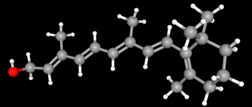
The cyclic structure and polyene chain of retinol form a strongly hydrophobic backbone that facilitates its entry into the lipid environment of the stratum corneum. At the same time, the extended conjugated double-bond system makes retinol susceptible to photoisomerization and oxidative degradation. Therefore, raw material stability, oxygen exposure within the formulation, the light-protective properties of the packaging, and storage conditions can all affect the amount of retinol actually retained in the product.[12]
1.2 Terminal Functional Groups Determine the Metabolic Positions of Different Retinoids
Retinol, retinal, and all-trans-retinoic acid share the same basic carbon skeleton, with their main differences located at the terminal end of the side chain.
Compound | Terminal Functional Group | Role in the Classical Retinoic Acid Pathway |
All-trans-retinol | —CH₂OH | Upstream metabolic precursor |
All-trans-retinal | —CHO | Intermediate metabolite |
All-trans-retinoic acid | —COOH | Major active ligand of the retinoic acid receptor (RAR) |
The core conversion pathway is:
All-trans-retinol (—CH₂OH) ⇌ All-trans-retinal (—CHO) → All-trans-retinoic acid (—COOH)
Retinyl esters are not obligatory intermediates in the formation of all-trans-retinoic acid, but instead represent a collateral metabolic form of retinol. A portion of the retinol entering cells can be esterified to form retinyl esters, thereby reducing the immediate reactivity of free retinol. Retinyl esters can also be hydrolyzed to release retinol again.[1]
2 How Retinol Is Converted into All-trans-Retinoic Acid
2.1 Metabolic Branching of Retinol after Entry into the Skin
Topically applied retinol must first be released from the formulation, pass through the stratum corneum, and enter metabolically active epidermal cells. Once inside the cells, retinol is not immediately and entirely converted into all-trans-retinoic acid. Instead, it may follow three possible pathways:
1. Remain as free retinol and bind to cellular retinol-binding proteins;
2. Undergo esterification to form retinyl esters, creating a temporary metabolic buffer;
3. Enter the sequential oxidation pathway to form retinal and all-trans-retinoic acid.
Together, these processes control the amount of intracellular retinol available for retinoic acid formation. Therefore, the labeled retinol concentration in a skincare product cannot be directly equated with the concentration of all-trans-retinoic acid ultimately generated within skin cells.[1]
2.2 First Step: Oxidation of Retinol to Retinal
The terminal primary alcohol group of retinol is oxidized to an aldehyde group, producing all-trans-retinal. This step involves enzymes with retinol dehydrogenase activity, including certain retinol dehydrogenases and short-chain dehydrogenases/reductases.
During the reaction, the oxidized forms of nicotinamide adenine dinucleotide and its phosphorylated counterpart are NAD⁺ and NADP⁺, respectively. In the oxidative direction, NAD⁺ or NADP⁺ can serve as electron acceptors, with the specific coenzyme preference depending on the enzyme involved. In vitro biochemical studies of human epidermal retinol dehydrogenase 2 have shown that this enzyme can catalyze the reversible conversion between all-trans-retinol and all-trans-retinal and has a marked preference for the NAD⁺/NADH coenzyme system.[2]
Retinol and retinal can undergo reversible conversion within the metabolic network. Retinal may either be further oxidized to all-trans-retinoic acid or be reduced back to retinol by reductases. This step therefore represents an important regulatory point controlling the amount of retinoic acid produced.
2.3 Second Step: Oxidation of Retinal to All-trans-Retinoic Acid
All-trans-retinal is subsequently catalyzed by members of the aldehyde dehydrogenase 1A family, with the aldehyde group being further oxidized to a carboxylic acid group to generate all-trans-retinoic acid (ATRA).
Compared with the reversible conversion between retinol and retinal, the conversion of retinal to ATRA proceeds predominantly in the direction of retinoic acid formation under physiological metabolic conditions. Once generated, ATRA generally enters pathways involving transport by binding proteins, nuclear receptor binding, or further oxidative clearance, rather than being extensively reduced back to retinal through the same pathway.
Experiments using cultured human keratinocytes have shown that exogenous all-trans-retinol can be metabolized into small amounts of ATRA and can mediate transcriptional responses through nuclear retinoic acid receptors.[11]
2.4 Why Biological Effects May Occur Even When Large Amounts of ATRA Are Not Detected
ATRA is a highly active cellular signaling molecule, and its effects do not depend on substantial accumulation within tissues. It may be generated locally and then rapidly bind to cellular retinoic acid-binding proteins or nuclear receptors before undergoing further metabolism. Its tissue concentration may also remain below the detection limit.
A short-term human skin study found that topical retinol could induce epidermal thickening and the expression of retinoid-responsive proteins, although no marked accumulation of free retinoic acid was detected in the tissue samples. These findings indicate that retinol can produce retinoic acid-like biological responses, but they do not establish that high concentrations of free ATRA must be maintained in the tissue over prolonged periods.[1]
3 Why Retinol Requires Metabolic Conversion
Retinol itself is not a high-affinity ligand for the retinoic acid receptor (RAR). Its classical transcriptional regulatory effects arise primarily from the ATRA generated through metabolism.[11]
The conversion of retinol into ATRA has three principal implications.
3.1 Controlling the Amount of Active Signal Generated
Cells can regulate the amount of substrate entering the nuclear receptor pathway through retinol esterification, the reversible conversion between retinol and retinal, and the oxidation of retinal to ATRA.
3.2 Separating Raw Material Uptake from Nuclear Receptor Activation
After retinol is taken up by the skin, it does not immediately produce the same signal as an equivalent amount of ATRA. Retinol must first undergo cellular uptake, binding, enzymatic conversion, and metabolic clearance. Its effects therefore generally develop more gradually.
3.3 Producing Different Degrees of Response in Different Skin Cells
Keratinocytes, fibroblasts, and other skin cells do not express identical levels of binding proteins, metabolic enzymes, or nuclear receptors. The same extracellular retinol concentration may therefore generate different levels of ATRA signaling in different cell types.
Accordingly, the actual biological activity of retinol depends on two consecutive processes: first, retinol must reach metabolically active skin cells; second, the cells must convert it into ATRA and generate an effective nuclear receptor signal.[1,3]
4 How ATRA Regulates Gene Transcription through RAR/RXR Heterodimers
4.1 What Are RAR and RXR?
RAR has three major subtypes: RARα, RARβ, and RARγ. In the human epidermis, RARγ is the predominant RAR subtype, while RARα is also expressed.[3]
The retinoid X receptor (RXR) likewise has three major subtypes: RXRα, RXRβ, and RXRγ. Both RAR and RXR are ligand-regulated nuclear receptors, but they perform different roles in classical ATRA signaling:
① ATRA primarily binds to RAR;
② RAR forms a heterodimer with RXR;
③ The RAR/RXR heterodimer participates in recognizing DNA regulatory sequences and organizing transcriptional complexes.
Therefore, the binding of ATRA to RAR is the principal ligand-recognition step in this pathway, while RXR primarily participates in transcriptional regulation as the dimerization partner of RAR.[3,4]
4.2 How RAR/RXR Regulates Gene Transcription
RAR/RXR heterodimers can recognize retinoic acid response elements (RAREs) in DNA. RAREs are located within the regulatory regions of certain retinoic acid-responsive genes.
After ATRA binds to RAR, the ligand-binding domain of RAR undergoes a conformational change, altering the receptor’s interactions with corepressors and coactivators. The resulting transcriptional complex further influences chromatin structure and RNA polymerase-associated processes, thereby changing the transcription levels of the corresponding genes.[4]
This process can be summarized as follows:
ATRA binds to RAR → The conformation of the RAR/RXR complex and the composition of coregulatory proteins change → Target-gene transcription changes → Cell proliferation, differentiation, and extracellular matrix metabolism are altered.
Activation of this transcriptional pathway does not mean that the expression of every associated gene is increased. RAR/RXR can directly regulate target genes containing RAREs and can also interact with other transcription factors and signaling pathways to reduce the expression of certain inflammation- or matrix degradation-related genes.
5 From Nuclear Receptor-Mediated Transcription to Changes in Skin Tissue
5.1 Epidermis: Regulation of Keratinocyte Proliferation and Differentiation
Epidermal keratinocytes are generated in the basal layer, after which they migrate upward and progressively complete differentiation. Nuclear receptor signaling generated by ATRA can regulate this cellular process, altering viable epidermal thickness, cellular proliferation status, and the expression of differentiation-related genes.
Human skin studies have shown that topical retinol can increase epidermal thickness and induce responsive molecules such as cellular retinol-binding proteins and cellular retinoic acid-binding proteins. Retinol and ATRA produce changes in the same general direction, although the responses induced by retinol are weaker overall.[1,10]
The effects of retinol on the epidermis arise primarily from the regulation of cellular programs rather than from directly disrupting the connections between corneocytes, as occurs with certain exfoliating agents. Changes in skin-surface smoothness and the appearance of fine lines result from the combined remodeling of epidermal structure and the dermal extracellular matrix.
5.2 Dermis: Regulation of Fibroblasts and the Extracellular Matrix
Dermal fibroblasts are responsible for synthesizing type I collagen, type III collagen, and various proteoglycans and glycosaminoglycans. After retinol is metabolized into ATRA, it can alter gene expression in fibroblasts and modify the surrounding extracellular matrix environment.
Comparative human skin studies have shown that both topical retinol and ATRA can increase the expression of the type I collagen α1-chain gene, COL1A1, and the type III collagen α1-chain gene, COL3A1, as well as increase type I and type III procollagen proteins. The changes produced by retinol are directionally consistent with those produced by ATRA, although their overall magnitude is lower.[10]
6 How Retinol Regulates Collagen Homeostasis at Both the Synthesis and Degradation Ends
The collagen content of the skin depends on the dynamic balance between formation and degradation:
Net collagen accumulation = New collagen formation − Existing collagen degradation
Naturally aged skin generally exhibits reduced procollagen synthesis, accompanied by increased expression or activity of matrix metalloproteinases. Acute ultraviolet exposure can induce AP-1 and multiple MMPs while suppressing procollagen transcription. In chronically photoaged skin, collagen synthesis and degradation may change simultaneously, but persistently enhanced matrix degradation still leads to collagen loss and fragmentation.[5–8]
The effects of retinol on collagen homeostasis are concentrated at two principal endpoints:
1. Increasing procollagen formation;
2. Reducing abnormal collagen degradation.
6.1 Synthesis End: Promoting the Formation of New Procollagen
Fibroblasts first synthesize procollagen polypeptide chains. Within the cell, procollagen undergoes hydroxylation, glycosylation, and triple-helix assembly before being secreted into the extracellular space. Its terminal propeptides are then removed, after which the molecules become further aligned and assembled into collagen fibrils.
Retinol is not an amino acid substrate for collagen synthesis. Instead, it generates ATRA and alters the cellular transcriptional state, thereby increasing the expression of collagen-related genes and procollagen proteins.
The pathway can be represented as follows:
Retinol → ATRA formation → RAR/RXR-mediated transcriptional regulation → Increased expression of COL1A1, COL3A1, and procollagen → A foundation is established for subsequent extracellular collagen formation.
A comparative human skin study showed that continuous retinol use increased the expression of COL1A1 and COL3A1, as well as type I and type III procollagen proteins.[10]
Another randomized, double-blind, vehicle-controlled study used 0.4% retinol in participants with a mean age of 87 years, applied up to three times weekly for 24 weeks. The results showed improvements in fine wrinkles and tactile roughness. In a small biopsy subgroup, increased type I procollagen immunostaining and glycosaminoglycan expression were also observed.[9]
Increased procollagen expression indicates that a new collagen synthesis program has been initiated. However, procollagen must still undergo secretion, terminal propeptide cleavage, fibril assembly, and crosslinking before a mature collagen fiber network can be formed.
6.2 Degradation End: Reducing Abnormal AP-1/MMP Signaling
Ultraviolet radiation can activate mitogen-activated protein kinase (MAPK) pathways in epidermal and dermal cells, increasing the expression of the c-Jun protein. c-Jun associates with other proteins to form the activator protein 1 (AP-1) transcription factor complex, which promotes the expression of multiple matrix metalloproteinases (MMPs).[5,6]
These include:
Enzyme | Principal Role in Collagen Degradation |
MMP-1 | Initiates the cleavage of type I and type III fibrillar collagen |
MMP-3 | Degrades certain extracellular matrix components and participates in the activation of other MMPs |
MMP-9 | Further processes denatured or already fragmented collagen |
The ultraviolet-related degradation process can be represented as follows:
Ultraviolet radiation → MAPK activation → Increased c-Jun/AP-1 → Increased MMP expression → Collagen cleavage and fragmentation
In vivo human skin studies have shown that ATRA pretreatment can suppress ultraviolet-induced increases in c-Jun and MMP expression and alleviate the ultraviolet-induced suppression of procollagen transcription.[5,6] These studies demonstrate that ATRA can suppress ultraviolet-induced c-Jun/AP-1- and MMP-related responses and reduce the suppression of procollagen transcription. Retinol can be metabolized into ATRA, and studies of naturally aged skin have also observed that topical retinol can reduce levels of collagen degradation-related MMPs. Therefore, the effects of retinol on this pathway can be attributed, at least in part, to the signaling and transcriptional regulation triggered after its metabolic conversion into ATRA.[7,11]
After being metabolized into ATRA, retinol primarily reduces abnormal MMP induction at the signaling and gene-transcription levels. It does not act as a small-molecule enzyme inhibitor that directly occupies the catalytic center of MMPs, nor does it halt normal extracellular matrix turnover.
6.3 Why the Synthesis and Degradation Ends Are Interrelated
Intact collagen fibers not only provide structural support but also provide fibroblasts with adhesion sites and mechanical tension.
When collagen is continuously cleaved and fragmented, fibroblasts have difficulty maintaining their normal spread morphology, cytoskeletal tension decreases, and their capacity for collagen synthesis may consequently weaken. Fragmented matrix can also promote oxidative stress and MMP-1 expression, creating a sustained cycle of matrix damage.[13]
By reducing abnormal collagen degradation, retinoic acid signaling can both preserve existing collagen and improve the structural environment surrounding fibroblasts, thereby providing conditions favorable for subsequent collagen synthesis and organization.
The major processes through which retinol progresses from metabolic conversion and nuclear receptor-mediated transcriptional regulation to epidermal renewal and changes in collagen homeostasis are summarized in the figure below.

Retinol can be converted through retinal into all-trans-retinoic acid. All-trans-retinoic acid binds to the retinoic acid receptor (RAR) and regulates transcription through RAR/RXR heterodimers, thereby influencing keratinocyte renewal and procollagen formation while reducing abnormal ultraviolet-related MMP induction. In this way, it participates in the regulation of collagen homeostasis at both the synthesis and degradation ends. The specific effects are influenced by the formulation, dose, skin condition, duration of use, and other factors.
7 From Retinol Raw Material to Actual Effects in the Skin
Whether retinol can produce a stable and reproducible skin response depends on a continuous sequence of processes:
Retinol content in the formulation → Amount actually retained after storage → Release from the formulation → Passage through the stratum corneum → Uptake by skin cells → Conversion into ATRA → ATRA binding to RAR and regulating RAR/RXR heterodimer-mediated transcription → Tissue remodeling
7.1 Raw Material and Formulation Stability
The conjugated polyene structure of retinol is readily affected by light, oxygen, and temperature. Degradation of retinol within a formulation reduces the amount of effective precursor reaching the skin.
7.2 Skin Delivery
Products with the same labeled concentration may differ in the proportion of retinol released from the product and entering the skin because of differences in solvents, emulsification systems, encapsulation methods, and formulation viscosity.
7.3 Capacity for Enzymatic Conversion
Retinol entering cells may undergo esterification, oxidation, or further metabolism. The amount of ATRA ultimately generated depends on the balance among retinol dehydrogenases, retinal reductases, aldehyde dehydrogenases, and clearance systems.
7.4 Time Required for Tissue Remodeling
Changes in gene expression mediated by RAR/RXR heterodimers generally occur before observable changes in skin structure and appearance. Newly synthesized procollagen must still undergo secretion, propeptide cleavage, fibril assembly, and crosslinking before gradually forming a mature collagen network. Therefore, short-term irritation, dryness, or desquamation cannot directly demonstrate that collagen levels in the skin have already increased.
Human studies support that specific retinol formulations can increase epidermal thickness, increase procollagen expression, and improve fine lines. However, these outcomes depend on the specific concentration, formulation, application site, frequency of use, and study population.[1,9,10]
8 Classification and Research Applications of Representative Chemicals Related to Retinol Metabolism, Nuclear Receptor Signaling, Collagen Homeostasis, and Stability Research
Note: The following products are intended for raw material analysis, cell-based experiments, enzymology studies, pathway validation, or in vitro models. This does not mean that all of them can be directly used in cosmetic formulations or for topical application in humans.
Table 1. Core Retinol Materials, Metabolites, and Esterified Derivatives
Classification | CAS No. | Aladdin Cat. No. | Name | Specification or Purity | Product Features and Applications |
Retinoic acid isomer and receptor ligand control | 5300-03-8 | 9-cis-Retinoic Acid | Moligand™, ≥98% (HPLC) | Used to compare retinoic acid geometric isomers, ligand selectivity for retinoic acid receptors and retinoid X receptors, and transcriptional responses. | |
Retinoic acid isomer control | 4759-48-2 | 13-cis-Retinoic Acid | Moligand™, ≥98% | Used for studies of retinoic acid isomerization, chromatographic separation, metabolite identification, and comparisons of the biological activities of different isomers. | |
Key metabolic intermediate | 116-31-4 | All-trans-Retinal | Moligand™, ≥98% | Used to study retinol oxidation, retinal reduction, and the retinal dehydrogenase-catalyzed formation of all-trans-retinoic acid. | |
Major active metabolite | 302-79-4 | Retinoic Acid | Moligand™, ≥98% | Used for retinoic acid receptor activation, retinoic acid response element-mediated transcription, keratinocyte differentiation, and collagen homeostasis studies. | |
Core retinol raw material | 68-26-8 | Retinol | Moligand™, ≥95% | Used for studies of retinol structure, photo-oxidative stability, skin delivery, enzymatic conversion, and retinoic acid-like biological responses. | |
Retinol solution for cell experiments | 68-26-8 | Retinol | Moligand™, 10 mM in DMSO | Used to study retinol dose responses, metabolic conversion, receptor dependence, and gene expression in cell culture. | |
High-purity retinol research material | 68-26-8 | Retinol | BioReagent, ≥97.5% (HPLC), ~3100 U/mg | Used for retinol quantification, metabolic kinetics, cellular uptake, and procollagen-related response studies. | |
Long-chain retinyl ester | 79-81-2 | Vitamin A Palmitate | 1,700,000 USP units/g | Used to study the storage form of retinyl esters, hydrolytic release, lipid-phase partitioning, and stability comparisons. | |
Retinyl propionate derivative | 7069-42-3 | Retinyl Propionate | ≥98%, mixture of isomers, stabilized with BHT, ~2500 U/mg | Used to study retinyl ester side-chain structure, isomer composition, skin partitioning, hydrolysis behavior, and stability. | |
Short-chain retinyl ester | 127-47-9 | Vitamin A Acetate | PharmPure™, USP, European Pharmacopoeia (Ph. Eur.) | Used for studies of retinyl ester structure, hydrolytic formation of retinol, analytical methods, and formulation stability. |
Table 2. Products for Studying Retinoic Acid Metabolism and RAR/RXR Nuclear Receptor Activation and Inhibition
Classification | CAS No. | Aladdin Cat. No. | Name | Specification or Purity | Product Features and Applications |
Retinoid X receptor agonist | 153559-49-0 | Bexarotene | Moligand™, ≥99% | Used to activate retinoid X receptors and to study nuclear receptor heterodimer-mediated transcription and comparisons with retinoic acid receptor pathways. | |
Pan-retinoic acid receptor antagonist | 171746-21-7 | AGN 193109, Pan-RAR Antagonist | Moligand™, ≥98% (HPLC) | Used to block retinoic acid receptor ligand responses and verify the receptor dependence of retinol- or retinoic acid-induced effects. | |
Pan-retinoic acid receptor inverse agonist | 215030-90-3 | BMS 493, Pan-RAR Inverse Agonist | Moligand™, ≥98% (HPLC) | Used to reduce the basal transcriptional activity of retinoic acid receptors and to study corepressor recruitment and receptor dependence. | |
Retinoid X receptor antagonist | 847239-17-2 | UVI 3003, RXR Antagonist | Moligand™, ≥98% (HPLC) | Used to inhibit retinoid X receptor activity and analyze its role in retinoic acid receptor/retinoid X receptor heterodimer-mediated transcription. | |
Retinoic acid receptor agonist | 71441-28-6 | Temarotene Acid | Moligand™, ≥98% | Used to activate retinoic acid receptors and study receptor-mediated cell differentiation, transcriptional responses, and collagen-related effects. | |
Nonselective aldehyde dehydrogenase inhibitor | 97-77-8 | Tetraethylthiuram Disulfide (TETD) | Moligand™, ≥97% | Used to interfere with retinal oxidation and evaluate the involvement of aldehyde dehydrogenases as a nonselective metabolic inhibition control. | |
Retinoic acid metabolism blocker | 115575-11-6 | Liarozole | Moligand™ | Used to inhibit the hydroxylation-dependent clearance of retinoic acid, prolong endogenous retinoic acid signaling, and study metabolic feedback regulation. | |
Aldehyde dehydrogenase inhibitor | 120-21-8 | 4-(Diethylamino)benzaldehyde | ≥98% | Used to inhibit the conversion of retinal to retinoic acid and verify the dependence of retinol-induced effects on metabolic activation. | |
Retinoic acid receptor α agonist | 102121-60-8 | AM580, RARα Agonist | ≥98% | Used to activate the retinoic acid receptor α subtype and study subtype-selective transcription and the functions of different receptor subtypes. | |
Selective retinoid X receptor agonist | 153559-76-3 | LG100268 | ≥98% | Used to selectively activate retinoid X receptors and study receptor ligand effects, heterodimer-mediated transcription, and pathway crosstalk. |
Table 3. Products for Studying Retinol Redox Coenzymes, Collagen Synthesis, and Maturation
Classification | CAS No. | Aladdin Cat. No. | Name | Specification or Purity | Product Features and Applications |
Cofactor for collagen hydroxylation and maturation | 50-81-7 | L-Ascorbic Acid | Anhydrous, Moligand™, ACS, ≥99% | Used to maintain iron in the reduced state at the active sites of collagen hydroxylases and support studies of procollagen hydroxylation, triple-helix stability, and secretion. | |
Coenzyme for retinol oxidation and retinoic acid formation | 53-84-9 | N657470 | β-Nicotinamide Adenine Dinucleotide (NAD) | Animal-origin-free, low endotoxin, Moligand™, for cell culture, ≥98% | Used to study retinol dehydrogenase and retinal dehydrogenase reactions, coenzyme dependence, and oxidation rates. |
Amino acid substrate for collagen synthesis | 147-85-3 | L-Proline | Animal-origin-free, USP, European Pharmacopoeia (Ph. Eur.), for cell culture, ≥99% | Used for fibroblast collagen synthesis cultures, amino acid substrate supplementation, and procollagen formation studies. | |
Divalent iron source for collagen hydroxylases | 7782-63-0 | Ferrous Sulfate Heptahydrate | Suitable for plant cell culture, ≥99% | Used to provide divalent iron for studies of collagen prolyl and lysyl hydroxylase activity and procollagen maturation. | |
Cosubstrate for collagen hydroxylases | 328-50-7 | K657647 | α-Ketoglutaric Acid | Moligand™, for cell culture, ≥99%, endotoxin ≤10 EU/g, total microbial count ≤100 cfu/g | Used to study divalent iron- and α-ketoglutarate-dependent collagen hydroxylase reactions, enzyme activity, and cosubstrate requirements. |
Analytical control for a characteristic collagen amino acid | 51-35-4 | L-Hydroxyproline | Moligand™, ≥99% | Used for hydroxyproline quantification, collagen content determination, and evaluation of collagen synthesis and degradation levels. | |
Retinol redox coenzyme | 53-59-8 | β-Nicotinamide Adenine Dinucleotide Phosphate | Moligand™, ≥90% | Used in short-chain dehydrogenase/reductase systems and for studies of retinol–retinal redox reactions and coenzyme selectivity. |
Table 4. Products for Studying Retinol Stability, MAPK/AP-1 Signaling, and Matrix Degradation
Classification | CAS No. | Aladdin Cat. No. | Name | Specification or Purity | Product Features and Applications |
Lipid-soluble antioxidant | 59-02-9 | D-α-Tocopherol | Moligand™, ≥97% (GC) | Used to study antioxidant protection of retinol in the oil phase, photo-oxidative stability, lipid free-radical reactions, and antioxidant combinations. | |
Phenolic antioxidant | 128-37-0 | 2,6-Di-tert-butyl-p-cresol (BHT) | Ultrapure grade, ≥99.5% (GC) | Used for oxidative protection and accelerated stability evaluation of retinol and retinyl ester raw materials, standard solutions, and formulations. | |
Lipid-soluble ascorbic acid derivative | 137-66-6 | L-Ascorbyl Palmitate | PharmPure™, USP | Used for oil-phase antioxidant protection, retinol formulation stability, and comparisons of the protective effects of different antioxidant combinations. | |
Broad-spectrum matrix metalloproteinase inhibitor | 142880-36-2 | GM 6001, Broad-Spectrum MMP Inhibitor | Moligand™, ≥98% | Used to inhibit matrix metalloproteinase activity, distinguish changes in enzyme expression from changes in enzyme activity, and study collagen and gelatin degradation. | |
Mitogen-activated protein kinase kinase inhibitor | 109511-58-2 | U0126, MKK Inhibitor | Moligand™, ≥98% | Used to inhibit upstream signaling of extracellular signal-regulated kinases and study ultraviolet-related AP-1 activation and matrix metalloproteinase induction. | |
Protein kinase C agonist and matrix degradation model inducer | 16561-29-8 | Phorbol 12-Myristate 13-Acetate (PMA) | Moligand™, ≥98% | Used to activate protein kinase C and downstream AP-1 signaling and establish models of matrix metalloproteinase expression and collagen degradation. | |
Amino-terminal kinase inhibitor | 129-56-6 | Anthra[1,9-cd]pyrazol-6(2H)-one | Moligand™, ≥98% | Used to inhibit amino-terminal kinase activity and analyze AP-1 formation, matrix metalloproteinase expression, and collagen degradation responses. |
Note: The products listed above are representative Aladdin research products. Additional product specifications, grades, and certificate of analysis information can be searched on the Aladdin website using the product name, CAS number, or catalog number.
References
[1] Kang S, Duell EA, Fisher GJ, et al. Application of retinol to human skin in vivo induces epidermal hyperplasia and cellular retinoid binding proteins characteristic of retinoic acid but without measurable retinoic acid levels or irritation. Journal of Investigative Dermatology. 1995;105(4):549-556. doi:10.1111/1523-1747.ep12323445.
[2] Lee SA, Belyaeva OV, Kedishvili NY. Biochemical characterization of human epidermal retinol dehydrogenase 2. Chemico-Biological Interactions. 2009;178(1-3):182-187. doi:10.1016/j.cbi.2008.09.019.
[3] Fisher GJ, Datta SC, Voorhees JJ. Retinoic acid receptor-γ in human epidermis preferentially traps all-trans retinoic acid as its ligand rather than 9-cis retinoic acid. Journal of Investigative Dermatology. 1998;110(3):297-300. doi:10.1046/j.1523-1747.1998.00112.x.
[4] Valcárcel R, Holz H, Jiménez CG, Barettino D, Stunnenberg HG. Retinoid-dependent in vitro transcription mediated by the RXR/RAR heterodimer. Genes & Development. 1994;8(24):3068-3079. doi:10.1101/gad.8.24.3068.
[5] Fisher GJ, Talwar HS, Lin J, et al. Retinoic acid inhibits induction of c-Jun protein by ultraviolet radiation that occurs subsequent to activation of mitogen-activated protein kinase pathways in human skin in vivo. Journal of Clinical Investigation. 1998;101(6):1432-1440. doi:10.1172/JCI2153.
[6] Fisher GJ, Datta S, Wang Z, et al. c-Jun-dependent inhibition of cutaneous procollagen transcription following ultraviolet irradiation is reversed by all-trans retinoic acid. Journal of Clinical Investigation. 2000;106(5):663-670. doi:10.1172/JCI9362.
[7] Varani J, Warner RL, Gharaee-Kermani M, et al. Vitamin A antagonizes decreased cell growth and elevated collagen-degrading matrix metalloproteinases and stimulates collagen accumulation in naturally aged human skin. Journal of Investigative Dermatology. 2000;114(3):480-486. doi:10.1046/j.1523-1747.2000.00902.x.
[8] Chung JH, Seo JY, Choi HR, et al. Modulation of skin collagen metabolism in aged and photoaged human skin in vivo. Journal of Investigative Dermatology. 2001;117(5):1218-1224. doi:10.1046/j.0022-202X.2001.01544.x.
[9] Kafi R, Kwak HSR, Schumacher WE, et al. Improvement of naturally aged skin with vitamin A (retinol). Archives of Dermatology. 2007;143(5):606-612. doi:10.1001/archderm.143.5.606.
[10] Kong R, Cui Y, Fisher GJ, et al. A comparative study of the effects of retinol and retinoic acid on histological, molecular, and clinical properties of human skin. Journal of Cosmetic Dermatology. 2016;15(1):49-57. doi:10.1111/jocd.12193.
[11] Kurlandsky SB, Xiao JH, Duell EA, Voorhees JJ, Fisher GJ. Biological activity of all-trans retinol requires metabolic conversion to all-trans retinoic acid and is mediated through activation of nuclear retinoid receptors in human keratinocytes. Journal of Biological Chemistry. 1994;269(52):32821-32827. doi:10.1016/S0021-9258(20)30065-X.
[12] Lee SC, Yuk HG, Lee DH, Lee KE, Hwang YI, Ludescher RD. Stabilization of retinol through incorporation into liposomes. Journal of Biochemistry and Molecular Biology. 2002;35(4):358-363. doi:10.5483/BMBRep.2002.35.4.358.
[13] Fisher GJ, Quan T, Purohit T, et al. Collagen fragmentation promotes oxidative stress and elevates matrix metalloproteinase-1 in fibroblasts in aged human skin. American Journal of Pathology. 2009;174(1):101-114. doi:10.2353/ajpath.2009.080599.
For more related articles, see below:
Beneath the Skin: Protein- and Peptide-Based Actives in Skincare and Aesthetic Medicine
Hyaluronidase: Unlocking a “Magic Enzyme” in Biomedicine
Trehalose: Physicochemical Characteristics, Manufacturing Processes, and Application Guidelines
Sources, Biological Functions, Isolation/Characterization, and Emerging Applications of Exosomes
Applicability Comparison of Vitronectin, Laminin and Fibronectin in Cell Culture Coating Systems
Ferulic Acid: Structural Features, Preparation Routes, and Key Points for Research Applications
Role of Sphingolipid Metabolic Remodeling in Apoptotic and Inflammatory Signaling
How to decipher the whitening code?
Niacin (Vitamin B3): Structural Features, Metabolic Roles, and Application Landscape
The "Six Key Checkpoints" of Skin Lightening
Tyrosinase: a key regulatory enzyme in melanin synthesis and its biological and applied significance
Enzymatic Hydroxylation Mechanism of Salicylate Conversion to Catechol


 危险品化学品经营许可证(带存储)