
Replication Mechanisms, Drug Targets, and Antiviral Intervention Strategies for Five Representative Human Viruses
Replication Mechanisms, Drug Targets, and Antiviral Intervention Strategies for Five Representative Human Viruses
SARS-CoV-2, human immunodeficiency virus, influenza virus, hepatitis C virus, and hepatitis B virus differ in genome composition, entry pathways, and replication strategies. Interventions targeting receptor binding, membrane fusion, nucleic acid replication, protein processing, genome integration, capsid assembly, and viral release represent major directions in antiviral drug development and mechanistic research.
Keywords: SARS-CoV-2; HIV; influenza virus; HCV; HBV; viral replication; viral protease; RNA polymerase; reverse transcriptase; antiviral drug
1 Viral Structure, Replication Modes, and Antiviral Targets
1.1 Basic Viral Structure
Viral particles generally consist of a DNA or RNA genome enclosed within a protein capsid. Some viruses also possess a lipid envelope derived from the host-cell membrane. Viral glycoproteins on the envelope recognize host receptors and mediate viral entry, whereas the capsid protects the viral genome and participates in uncoating, intracellular transport, viral assembly, and maturation. Viruses lack independent energy metabolism, protein-translation machinery, and complete replication systems and must enter host cells and use host ribosomes, membrane structures, nucleotides, and energy resources to replicate.
1.2 Viral Genomes and Replication Modes
Viral genomes can be divided into DNA and RNA genomes and further classified as single-stranded, double-stranded, positive-sense, negative-sense, or segmented genomes. Positive-sense RNA can directly function as messenger RNA for protein translation, whereas negative-sense RNA must first be converted into complementary positive-sense RNA by a viral RNA-dependent RNA polymerase. Retroviruses first reverse-transcribe RNA into DNA and then integrate viral DNA into the host chromosome. Although HBV is a DNA virus, its replication requires transcription of a pregenomic RNA intermediate, which is subsequently reverse-transcribed by the viral polymerase to generate new viral DNA.
1.3 Major Stages Targeted by Antiviral Drugs
(1) Viral Attachment and Entry
Viruses first recognize host-cell receptors and coreceptors through envelope glycoproteins or capsid proteins. Blocking interactions between viral ligands and host receptors, inhibiting host-protease activation of viral glycoproteins, or interfering with fusion between the viral and cellular membranes can reduce viral entry.
(2) Viral Nucleic Acid Replication
RNA viruses generally depend on viral RNA-dependent RNA polymerases, whereas retroviruses and HBV depend on reverse transcriptase or viral polymerases with reverse-transcription activity. Nucleoside or nucleotide analogues can competitively enter viral nucleic acid synthesis and cause chain-extension arrest, delayed termination, or accumulation of mutational errors.
(3) Viral Protein Processing
Some RNA viruses and retroviruses initially translate large polyproteins, which are subsequently cleaved by viral proteases into mature functional proteins. Inhibition of viral proteases prevents maturation of replication enzymes, structural proteins, and accessory proteins, thereby interrupting viral replication.
(4) Assembly, Maturation, and Release
Viral nucleic acids and structural proteins must assemble into nucleocapsids in a defined sequence and undergo envelope acquisition, proteolytic maturation, or budding from host membranes to form infectious particles. Capsid modulators, maturation inhibitors, and viral-release inhibitors can act at this stage.
2 Severe Acute Respiratory Syndrome Coronavirus 2
2.1 Viral Structure and Genome
Severe acute respiratory syndrome coronavirus 2 (SARS-CoV-2) belongs to the genus Betacoronavirus within the family Coronaviridae and is an enveloped positive-sense single-stranded RNA virus. Its major structural proteins include spike protein S, membrane protein M, envelope protein E, and nucleocapsid protein N. Nonstructural proteins include the 3C-like protease (3CLpro), also known as the main protease (Mpro), the papain-like protease (PLpro), helicase, and RNA-dependent RNA polymerase (RdRp). The S protein mediates receptor recognition and membrane fusion, the N protein binds genomic RNA, and the M and E proteins participate in viral-particle assembly and budding.

Figure 1 Schematic representation of SARS-CoV-2 virion structure
2.2 Viral Entry and Replication
The receptor-binding domain of the S protein first recognizes angiotensin-converting enzyme 2 on the host-cell surface. The S protein is then cleaved by host proteases and undergoes conformational changes that promote fusion of the viral envelope with the plasma membrane or endosomal membrane. After viral RNA is released into the cytoplasm, it is translated directly to generate replicase polyproteins. The 3CLpro and PLpro proteases cleave these polyproteins into mature nonstructural proteins, which assemble into the replication-transcription complex. RdRp synthesizes new genomic and subgenomic RNAs. The membrane structural proteins S, M, and E are synthesized on ER-associated ribosomes and enter the ER–Golgi intermediate compartment, whereas the N protein is synthesized in the cytosol and forms ribonucleoprotein complexes with genomic RNA. Newly formed virions assemble mainly at the ER–Golgi intermediate compartment and are subsequently released through intracellular vesicular transport.
2.3 Major Drug Targets
(1) S Protein and Viral Entry
Intervention against the receptor-binding region of the S protein, the interaction interface with host ACE2, or conformational changes involved in membrane fusion can reduce viral attachment to host cells. Such strategies include receptor-binding blockers, fusion-inhibitory peptides, and S protein-targeting antibodies.
(2) 3CLpro and PLpro
The 3CLpro and PLpro proteases cleave viral replicase polyproteins and are essential for coronavirus replication. The relatively well-defined substrate-recognition site of 3CLpro makes it suitable for structure-based inhibitor screening. In addition to processing viral polyproteins, PLpro may affect host ubiquitination and interferon-related signaling.
(3) RNA-Dependent RNA Polymerase
RdRp is the core enzyme responsible for viral RNA replication. After entering cells, nucleoside or nucleotide analogues are converted into active triphosphate forms, compete with natural nucleotides, and cause extension arrest or delayed termination during viral RNA synthesis.
(4) Drug Repurposing and In Vitro Screening
Lopinavir, ritonavir, saquinavir, nelfinavir, carfilzomib, and other compounds have been used in virtual screening, molecular docking, or drug-repurposing studies targeting coronavirus proteases. Computational studies identified nelfinavir as a potential inhibitor of the SARS-CoV-2 main protease, but predicted binding affinity cannot replace enzymatic, cell-infection, and in vivo efficacy validation. Remdesivir and chloroquine diphosphate showed in vitro inhibitory activity in early cell-infection systems. Remdesivir mainly targets viral RdRp, whereas chloroquine diphosphate may affect endosomal and lysosomal environments as well as multiple host-cell processes.
2.4 SARS-CoV-2-Related Products
Product Name | CAS No. | Major Target or Mechanism | Research Positioning |
Lopinavir | HIV protease inhibitor previously investigated for coronavirus protease drug repurposing | Early virtual screening, in vitro research, and drug-repurposing studies | |
Ritonavir | HIV protease inhibitor that also inhibits CYP3A-mediated drug metabolism | Early coronavirus studies in combination with Lopinavir | |
Saquinavir | HIV protease inhibitor previously used in 3CLpro structural docking studies | Virtual screening of coronavirus protease inhibitors | |
Carfilzomib | Irreversible proteasome inhibitor | Early computational screening and in vitro candidate-compound research | |
Nelfinavir | HIV protease inhibitor previously used in computational screening against coronavirus main protease | Drug repurposing, molecular docking, and viral-replication inhibition studies | |
Remdesivir | Adenosine nucleotide analogue prodrug that interferes with RdRp-mediated viral RNA synthesis | Coronavirus polymerase, viral replication, and pharmacological studies | |
Chloroquine Diphosphate | Affects endosomal and lysosomal environments and multiple host-cell processes | Early in vitro SARS-CoV-2 infection studies |
3 Human Immunodeficiency Virus
3.1 Viral Structure and Cell Entry
Human immunodeficiency virus (HIV) is a retrovirus whose particles contain two copies of a positive-sense single-stranded RNA genome. Its envelope glycoprotein complex consists of gp120 and gp41. Gp120 first binds CD4 on the host-cell surface and subsequently interacts with the CCR5 or CXCR4 coreceptor. Receptor binding induces conformational changes in the envelope glycoprotein, allowing the gp41 fusion peptide to insert into the host-cell membrane. Gp41 then folds into a six-helix bundle, bringing the viral envelope and cell membrane into proximity and completing membrane fusion.
3.2 Reverse Transcription and Genome Integration
After the viral core enters the cytoplasm, uncoating occurs. Reverse transcriptase uses viral RNA as a template to synthesize complementary DNA and subsequently generates double-stranded viral DNA. Viral DNA forms a preintegration complex with integrase and other viral and host proteins and is transported into the nucleus. Integrase catalyzes viral-DNA end processing and strand transfer, inserting viral DNA into the host chromosome to form a long-lived provirus. The provirus uses the host transcriptional machinery to produce new viral RNAs, structural proteins, and viral enzymes.
3.3 Viral Assembly and Maturation
HIV genomic RNA, Gag polyprotein, Gag-Pol polyprotein, and envelope proteins assemble near the plasma membrane, and immature viral particles leave the cell by budding. Viral protease subsequently cleaves the Gag and Gag-Pol polyproteins, allowing matrix protein, capsid protein, nucleocapsid protein, reverse transcriptase, integrase, and protease to rearrange and form infectious mature particles. HIV can be targeted at multiple stages, including attachment, coreceptor binding, membrane fusion, reverse transcription, integration, proteolytic processing, and maturation. Consequently, related drugs are generally used in combinations that act on different targets.
3.4 HIV-Related Products
Product Name | CAS No. | Major Target or Mechanism | Research Positioning |
Fostemsavir | Prodrug of Temsavir that binds HIV-1 gp120 and inhibits viral attachment | HIV attachment and gp120 functional research | |
Peptide T | Synthetic octapeptide derived from an HIV gp120-related sequence | HIV receptor-binding and neuroimmune research | |
Plerixafor | CXCR4 antagonist | CXCR4-tropic HIV entry and coreceptor-mechanism research | |
Maraviroc | CCR5 antagonist that blocks entry of CCR5-tropic HIV | HIV entry inhibition and CCR5 functional research | |
Cenicriviroc | Dual CCR2/CCR5 antagonist | HIV entry, inflammation, and chemokine-receptor research | |
Enfuvirtide | Binds gp41 and prevents six-helix bundle formation and membrane fusion | HIV membrane-fusion and gp41 functional research | |
Zidovudine | Nucleoside analogue reverse-transcriptase inhibitor that causes viral DNA chain termination | HIV reverse transcription and nucleoside-analogue research | |
Stavudine | Nucleoside analogue reverse-transcriptase inhibitor | HIV reverse-transcription and resistance-mechanism research | |
Lamivudine | Cytidine analogue that inhibits viral reverse transcriptase | HIV and HBV polymerase research | |
Zalcitabine | Dideoxycytidine reverse-transcriptase inhibitor | Historical HIV reverse-transcriptase inhibition studies | |
Nevirapine | Non-nucleoside reverse-transcriptase inhibitor that binds an allosteric site on HIV-1 reverse transcriptase | HIV reverse-transcriptase and resistance research | |
Efavirenz | Non-nucleoside reverse-transcriptase inhibitor | HIV-1 reverse-transcription and allosteric-inhibition research | |
Etravirine | Second-generation non-nucleoside reverse-transcriptase inhibitor | NNRTI-resistance mutations and viral-replication studies | |
Raltegravir | Integrase strand-transfer inhibitor | HIV integration reactions and integrase-inhibition research | |
Dolutegravir | Second-generation integrase strand-transfer inhibitor | HIV integrase, resistance, and pharmacological research | |
Bictegravir | Second-generation integrase strand-transfer inhibitor | HIV-1 integration and resistance-mechanism research | |
Lopinavir | HIV protease inhibitor | Viral polyprotein processing and maturation studies | |
Ritonavir | HIV protease inhibitor and drug-metabolism inhibitor | HIV protease and combination-drug research | |
Saquinavir | HIV protease inhibitor | HIV protease structure and enzymatic-activity research | |
Nelfinavir | HIV protease inhibitor | HIV protein processing and drug-repurposing studies | |
Indinavir | HIV protease inhibitor | HIV protease and viral-maturation research | |
Amprenavir | HIV protease inhibitor | Protease catalysis and resistance-mutation research | |
Bevirimat | Interferes with Gag processing and inhibits viral-particle maturation | HIV maturation-inhibitor research |
4 Influenza Virus
4.1 Viral Structure and Classification
Influenza viruses belong to the family Orthomyxoviridae and are enveloped viruses with segmented negative-sense RNA genomes. Their envelopes mainly contain hemagglutinin (HA) and neuraminidase (NA), and influenza A virus also contains the M2 proton channel. Internal components include nucleoprotein NP, matrix protein M1, and an RNA polymerase complex composed of PB1, PB2, and PA. Influenza viruses are divided into types A, B, C, and D according to the antigenicity of their nucleoprotein and matrix protein. Influenza A viruses are further classified into subtypes according to HA and NA antigen types.
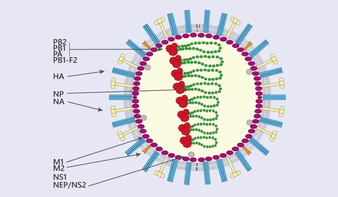
Figure 2 Schematic representation of influenza virus structure and genome organization
4.2 Viral Entry, Replication, and Release
HA recognizes sialic acid receptors on the host-cell surface and mediates viral endocytosis. Endosomal acidification induces conformational changes in HA and promotes fusion of the viral envelope with the endosomal membrane. The M2 proton channel promotes acidification within the viral particle, allowing viral ribonucleoprotein complexes to dissociate from M1 and be released into the cytoplasm. Viral ribonucleoprotein complexes then enter the nucleus, where transcription and genome replication are mediated by the viral polymerase. Newly synthesized viral RNA, nucleoprotein, polymerase, and envelope proteins assemble near the plasma membrane. NA cleaves sialic acid residues from the cell surface, promoting release of progeny virions from host cells.
4.3 Major Drug Targets
(1) M2 Proton Channel
Amantadine and rimantadine bind the M2 proton channel of influenza A virus and inhibit internal acidification and viral uncoating. Amino acid substitutions in M2 can markedly reduce drug binding and confer resistance.
(2) Neuraminidase
Zanamivir, oseltamivir, and laninamivir occupy the NA catalytic site and inhibit cleavage of sialic acid residues, preventing newly formed virions from being released efficiently from the cell surface and reducing spread to neighboring cells.
(3) Viral RNA Polymerase
After entering cells, favipiravir is converted into an active nucleoside triphosphate form that is recognized and incorporated by the viral RNA polymerase. This can inhibit RNA-chain elongation or increase the mutational burden of the viral genome.
(4) Membrane Fusion and Host Signaling
Arbidol can affect viral-envelope protein conformation and the membrane-fusion process. Influenza infection also activates MAPK, NF-κB, PI3K/AKT, and interferon-related signaling pathways. These host pathways regulate viral gene expression, inflammation, cell survival, and viral release and are therefore important directions in host-targeted antiviral research.
4.4 Influenza Virus-Related Products
Product Name | CAS No. | Major Target or Mechanism | Research Positioning |
Amantadine Hydrochloride | Blocks the influenza A virus M2 proton channel and inhibits uncoating | M2 channel, viral uncoating, and resistance-mutation research | |
Rimantadine | M2 proton-channel inhibitor | Influenza A virus M2 function and resistance research | |
Zanamivir | Neuraminidase inhibitor that prevents progeny-virus release | Influenza A and B virus NA research | |
Oseltamivir Phosphate | Oral prodrug converted metabolically into an active carboxylate form | Influenza NA inhibition and pharmacological research | |
Oseltamivir Acid | Active metabolite of Oseltamivir that directly inhibits NA | NA enzymatic activity, binding, and resistance research | |
Laninamivir | Long-acting neuraminidase inhibitor | Influenza-virus release and prolonged-inhibition studies | |
Arbidol Hydrochloride | Interferes with viral-envelope protein conformation and membrane fusion | Enveloped-virus entry and membrane-fusion research | |
Favipiravir | Metabolized into an active nucleoside triphosphate that inhibits viral RdRp | Influenza polymerase and RNA-replication studies |
5 Hepatitis C Virus
5.1 Viral Structure and Cell Entry
Hepatitis C virus (HCV) belongs to the family Flaviviridae and is an enveloped positive-sense single-stranded RNA virus that primarily infects hepatocytes. Viral particles sequentially interact with host factors including scavenger receptor class B type I, CD81, Claudin-1, and Occludin and enter cells through clathrin-mediated endocytosis. Endosomal acidification promotes fusion of the viral envelope with the endosomal membrane, and uncoating releases genomic RNA into the cytoplasm. Because HCV RNA itself functions as messenger RNA, it can directly initiate viral-protein translation.
5.2 Polyprotein Processing and RNA Replication
The HCV genome encodes a large polyprotein that is subsequently cleaved by host proteases and viral NS2 and NS3/4A proteases to generate mature core protein, E1, E2, p7, NS2, NS3, NS4A, NS4B, NS5A, and NS5B. NS3/4A processes the viral polyprotein and can also interfere with host innate immune signaling. NS4B induces formation of membranous replication platforms, NS5A participates in replication-complex organization, RNA replication, and viral-particle formation, and NS5B functions as the RNA-dependent RNA polymerase that catalyzes viral RNA synthesis.
5.3 Viral Assembly and Direct-Acting Targets
Newly synthesized HCV RNA and core protein undergo initial assembly near lipid droplets and coordinate with host very-low-density lipoprotein synthesis and secretion to form infectious lipoviral particles. HCV direct-acting antiviral compounds are mainly classified as NS3/4A protease inhibitors, NS5A inhibitors, and NS5B polymerase inhibitors. Host factors such as Cyclophilin A can also serve as indirect intervention targets. Combining compounds from multiple classes simultaneously suppresses viral protein processing, replication-complex formation, and RNA-chain elongation and reduces the effect of individual resistance mutations.
5.4 HCV-Related Products
Product Name | CAS No. | Major Target or Mechanism | Research Positioning |
Boceprevir | Reversibly binds HCV NS3 protease and inhibits polyprotein processing | First-generation NS3 protease-inhibitor and resistance research | |
Telaprevir | Inhibits NS3/4A protease | HCV protease structure, catalysis, and resistance studies | |
Paritaprevir | NS3/4A protease inhibitor | HCV polyprotein processing and combination-treatment research | |
Simeprevir | NS3/4A protease inhibitor | HCV protease and viral-replication studies | |
Grazoprevir | Second-generation NS3/4A protease inhibitor | HCV protease, genotype differences, and resistance research | |
Asunaprevir | NS3 protease inhibitor | HCV NS3 enzymatic activity and drug-sensitivity studies | |
Elbasvir | NS5A inhibitor | HCV replication-complex and combination-inhibition research | |
Ledipasvir | NS5A inhibitor | HCV RNA replication and viral-particle formation studies | |
Ombitasvir | NS5A inhibitor | Replication studies involving different HCV genotypes | |
Daclatasvir | NS5A inhibitor | NS5A function, viral replication, and resistance studies | |
Beclabuvir | NS5B non-nucleoside allosteric inhibitor | HCV polymerase conformation and replication studies | |
Sofosbuvir | Nucleotide analogue prodrug that inhibits NS5B and causes RNA-chain termination | HCV RNA polymerase and viral-replication research | |
Dasabuvir | NS5B non-nucleoside allosteric inhibitor | HCV polymerase allosteric-regulation research | |
Alisporivir | Inhibits Cyclophilin A-dependent HCV replication | Host-targeted anti-HCV mechanism research |
6 Hepatitis B Virus
6.1 Viral Genome and cccDNA
Hepatitis B virus (HBV) belongs to the family Hepadnaviridae and is an enveloped virus with a partially double-stranded relaxed circular DNA genome. After the virus enters hepatocytes, the nucleocapsid transports relaxed circular DNA into the nucleus. Host repair enzymes remove the viral polymerase and RNA primer, fill the DNA gaps, and generate covalently closed circular DNA (cccDNA). cccDNA persists as a minichromosome and serves as the principal template for continuous viral-RNA transcription. Its long-term stability is a major molecular basis for persistent HBV infection and viral rebound after treatment discontinuation.
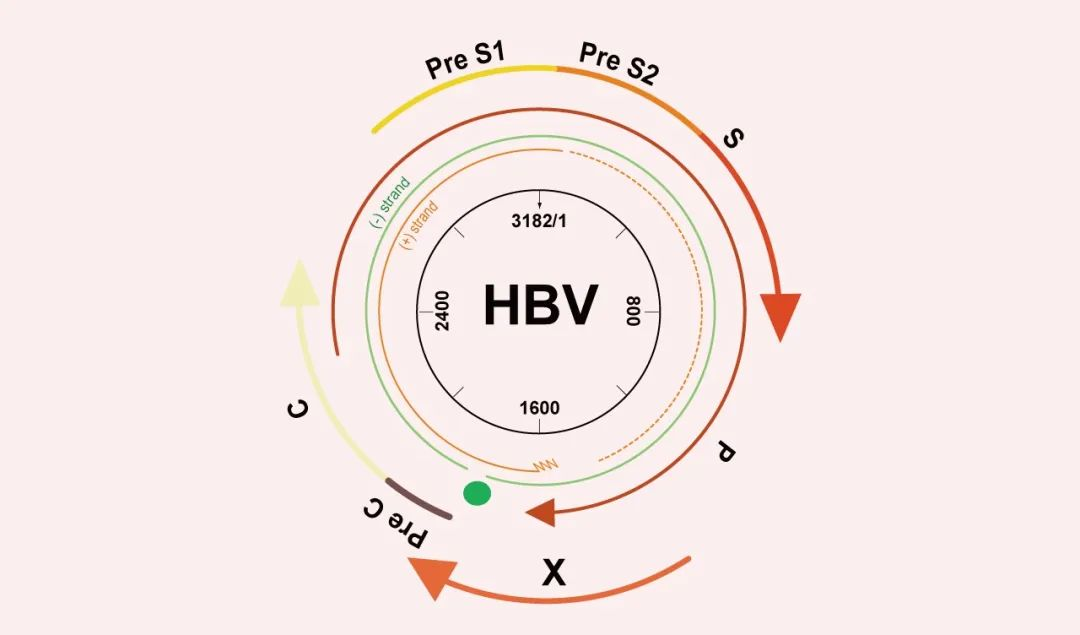
Figure 3 Schematic representation of HBV genome organization and functional regions
6.2 Pregenomic RNA and Reverse Transcription
cccDNA is transcribed into pregenomic RNA, surface-antigen RNAs, and other viral transcripts. Pregenomic RNA functions both as a translation template for core protein and polymerase and as a genome precursor packaged together with viral polymerase into newly forming nucleocapsids. Within the nucleocapsid, the viral polymerase first synthesizes negative-strand DNA using pregenomic RNA as the template while degrading the RNA template. It then synthesizes an incomplete positive-strand DNA, ultimately generating a new relaxed circular DNA genome.
6.3 Viral Particles and Antigen Release
Nucleocapsids containing mature viral DNA acquire envelopes composed of large, middle, and small surface proteins and are released as infectious Dane particles. Some nucleocapsids return to the nucleus and replenish the cccDNA pool. Infected cells also release large quantities of spherical or filamentous subviral particles lacking complete nucleocapsids. These particles mainly consist of HBsAg and are the principal source of the abundant circulating surface antigen detected in infected individuals.
6.4 Major Drug Targets
(1) HBV Polymerase
Nucleotide analogues such as Tenofovir and Adefovir are converted into active forms within cells, compete with natural substrates, and are incorporated by the viral polymerase into newly synthesized DNA, thereby inhibiting reverse transcription and viral-DNA synthesis.
(2) Core Protein and Capsid Assembly
Core protein allosteric modulators bind the core-protein dimer interface or capsid allosteric pockets, alter capsid-assembly kinetics, induce formation of aberrant capsids, or prevent proper packaging of pregenomic RNA and polymerase.
(3) Viral RNA and HBsAg Expression
Interference with HBV RNA tailing, stability, and degradation can reduce viral-transcript levels and subsequently decrease expression of HBsAg and other viral proteins. Research compounds such as RG7834 are mainly used to investigate PAPD5/PAPD7-related RNA-stability mechanisms.
(4) Host Immune Receptors
TLR7 and TLR8 agonists can stimulate dendritic cells, monocytes, and other immune cells to produce cytokines and enhance innate and subsequent adaptive immune responses. Their antiviral effects must be evaluated together with the intensity of immune activation, hepatic inflammation, and the safety range.
6.5 HBV-Related Products
Product Name | CAS No. | Major Target or Mechanism | Research Positioning |
RG7834 | Interferes with PAPD5/PAPD7-related RNA tailing and stability, reducing HBV RNA and HBsAg expression | HBV RNA-stability and surface-antigen suppression studies | |
Tenofovir | Nucleotide analogue that inhibits HBV polymerase-mediated reverse transcription | HBV polymerase, viral-DNA synthesis, and resistance research | |
Adefovir | Adenine nucleotide analogue that inhibits viral reverse transcription | HBV polymerase and nucleotide-analogue research | |
Vesatolimod | TLR7 agonist that enhances host innate immune responses | HBV immunomodulation and host-targeted research | |
Selgantolimod | TLR8 agonist that promotes innate and cellular immune responses | HBV- and HIV-related immunomodulatory research |
7 Comparison of Targeted Strategies for the Five Viruses
Virus | Genome Type | Major Drug Targets | Major Intervention Characteristics | Key Research Challenges |
SARS-CoV-2 | Enveloped positive-sense single-stranded RNA | S protein, 3CLpro, PLpro, and RdRp | Blocks viral entry, protein processing, and RNA replication | Viral evolution, target mutations, and therapeutic time window |
HIV | Enveloped retrovirus with an RNA genome | gp120, CCR5, CXCR4, gp41, reverse transcriptase, integrase, and protease | Multitarget intervention covering entry, reverse transcription, integration, and maturation | Proviral integration, latent reservoirs, and resistance |
Influenza virus | Enveloped segmented negative-sense RNA | HA, M2, NA, and viral polymerase | Interferes with uncoating, RNA replication, membrane fusion, and viral release | Antigenic drift, reassortment, and drug resistance |
HCV | Enveloped positive-sense single-stranded RNA | NS3/4A, NS5A, NS5B, and Cyclophilin A | Combined blockade of replication using direct-acting or host-targeted compounds | Genotype differences and resistance-associated variants |
HBV | Enveloped partially double-stranded DNA virus that reverse-transcribes through an RNA intermediate | Polymerase, core protein, capsid, viral RNA, and host immunity | Inhibits reverse transcription, capsid assembly, and viral-antigen expression | Persistence of cccDNA and immune tolerance |
The five viruses differ substantially in receptor recognition, genome replication, and viral-particle maturation. Major intervention stages for SARS-CoV-2 include S protein-mediated entry, viral-protease processing, and RdRp-mediated RNA replication. HIV can be inhibited at multiple stages, including attachment, membrane fusion, reverse transcription, integration, and maturation. Influenza antiviral drugs mainly target the M2 proton channel, neuraminidase, viral polymerase, and membrane-fusion process. HCV can be blocked at multiple steps through direct-acting targets including NS3/4A, NS5A, and NS5B. HBV treatment research must simultaneously address reverse transcription, capsid assembly, viral-RNA stability, and the persistence of cccDNA.
References
[1] Xu Z, et al. Nelfinavir was predicted to be a potential inhibitor of 2019-nCoV main protease by an integrative approach combining homology modelling, molecular docking and binding free energy calculation. bioRxiv.
[2] Wang M, et al. Remdesivir and chloroquine effectively inhibit the recently emerged novel coronavirus (2019-nCoV) in vitro. Cell Res. 2020;30:269-271.
[3] Wilen CB, Tilton JC, Doms RW. HIV: Cell Binding and Entry. Cold Spring Harb Perspect Med. 2012;2(8):a006866.
[4] Hutchinson EC. Influenza Virus. Trends Microbiol. 2018;26(9):809-810.
[5] Ludwig S, et al. Influenza-Virus-Induced Signaling Cascades: Targets for Antiviral Therapy? Trends Mol Med. 2003;9(2):46-52.
For more related articles, please see below:
[1] Hepatitis A virus RNA extraction
[2] Encephalitis B virus RNA extraction
[3] Influenza virus RNA extraction
[5] Pathogenesis, Virological Characteristics, and Laboratory Detection Strategies for Nipah Virus (NiV)


 危险品化学品经营许可证(带存储)