
Structural Features, Engineering Design, and Applications of Nanobodies in Tumor Diagnosis and Therapy
Structural Features, Engineering Design, and Applications of Nanobodies in Tumor Diagnosis and Therapy
Nanobodies have the characteristics of small molecular size, high epitope accessibility, and flexible engineering potential, and can be used in tumor molecular imaging, targeted delivery, immune regulation, and engineered cell therapy.
Keywords: nanobody; VHH; heavy-chain-only antibody; multivalency; multispecificity; tumor imaging; targeted therapy; drug delivery; immunotherapy; antibody library
1 Source, Structure, and Molecular Features of Nanobodies
1.1 Biological Source of Nanobodies
Nanobodies usually refer to the variable domain of camelid heavy-chain-only antibodies (VHH). Camelid heavy-chain-only antibodies naturally lack light chains and the CH1 domain of heavy chains, and a single VHH can independently complete antigen recognition. Their molecular weight is usually about 12-15 kDa. The variable domain of shark new antigen receptor antibodies, VNAR, also belongs to single-domain antibodies, but its evolutionary origin, structural composition, and antigen-binding mode differ from VHH. In tumor research, nanobodies usually mainly refer to VHH.
1.2 Structural Composition of VHH
VHH retains a typical immunoglobulin fold and is composed of framework regions and three complementarity-determining regions.
(1) Framework regions
The framework residues in VHH that originally contact the light chain are relatively hydrophilic, which can reduce aggregation caused by exposed hydrophobic surfaces after light chain absence and support independent folding of a single domain.
(2) CDR1 and CDR2
CDR1 and CDR2 participate in initial antigen surface recognition and binding interface formation. Their sequence and spatial conformation affect VHH specificity and affinity.
(3) CDR3
CDR3 is usually longer than the CDR3 of conventional antibody heavy-chain variable regions and can enter enzyme active pockets, receptor grooves, and other spatially restricted epitopes. Some VHHs also contain additional disulfide bonds connecting CDR3 with CDR1 or framework regions, helping stabilize the antigen-binding conformation.
1.3 Main Molecular Features of Nanobodies
(1) Small molecular size
A smaller hydrodynamic radius facilitates tissue diffusion and recognition of hidden epitopes, but also increases glomerular filtration and rapid clearance.
(2) Relatively simple expression and engineering
VHH does not require heavy-chain and light-chain pairing. It can be expressed in bacteria, yeast, or mammalian cells and is convenient for tandem fusion, domain fusion, site-directed mutagenesis, and chemical conjugation.
(3) Clone-dependent stability
Some VHHs have good thermal stability, solubility, and refolding ability, but aggregation tendency, freeze-thaw stability, and formulation tolerance differ markedly among clones and still need to be verified individually.
(4) Natural lack of Fc function
A single VHH cannot naturally mediate Fcγ receptor binding, complement activation, or FcRn recycling, so it does not have the long circulation and Fc effector functions of intact IgG. Functional supplementation is required according to the intended application.
2 Performance Differences Between Nanobodies and Traditional Monoclonal Antibodies
Comparison Item | Nanobody | Traditional Monoclonal Antibody |
Basic structure | Single VHH domain | IgG composed of two heavy chains and two light chains |
Molecular weight | Usually about 12-15 kDa | Usually about 150 kDa |
Epitope accessibility | More likely to recognize grooves, clefts, and hidden epitopes | More suitable for forming larger antigen-binding interfaces |
Distribution in solid tumors | Faster diffusion, but may be limited by high-affinity capture | Relatively slower penetration and longer tissue retention |
Blood half-life | Usually short in monovalent form | FcRn recycling supports longer circulation |
Fc-mediated effects | Naturally absent | Can mediate ADCC, ADCP, and complement effects |
Engineering features | Convenient for tandem linkage, fusion, and site-specific conjugation | Mature Fc engineering and industrial production systems |
Main application tendency | Rapid imaging, multispecific construction, and targeted delivery | Long-term blockade, systemic therapy, and immune effector recruitment |
3 Multivalent, Multispecific, and Functional Conjugation Design of Nanobodies
The core of nanobody engineering is to combine binding valency, target number, half-life, and functional payload according to target distribution, mechanism of action, and expected in vivo exposure.
(1) Multivalent design
Tandem fusion of two or more identical VHHs can increase apparent binding strength and receptor occupancy and enhance selectivity toward cells with high antigen density. Multivalent binding may also induce receptor clustering, internalization, or abnormal activation, so specific formats need to be evaluated together with target function.
(2) Multi-epitope design
Combining VHHs that recognize different epitopes of the same target can reduce escape caused by single-epitope masking or mutation and may enhance receptor blockade and internalization.
(3) Multispecific design
Combining recognition modules for different tumor antigens can expand coverage of tumor heterogeneity. Connecting tumor antigen-targeting VHHs with CD3, CD16, or immune checkpoint-binding modules can recruit T cells, recruit NK cells, or concentrate immune modulation in the tumor area.
(4) Half-life extension design
Albumin-binding VHHs, Fc fusion, or other structures that increase hydrodynamic radius can reduce renal clearance and prolong circulation time. Imaging probes usually need rapid reduction of blood background, while sustained therapeutic molecules require longer receptor occupancy, so their pharmacokinetic designs differ.
(5) Functional conjugation design
Nanobodies can be linked to fluorescent dyes, radionuclides, cytotoxic drugs, protein toxins, photosensitizers, or nanocarriers. Site-specific conjugation helps control modification site and payload number, reducing interference of random labeling with the antigen-binding interface.
4 Applications of Nanobodies in Tumor Molecular Imaging and Diagnosis
4.1 Tumor Molecular Imaging
(1) PET and SPECT imaging
Nanobodies can be linked to positron- or single photon-emitting radionuclides for detecting tumor antigen expression, lesion distribution, and interlesional heterogeneity. The radionuclide half-life should match the rapid distribution and clearance characteristics of nanobodies to reduce non-target tissue radiation caused by unbound probes and kidney uptake.
(2) Fluorescence imaging
Fluorescently labeled nanobodies can be used for cell imaging, tissue sections, multiplex marker analysis, and intraoperative navigation. Near-infrared dyes have lower tissue autofluorescence and are more suitable for in vivo tumor boundary recognition.
(3) Multimodal imaging
Integrating radioactive and fluorescent labels into the same nanobody can combine preoperative whole-body localization with intraoperative local navigation, but dual modification should avoid affecting binding activity and in vivo distribution.
4.2 Pathological Detection and Body Fluid Biomarker Analysis
Nanobodies are small and have good epitope accessibility, making them suitable as recognition molecules in immunohistochemistry, immunofluorescence, and biosensors, especially for detecting epitopes that are spatially restricted or easily masked from traditional antibodies. Immobilizing VHHs on electrochemical, optical, or microfluidic platforms can also be used to detect tumor antigens, exosomes, and other biomarkers in body fluids. Detection performance depends not only on affinity, but also on immobilization orientation, surface density, sample matrix, and antigen conformation.
4.3 Interpretation of Imaging and Diagnostic Results
High probe uptake in tumors may result from specific binding, but may also be influenced by vascular permeability, local blood flow, and nonspecific retention. Imaging studies should jointly evaluate tumor uptake value, tumor-to-blood ratio, tumor-to-muscle ratio, major organ distribution, and time-dependent changes, and should include non-binding VHHs, target-negative models, and competitive blocking groups. Enhanced fluorescence or radioactive signal cannot be directly equated with increased target expression and still needs confirmation through histological or protein detection.
5 Applications of Nanobodies in Tumor-Targeted Therapy and Drug Delivery
5.1 Tumor-Targeted Therapeutic Formats
(1) Receptor and ligand blockade
Nanobodies can bind tumor cell surface receptors, ligands, or enzymatic regions to block ligand binding, receptor dimerization, and downstream signaling. When monovalent VHH has a short duration of action, multivalency, dual-epitope binding, or half-life extension can improve blocking capacity.
(2) Nanobody-drug conjugates
VHHs can deliver cytotoxic drugs to tumor cells. Suitable targets should be highly expressed in tumors, expressed at low levels in critical normal tissues, and internalized after binding. The linker also needs to be stable in circulation and release the payload in the endosomal, lysosomal, or cytoplasmic environment.
(3) Nanobody-toxin fusion proteins
Fusing VHHs with protein toxins or toxin catalytic domains can use target-mediated endocytosis to kill tumor cells. These molecules have high potency, but low-level target expression in normal tissues and nonspecific uptake may also cause significant toxicity.
(4) Radionuclide-targeted therapy
Therapeutic α or β radionuclides can be delivered to tumor tissues through nanobodies, using radiation to kill target cells and neighboring cells. Kidney reabsorption and retention of radioactive metabolites are important limitations.
(5) Targeted delivery by nanocarriers
Nanobodies can modify liposomes, polymer particles, or other carriers for delivery of chemotherapeutic drugs, nucleic acids, and photosensitizers. The overall particle size, surface charge, and protein corona of the carrier will redefine in vivo distribution, so the high penetration of free VHH cannot be directly extrapolated to large carriers.
5.2 Key Evaluation Factors for Targeted Delivery
Drug delivery efficacy needs to consider target expression, internalization speed, intracellular trafficking, linker stability, payload release, and normal tissue uptake simultaneously. Observing nanobody binding to the cell surface alone does not prove that the payload can enter the appropriate intracellular compartment. In vitro killing results alone also cannot replace evaluation of serum stability, free payload, and major organ distribution.
6 Role of Nanobodies in Tumor Immune Regulation and Cell Therapy
6.1 Tumor Immune Regulation Strategies
(1) Immune checkpoint blockade
Nanobodies can block inhibitory receptor-ligand interactions between tumor cells and immune cells, and can also be combined with tumor antigen-recognition modules to increase the local concentration of immunoregulatory molecules in tumors.
(2) T cell recruitment
After a tumor antigen-targeting VHH is linked with a CD3-binding module, it can bind tumor cells and T cells simultaneously, inducing immune synapse formation and target cell killing.
(3) NK cell recruitment
Combining tumor-targeting VHHs with CD16 or other NK cell receptor-binding modules can promote NK cell proximity to and killing of tumor cells.
(4) Tumor microenvironment regulation
Nanobodies can target immunosuppressive cytokines, chemokines, myeloid cells, or abnormal stromal components to alter immune cell infiltration and local immunosuppressive status.
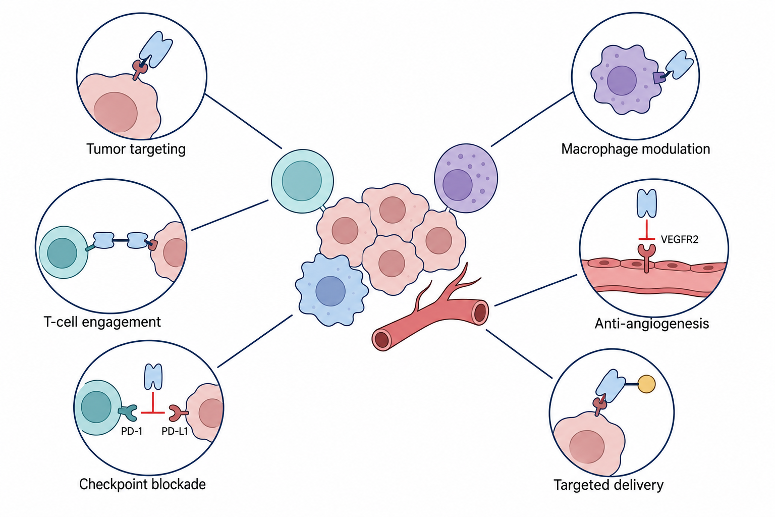
Figure 1. Schematic of Nanobody Applications in Tumor-Targeted Therapy, Drug Delivery, and Immune Microenvironment Modulation
6.2 Applications of Nanobodies in Engineered Cells
VHHs can replace traditional single-chain variable fragments as the extracellular recognition domains of CAR-T or CAR-NK cells. Their simple structure and low risk of interchain mismatch facilitate construction of dual-target and multitarget CARs. VHHs can also be expressed intracellularly as intracellular nanobodies to interfere with oncogenic proteins, fusion proteins, or signaling complexes, and can be connected with protein degradation modules to reduce target protein levels.
High binding capacity of nanobodies in the free state does not mean they will necessarily have ideal function in CAR or multispecific formats. Epitope position, hinge length, transmembrane region, co-stimulatory domain, and immune synapse geometry all affect final activity. Excessively strong or sustained effector cell activation may also cause cytokine release, exhaustion, and normal tissue damage.
7 Nanobody Library Construction, Screening, and Preparation Technologies
7.1 Nanobody Library Types
(1) Immune library
Immune libraries are derived from camelids immunized with target antigens. They usually make it easier to obtain specific clones that have undergone in vivo affinity maturation, but require an animal immunization cycle.
(2) Naïve library
Naïve libraries are derived from individuals not immunized with a specific antigen and can be used for screening multiple targets, but usually require a larger effective library size.
(3) Synthetic and semi-synthetic libraries
Synthetic and semi-synthetic libraries artificially design CDR diversity based on stable VHH frameworks. They can reduce animal dependence and improve sequence controllability. Their performance depends on framework selection, amino acid distribution, and actual functional diversity.
7.2 Nanobody Screening Methods
(1) Phage display
Phage display enriches binding clones through multiple rounds of antigen adsorption, washing, and amplification. It is a mature technology suitable for large-scale libraries.
(2) Yeast display
Yeast display can be combined with flow cytometry sorting to evaluate both VHH surface expression and antigen-binding ability.
(3) Ribosome display and mRNA display
These methods are not limited by cell transformation efficiency and can establish larger in vitro libraries, but subsequent clone expression and functional validation requirements are higher.
(4) Cell-based screening
When targeting membrane receptors, target-positive cells can be used for screening, and target-negative cells can be used to subtract nonspecific clones, improving recognition of native conformational epitopes.
7.3 Affinity Maturation, Humanization, and Preparation
Affinity maturation can improve binding capacity through CDR site-directed mutagenesis, error-prone PCR, and secondary library screening, but specificity, dissociation rate, expression level, and aggregation risk should be evaluated simultaneously. Humanization mainly reduces potential immunogenicity by adjusting VHH framework residues while preserving key sites that maintain independent folding and CDR conformation. Excessive replacement may reduce stability and affinity and needs to be corrected through back-mutation and experimental validation.
Nanobodies can be expressed in bacteria, yeast, or mammalian cells. After preparation, purity, monomer ratio, molecular weight, aggregation, endotoxin, binding affinity, and biological activity need to be tested. Multivalent, fusion, or conjugated molecules also require evaluation of structural integrity, labeling efficiency, serum stability, and binding ability after engineering.
8 Pharmacokinetics, Safety, and Technical Challenges in Clinical Translation of Nanobodies
Clinical translation of nanobodies needs to address in vivo exposure, safety, target selection, and production consistency simultaneously.
(1) Rapid clearance and kidney uptake
Monovalent VHHs are usually rapidly filtered through the glomerulus and may be reabsorbed in proximal renal tubules. Rapid clearance is suitable for imaging, but reduces sustained exposure of therapeutic molecules and increases renal accumulation of radioactive or toxic payloads.
(2) Immunogenicity
Non-human frameworks, CDRs, artificial linkers, and newly formed structural interfaces may all induce anti-drug antibodies. Humanization can reduce some risks, but cannot completely eliminate immunogenicity.
(3) Normal tissue toxicity
High affinity, multivalency, and cytotoxic payloads can amplify damage caused by low-level target expression in normal tissues. Therefore, tumor expression proportion, normal tissue distribution, soluble antigen, and target internalization need to be comprehensively evaluated.
(4) Tumor heterogeneity and antigen escape
A single target may not cover all tumor cells. Dual-target or multitarget design can reduce escape risk, but also increases the complexity of normal tissue cross-binding and mechanism-of-action analysis.
(5) Molecular stability and aggregation
Multivalency, Fc fusion, and hydrophobic payload conjugation may increase aggregation, degradation, and decreased expression level. A quality control system capable of distinguishing monomers, aggregates, degradation products, and different conjugation forms needs to be established.
(6) Dose and mechanism-of-action validation
Clinical dose cannot be calculated only from in vitro affinity. Pharmacokinetics, tissue distribution, receptor occupancy, and pharmacodynamic biomarkers also need to be considered to distinguish insufficient exposure, target independence, and downstream resistance.
9 Products Related to Nanobody Research
Cat. No. | Product Name | Grade & Purity | Research Stage | Main Application |
VHH Human IgG1-Fc, anti-HEL | Carrier-free, recombinant, ExactAb™, low endotoxin, azide-free, validated, PBS Only, ≥95%(SDS-PAGE), see COA | VHH-Fc fusion design | Used as an Fc-fused nanobody model to study molecular bivalency, Fc function introduction, and half-life extension | |
Human IgG1-VHH (AF647), anti-HEL | ExactAb™, validated, Ex:650 nm, Em:668 nm, 0.5 mg/mL | Far-red labeled VHH | Used to observe VHH binding, cellular localization, and tissue distribution, and serves as a fluorescently labeled nanobody model | |
Human IgG1-VHH (Biotin), anti-HEL | ExactAb™, validated, 0.5 mg/mL | Biotin-labeled VHH | Used in ELISA, affinity capture, and streptavidin detection systems to evaluate nanobody binding performance | |
Human IgG1-VHH (FITC), anti-HEL | ExactAb™, validated, Ex:498 nm, Em:517 nm, 0.5 mg/mL | Green fluorescent labeled VHH | Used in flow cytometry and fluorescence imaging to verify antigen-binding ability after VHH labeling | |
EZ-HiFi Seamless Cloning Kit | BioReagent, Suitable for molecular biology, sterile | Nanobody construction | Used for VHH sequence cloning, linker insertion, and construction of multivalent or multispecific expression vectors | |
Rapid Multi-fragment DNA Seamless Cloning Premix | BioReagent, 2× | Multimodule sequence assembly | Used for one-step assembly of multiple VHHs, Fc, tags, or functional domains | |
Ultra-Universal One Step Seamless Cloning Mix |
| VHH vector construction | Used for rapid assembly of nanobody expression vectors, fusion proteins, and engineered formats | |
BDP FL NHS Ester | Ex:502 nm, Em:511 nm, ≥95% | Nanobody fluorescent conjugation | Labels VHHs or VHH fusion proteins through active ester chemistry for green fluorescence imaging and binding detection | |
Cyanine 3, SE | ≥95%(HPLC) | Nanobody fluorescent conjugation | Used for orange-red labeling of nanobodies and multichannel imaging | |
Cyanine 5, SE | Ex:647 nm, Em:665 nm, ≥95%(HPLC) | Far-red conjugation | Used for cell, tissue, and in vivo distribution imaging; affinity and nonspecific binding need to be revalidated after labeling | |
Cypate (Near-infrared fluorescent dye) | BioReagent, biological stain, suitable for fluorescence analysis, for microscopy, ≥97% | Near-infrared imaging design | Used to construct near-infrared fluorescent nanobody probes for studying tumor localization and tissue distribution | |
Human BCMA/TNFRSF17 ELISA Kit | BioReagent | BCMA target quantification | Detects BCMA levels in samples and provides target information for BCMA-targeted nanobody or engineered cell research | |
Human Carcinoembryonic Antigen (CEA) ELISA Kit | BioReagent, for Enzyme immunoassay(ELISA) | CEA target quantification | Detects soluble CEA for sample stratification and result validation in CEA-targeted nanobody models | |
Human Carcinoembryonic Antigen Related Cell Adhesion Molecule 6 (CEACAM6) ELISA Kit | BioReagent | CEACAM6 target quantification | Used for CEACAM6-related tumor biomarker research and sample detection in targeted molecule development | |
Human Carbohydrate Antigen 125 (CA125) ELISA Kit | BioReagent | Tumor biomarker detection | Detects CA125 levels and assists in evaluating related tumor models and diagnostic research samples | |
Human Cytotoxic T-Lymphocyte Associated Antigen 4 (CTLA4) ELISA Kit | BioReagent | Immune checkpoint detection | Detects CTLA4 levels and assists in immune checkpoint nanobody and immune regulation research | |
Human Melanoma-associated Antigen 3 (MAGE-A3) ELISA Kit | BioReagent | Tumor antigen detection | Used for MAGE-A3-related tumor sample evaluation and target antigen research | |
Endotoxin Removal Resin | BioReagent, 50% v/v | Nanobody sample purification | Removes endotoxin from recombinant nanobody or fusion protein preparations, suitable for pretreatment before cell and animal functional experiments | |
Endotoxin Removal Reagent | BioReagent, Suitable for molecular biology, for DNA and RNA applications | Nucleic acid sample endotoxin control | Reduces endotoxin interference in nanobody expression plasmids or nucleic acid delivery samples | |
Endo-free Plasmid Maxi Kit | BioReagent, for DNA and RNA applications | Expression vector preparation | Prepares low-endotoxin VHH expression plasmids for cell transfection and engineered cell construction | |
Anti-Flag Nanobody Magnetic Beads | BioReagent, magnetic bead content: ≥10 mg/mL; bead diameter: 2 μm | Tagged protein enrichment | Used for immunoprecipitation and purification of Flag-tagged VHHs, fusion nanobodies, and related protein complexes | |
Anti-GFP Nanobody Magnetic Beads | BioReagent, magnetic bead content: ≥10%(V/V); bead diameter: 10-30 μm | GFP fusion protein enrichment | Used for GFP-fused VHH expression validation, affinity purification, and protein interaction analysis | |
Anti-HA Nanobody Magnetic Beads | BioReagent, magnetic bead content: ≥10 mg/mL; bead diameter: 2 μm | HA-tagged protein enrichment | Used for immunoprecipitation of HA-tagged nanobodies or engineered fusion proteins | |
Anti-V5 Nanobody Magnetic Beads | BioReagent, magnetic bead content: ≥10 mg/mL; bead diameter: 2 μm | V5-tagged protein enrichment | Used for expression validation, purification, and interaction research of V5-tagged VHHs | |
Anti-mCherry Nanobody Magnetic Beads | BioReagent, magnetic bead content: ≥10%(V/V) | Fluorescent fusion protein enrichment | Used for immunoprecipitation and purification of mCherry-fused nanobodies or related complexes | |
Flag-tag Protein IP Assay Kit (Nanobody Magnetic Beads Method) | BioReagent, ready-to-use, for IP | Flag fusion protein immunoprecipitation | Used for rapid enrichment and interaction validation of Flag-tagged VHHs and their binding complexes | |
GFP-tag IP/Co-IP (Nanobody Magnetic Beads) | BioReagent, for IP | GFP fusion protein co-precipitation | Used to analyze expression, stability, and protein interactions of GFP-fused nanobodies | |
HA IP/Co-IP Kit (Nanobody Magnetic Beads) | BioReagent, for IP | HA fusion protein co-precipitation | Used for interaction analysis of HA-tagged VHHs and engineered fusion proteins | |
RFP-Tag IP/Co-IP Kit (Nanobody Magnetic Beads) | BioReagent, for IP | RFP fusion protein co-precipitation | Used for purification and protein complex research of red fluorescent protein-fused VHHs | |
V5-tag IP/Co-IP Kit (Nanobody Magnetic Beads) | BioReagent, ready-to-use, for IP | V5 fusion protein immunoprecipitation | Used for expression validation and interaction research of V5-tagged nanobodies and fusion constructs |
The advantage of nanobodies lies not only in their small molecular size, but also in the ability to design multivalency, multispecificity, half-life, and functional payloads around target features and mechanisms of action. Only by jointly evaluating target selection, in vivo distribution, binding and functional activity, preparation quality, and safety can their structural advantages be translated into reliable tumor diagnostic and therapeutic effects.
For more related articles, please see below:
[2] Principles, Experimental Design, and Application Guide for IP/Co-IP Tag Immunomagnetic Beads
[5] Immunoprecipitation Kits: Principles, System Composition, and Typical Applications


 危险品化学品经营许可证(带存储)