
Structure–Property Relationships of Anthraquinones and Related Anthranoid Glycosides: Redox Behavior, Substituent Effects, and Biotransformation
Structure–Property Relationships of Anthraquinones and Related Anthranoid Glycosides: Redox Behavior, Substituent Effects, and Biotransformation
Anthraquinone compounds are widely found in plant natural products, dyes, and organic redox materials. Different anthraquinone derivatives can exhibit marked differences in color, reduction potential, acid–base properties, dissolution behavior, and biotransformation. These properties are governed by the same set of structural factors: the quinoid 9,10-dicarbonyl group determines the fundamental electronic properties; peripheral substituents alter electron distribution, hydrogen bonding, and ionization states; and glycosyl groups further modify polarity, bond stability, and modes of biotransformation.
1 Basic Structures of Anthraquinones and Related Anthracene Derivatives
1.1 From Anthracene to 9,10-Anthraquinone
Anthracene consists of three linearly fused six-membered carbon rings and has the molecular formula C₁₄H₁₀. Anthraquinone (AQ) usually refers to 9,10-anthraquinone, also known as anthracene-9,10-dione, with the molecular formula C₁₄H₈O₂.[1]
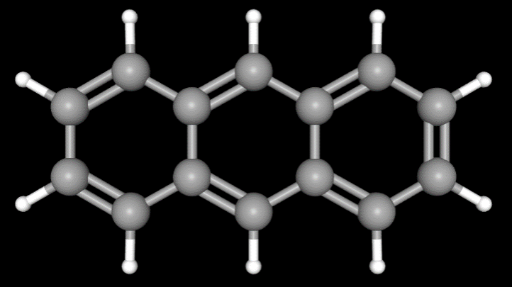

The principal structural difference between the two compounds lies in the central six-membered ring:
Anthracene: C9 and C10 are C—H carbon centers
9,10-Anthraquinone: C9 and C10 each form a C=O group
Anthraquinone therefore contains two carbonyl groups positioned opposite each other. In the conventional valence-bond representation, the central ring has a characteristic quinoid dicarbonyl structure, whereas the two outer rings retain benzene-like structural features. Electronic coupling remains between the p orbitals of these regions, forming an extended π-electron system.
Introduction of the two carbonyl groups alters the electronic structure of the original anthracene framework, giving 9,10-anthraquinone more pronounced electron-accepting and redox properties than anthracene. The color, reduction behavior, and substituent effects discussed below all arise from this structural change.
1.2 Anthraquinones, Anthrones, and Dianthrones
Anthracene-derived compounds commonly encountered in natural-product research do not all have the same oxidation state. These structures can be classified according to their core frameworks:
Type | Core structural feature | Representative compounds |
9,10-Anthraquinone | Carbonyl groups at both C9 and C10 | Anthraquinone, emodin, chrysophanol, aloe-emodin, rhein |
Anthraquinone O-glycoside | Retains the 9,10-anthraquinone core; the sugar is connected to a phenolic hydroxyl group through an oxygen atom | Emodin-8-O-β-D-glucoside |
Anthrone | Contains one carbonyl group and the corresponding dihydroanthracene structure | Aloe-emodin anthrone |
Anthrone-type C-glycoside | The sugar is attached to the anthrone core through a C—C bond | Aloin A |
Dianthrone glycoside | Two anthrone-type units are connected through a C—C bond and contain glycosyl groups | Sennoside A, sennoside B |
Emodin-8-O-β-D-glucoside is a structurally well-defined anthraquinone O-glycoside.[7] Aloin A, in contrast, has an anthrone core, and its glucose is attached through a C—C bond; it is therefore classified as an anthrone-type C-glycoside.[10,11] Sennoside A has a dianthrone diglucoside structure.[12]
2 How the 9,10-Dicarbonyl Group Confers Redox Properties on Anthraquinone
2.1 Conjugated Carbonyl Groups Form an Electron-Accepting Structure
The oxygen atom in a C=O bond is highly electronegative, giving the carbonyl group pronounced electronic polarization. At the same time, the two carbonyl groups of anthraquinone interact with the surrounding π system, giving the molecule low-energy unoccupied orbitals with π* character that can accept external electrons.
After anthraquinone accepts an electron, the newly introduced negative charge is not confined entirely to a single carbonyl oxygen. Instead, it can be redistributed throughout the quinoid structure. This electron delocalization helps stabilize the reduced states and provides an important structural basis for the reversible or quasi-reversible reduction of quinones.[2,3]
In aprotic media, 9,10-anthraquinone can undergo two consecutive one-electron reduction processes. It first accepts one electron to form the anthraquinone radical anion (AQ•⁻) and then accepts a second electron to form the anthraquinone dianion (AQ²⁻). This stepwise reduction reflects the ability of the anthraquinone dicarbonyl group and its conjugated electronic structure to stabilize different reduced states.[3]
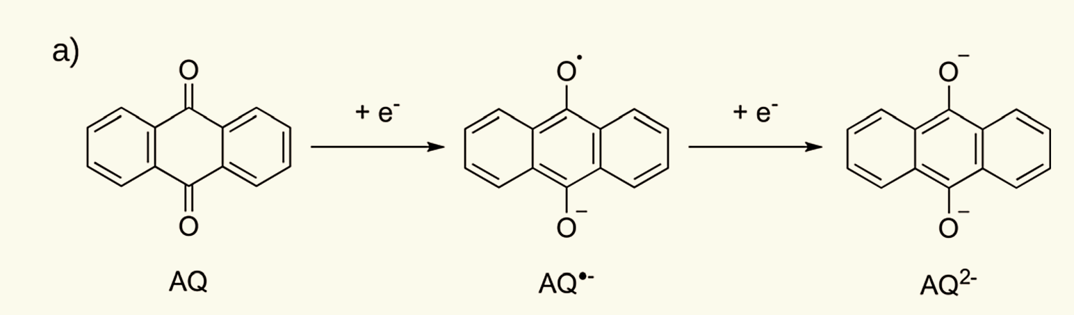
Stepwise One-Electron Reduction of 9,10-Anthraquinone
9,10-Anthraquinone (AQ) accepts one electron to form the anthraquinone radical anion (AQ•⁻) and then accepts a second electron to form the anthraquinone dianion (AQ²⁻). Source: Gallmetzer JM, Kröll S, Werner D, et al. Physical Chemistry Chemical Physics, 2022, 24: 16207–16219, Fig. 1(a), CC BY 4.0.
2.2 Stepwise One-Electron Reduction in Aprotic Media
In aprotic media such as acetonitrile, 9,10-anthraquinone can exhibit two consecutive one-electron reduction processes.[2]
First one-electron reduction:
AQ + e⁻ ⇌ AQ•⁻
The anthraquinone radical anion is formed.
Second one-electron reduction:
AQ•⁻ + e⁻ ⇌ AQ²⁻
Acceptance of a second electron produces the dianion.
Cyclic voltammetry can reveal two reduction processes corresponding to the formation of AQ•⁻ and AQ²⁻. Chemically, this means that the anthraquinone dicarbonyl group can accept electrons consecutively, while the conjugated structure stabilizes the different degrees of reduction.[2]
2.3 Hydrogen Bonding and Proton Participation Alter the Reduction Pathway
When hydrogen-bond donors or proton donors, such as alcohols, phenols, and carboxylic acids, are added to the system, the negative charge generated by reduction can receive additional stabilization through hydrogen bonding or protonation. Experiments show that, as the donor interaction becomes stronger, the two reduction waves of anthraquinone can shift toward more positive potentials, move closer together, and under certain conditions appear as a two-electron reduction process.[2]
Under acidic aqueous conditions, electron transfer can also become coupled with proton transfer, resulting in proton-coupled electron transfer (PCET).
The overall reduction of 9,10-anthraquinone-2,7-disulfonic acid in aqueous sulfuric acid can be represented as follows:
AQDS + 2e⁻ + 2H⁺ ⇌ AQDSH₂
Here, AQDS denotes 9,10-anthraquinone-2,7-disulfonic acid. Huskinson et al. showed that this compound undergoes a rapid and reversible two-electron–two-proton reduction in sulfuric acid on a glassy carbon electrode.[13]
The number of participating protons does not remain constant under all pH conditions. The potential–pH relationship of this system indicates that different overall processes, including two-electron/two-proton, two-electron/one-proton, and two-electron/no-proton processes, can occur under different acid–base conditions.[13]
The redox behavior of anthraquinone is therefore governed jointly by three factors:
Factor | Effect on the reduction process |
Quinoid 9,10-dicarbonyl group | Provides the principal electron-accepting structure |
Conjugated π system | Distributes the electron density generated by reduction and stabilizes the reduced states |
Hydrogen bonding, protons, and substituents | Alter the relative stability of the oxidized and reduced states, thereby regulating the reduction potential and reaction pathway |
3 Why Hydroxy and Other Substituents Impart Color to Many Anthraquinone Derivatives
3.1 Color Arises from Changes in Electronic Transition Energies
For a molecule to display visible color, its electronic absorption must extend into the visible region.
9,10-Anthraquinone contains a π-electron system and carbonyl groups, but the principal absorption of unsubstituted anthraquinone in solution does not extend into the visible region as prominently as that of hydroxyanthraquinones. After hydroxy or other substituents are introduced, their electronic interactions with the anthraquinone π system can alter the energy gap between occupied and unoccupied orbitals, shifting low-energy electronic transitions toward the visible region.[4]
Alizarin (1,2-dihydroxyanthraquinone) is a representative example. In acidic solution, alizarin exhibits pronounced visible absorption at approximately 425 nm, and this low-energy absorption is primarily associated with a π→π* transition. Purpurin (1,2,4-trihydroxyanthraquinone) contains one additional hydroxyl group compared with alizarin and consequently displays a markedly different visible absorption spectrum.[4]
Hydroxyl groups therefore do not merely increase molecular polarity; they also directly alter the electron distribution within the conjugated anthraquinone system.
3.2 Intramolecular Hydrogen Bonding Further Modulates the Electronic Structure
When a phenolic hydroxyl group is located adjacent to a carbonyl group, an O—H···O=C intramolecular hydrogen bond can form.
Spectroscopic studies of alizarin have shown that one of its phenolic hydroxyl groups can form a strong intramolecular hydrogen bond with the carbonyl group at C9. Researchers observed the corresponding O—H stretching features using fluorescence excitation, laser-induced fluorescence, and fluorescence-detected infrared spectroscopy.[5]
Intramolecular hydrogen bonding alters the electron density around the hydroxyl and carbonyl groups while also restricting the local molecular conformation. Solvent hydrogen bonding, phenolic hydroxyl dissociation, and metal-ion coordination can further alter the energy differences between electronic states. Consequently, the same hydroxyanthraquinone can display different absorption maxima and colors in different solvents or under different pH and coordination conditions.
The coloration of anthraquinone derivatives can be summarized as follows:
The conjugated carbonyl groups of anthraquinone establish the fundamental electronic structure → hydroxy and other substituents alter electron density and orbital energy levels → intramolecular hydrogen bonding and ionization states further modulate electronic transition energies → visible absorption and color change.
Chrysophanol, emodin, aloe-emodin, and rhein all retain the 9,10-anthraquinone core but differ in the number of hydroxyl groups and the functional groups attached to their side chains.[6]
Compound | Principal substitution pattern | Resulting chemical difference |
Chrysophanol | 1,8-Dihydroxy, 3-methyl | Contains two phenolic hydroxyl groups; the methyl group itself does not participate in common acid–base ionization or conventional hydrogen bonding |
Aloe-emodin | 1,8-Dihydroxy, 3-hydroxymethyl | The hydroxymethyl group introduces an additional alcoholic hydroxyl group and increases the capacity for intermolecular hydrogen bonding |
Rhein | 1,8-Dihydroxy, 3-carboxyl | The carboxyl group can undergo acid–base dissociation, causing the charge state to vary with pH |
Emodin | 1,3,8-Trihydroxy, 6-methyl | The additional phenolic hydroxyl group further alters electronic effects, hydrogen bonding, and ionization behavior |
4.1 Methyl, Hydroxymethyl, and Carboxyl Groups Produce Different Intermolecular Interactions
Chrysophanol, aloe-emodin, and rhein contain methyl, hydroxymethyl, and carboxyl groups, respectively. These three functional groups exhibit distinctly different chemical properties.
The methyl group (—CH₃) consists mainly of C—C and C—H bonds. It does not act as a conventional hydrogen-bond donor or acceptor and does not undergo ionization under ordinary acid–base conditions.
The hydroxymethyl group (—CH₂OH) introduces an alcoholic hydroxyl group. The hydroxyl group can serve as a hydrogen-bond donor, while its oxygen atom can serve as a hydrogen-bond acceptor, thereby increasing interactions with water and other polar molecules.
In addition to its hydrogen-bonding capacity, the carboxyl group (—COOH) can undergo acid–base dissociation:
—COOH ⇌ —COO⁻ + H⁺
When the carboxyl group is converted into a carboxylate, the molecule carries a negative charge, producing marked changes in hydration, ionic interactions, and partitioning behavior in different solvents.
The important differences among these three functional groups include their ability to form hydrogen bonds, undergo ionization, and form charged species to different extents at a given pH.
4.2 Hydroxyl-Group Position Affects Electronic Effects and Intramolecular Hydrogen Bonding
Emodin has a 1,3,8-trihydroxy substitution pattern. Phenolic hydroxyl groups at different positions do not have identical effects.
Hydroxyl groups located adjacent to anthraquinone carbonyl groups have the geometric arrangement required to form intramolecular O—H···O=C hydrogen bonds. Hydroxyl groups at other positions influence molecular properties mainly through conjugation, acid–base dissociation, and intermolecular hydrogen bonding.
Electrochemical studies show that even when the functional-group type is unchanged, placing a hydroxyl or amino group at different positions on anthraquinone can produce different reduction potentials.[3]
The properties of anthraquinone derivatives are therefore jointly determined by the type, number, and position of their functional groups and cannot be predicted solely from the number of hydroxyl groups.
Actual aqueous solubility likewise cannot be inferred directly from the assumption that “more hydroxyl groups produce higher solubility.” Dissolution is governed simultaneously by solvation, ionization, intramolecular hydrogen bonding, intermolecular hydrogen bonding in the crystal, and π-stacking. Even if a molecule interacts more strongly with water, enhanced stabilization of its crystal structure may prevent its aqueous solubility from increasing in parallel with the number of functional groups.
5 How Glycosylation Alters the Properties of Anthracene-Derived Compounds
5.1 Anthraquinone O-Glycosides: Changes in Polarity and Glycosidic-Bond Hydrolysis
In anthraquinone O-glycosides, the anomeric carbon of the sugar is connected through an oxygen atom to a phenolic hydroxyl group on the anthraquinone.
Rhein-8-O-β-D-glucoside, chrysophanol-8-O-β-D-glucoside, aloe-emodin-8-O-β-D-glucoside, and emodin-8-O-β-D-glucoside have been isolated and identified from the roots of Rheum palmatum.[7]
Glucose contains multiple hydroxyl groups. Glycosylation therefore generally increases the number of hydrogen-bonding sites, polar surface area, and modes of solvation while also increasing molecular size. Glycosides and their corresponding aglycones consequently often differ in chromatographic retention, phase partitioning, and biological-membrane permeability.
O-Glycosides also have an important structural characteristic: the core and sugar are connected by a hydrolyzable O-glycosidic bond.
Experimental studies have shown that, under pressurized liquid extraction conditions using methanol/water systems, emodin-8-O-β-D-glucoside, chrysophanol-8-O-β-D-glucoside, and physcion-8-O-β-D-glucoside can undergo hydrolysis to form their corresponding aglycones. Increasing the extraction temperature or the proportion of water in the system accelerates this process.[8]
Studies of anthraquinone glycosides from madder root also show that these glycosides can be converted into the corresponding products with fewer glycosyl groups or into their aglycones through acid or enzymatic hydrolysis.[9]
O-Glycosylation therefore introduces chemical changes at two levels:
① The glycosyl group increases the number of hydroxyl groups and polar interactions;
② The O-glycosidic bond introduces a reaction site that can undergo chemical or enzymatic cleavage.
5.2 C-Glycosides: The Sugar and Core Are Connected Through a C—C Bond
The principal structural difference between C-glycosides and O-glycosides lies in the linkage between the sugar and the core.
O-Glycosides contain a C—O—C linkage, whereas in C-glycosides, the anomeric carbon of the sugar forms a direct C—C bond with a carbon atom of the core. This linkage does not undergo the ordinary acetal hydrolysis pathway of an O-glycosidic bond; different chemical or enzymatic reactions are therefore required to cleave the connection between the core and the sugar.
Aloin A, also known as barbaloin, is a representative example. It is an aloe-emodin anthrone-type C-glucoside rather than a 9,10-anthraquinone O-glucoside.[11]
Early cell-free biosynthesis experiments further demonstrated that glucose from uridine diphosphate glucose can be transferred to aloe-emodin anthrone, forming the C—C glycosidic bond of aloin.[10]
The C—C linkage increases the resistance of the core–sugar connection to conventional glycosidic-bond hydrolysis. This does not mean that the entire molecule remains stable under all conditions; the anthrone core itself can still undergo oxidation, reduction, or other structural transformations.
5.3 Dianthrone Glycosides: The Structure and Intestinal Transformation of Sennoside A
Sennoside A consists of two connected anthrone-type units that form a dianthrone structure and contains two β-D-glucosyl groups. Its molecular formula is C₄₂H₃₈O₂₀.[12]
The intestinal transformation of sennoside A involves hydrolysis of its O-glycosidic bonds and reductive cleavage of the dianthrone structure. These two types of reactions can occur in different sequences. Reported pathways include initial hydrolysis to form sennidin A followed by reduction, as well as initial reduction to form 8-glucosylrheinanthrone followed by hydrolysis, ultimately producing rhein anthrone. Hydrolases and reductases produced by intestinal microorganisms participate in these respective steps.[14]
A 2025 study identified the NfrA protein with sennoside A-reducing activity from Bifidobacterium pseudocatenulatum and verified its reductive activity using the recombinant protein.[14] A subsequent study identified different bacterial guilds with similar sennoside A-reducing activity in the human gut microbiota, together with a new related reductase.[15]
Sennoside A illustrates another important factor in the study of glycoside structures: the mode of glycosyl attachment determines certain bond-cleavage reactions, while the oxidation state of the core determines subsequent redox reactions; together, these factors influence the metabolites ultimately formed.
6 Typical Research Applications of Anthraquinone Structures
Research area | Structural property utilized | Representative research finding |
Aqueous organic redox flow batteries | The quinoid dicarbonyl group can undergo reversible electron–proton transfer | 9,10-Anthraquinone-2,7-disulfonic acid undergoes rapid two-electron–two-proton reduction[13] |
Photoinduced charge accumulation | Anthraquinone has electron-accepting ability, and its reduction can be coupled with proton transfer | A copper(I)–anthraquinone molecular system achieves photochemical two-electron accumulation on the anthraquinone unit[16] |
Natural-product metabolism research | Glycosidic bonds and reducible cores provide defined sites for biotransformation | Sennoside A reductases have been identified through chemical-probe, proteomic, and recombinant-enzyme experiments[14,15] |
6.1 Redox Energy Storage
Huskinson et al. used 9,10-anthraquinone-2,7-disulfonic acid to construct an aqueous organic redox flow battery. The anthraquinone moiety of this molecule performs the redox reaction, while the sulfonic acid groups substantially increase its aqueous solubility. The study also showed that structural modification of the peripheral groups on anthraquinone can simultaneously regulate the reduction potential and solubility.[13]
This application relies on the fundamental chemical ability of the anthraquinone dicarbonyl group to accept electrons reversibly, while peripheral substituents regulate the practical operating conditions.
6.2 Photoinduced Electron Transfer and Charge Accumulation
Xie et al. constructed a molecular system containing a copper(I) light-absorbing unit and an anthraquinone electron acceptor. Under irradiation and in the presence of a sacrificial electron donor, the system accumulated two electrons on the anthraquinone moiety and formed a two-electron-reduced, singly protonated photoproduct confirmed by X-ray crystallography.[16]
This result demonstrates that the electron-accepting ability of anthraquinone can be driven not only by an electrode potential but also be used for intramolecular charge transfer and electron storage following photoexcitation.
6.3 From Metabolite Tracking to Identification of Specific Enzymes
Research on sennoside A further demonstrates that structural information on anthracene-derived natural products can be used directly to investigate metabolic mechanisms.
Once a molecule is known to contain hydrolyzable glycosidic bonds and a reducible core, the corresponding metabolites can be tracked and the microbial enzymes responsible for specific structural transformations can be identified. Chemical probes developed for sennoside A have linked its reduction to specific bacteria and reductases, advancing the research from “detecting metabolites” to “identifying the enzymes that catalyze the transformation.”[14,15]
7 Structure–Property Relationships of Anthraquinone Compounds
The principal properties of anthraquinone compounds can be organized into four structural levels.
Structural level | Principal role | Effect on properties |
Quinoid 9,10-dicarbonyl group | Accepts electrons and forms different reduced states | Determines the fundamental redox behavior |
Conjugated π system | Regulates electron delocalization and electronic transitions | Contributes to stabilization of redox states and ultraviolet–visible absorption |
Peripheral functional groups such as hydroxyl and carboxyl groups | Alter electronic effects, hydrogen bonding, and ionization | Regulate reduction potential, color, acid–base properties, and solvation |
O-Glycosyl or C-glycosyl groups | Alter polarity, molecular size, and linkage stability | Alter the mode of hydrolysis, molecular partitioning, and biotransformation pathways |
The 9,10-dicarbonyl group in the anthraquinone core enables these compounds to accept electrons and participate in redox reactions. Peripheral substituents such as hydroxyl groups alter the electronic structure through conjugation, hydrogen bonding, and acid–base equilibria and can therefore further regulate absorption spectra and reduction potentials. Functional groups such as methyl, hydroxymethyl, and carboxyl groups primarily alter hydrogen bonding, ionization, and solvation behavior.
Glycosylation introduces additional structural factors. O-Glycosides contain hydrolyzable O-glycosidic bonds, whereas C-glycosides form C—C linkages with distinct stability and reaction patterns. When the core is additionally present in an anthrone or dianthrone state, glycoside hydrolysis can become linked with redox transformations of the core.
8 Chemicals Related to Research on the Structures, Redox Behavior, and Biotransformation of Anthraquinones and Related Anthranoid Glycosides
Table 1. Anthraquinone Parent Core, Hydroxy-Substituted Anthraquinones, and Naturally Occurring Substituted Anthraquinones
Classification | CAS Number | Aladdin Cat. No. | Name | Specification or Purity | Product Features and Applications |
Anthracene parent-structure reference | 120-12-7 | Anthracene | ≥99%(GC) | Carbonyl-free fused aromatic hydrocarbon reference; used to compare the anthracene and anthraquinone frameworks, carbonyl introduction, and electronic properties before and after oxidative conversion | |
9,10-Anthraquinone parent core | 84-65-1 | Anthraquinone | Melting-point standard | Parent anthraquinone containing a 9,10-dicarbonyl group; used for melting-point apparatus calibration, thermal analysis, and structural comparison of the anthraquinone core | |
1,2-Dihydroxyanthraquinone | 72-48-0 | Alizarin (1,2-DHAQ) | AR, Moligand™ | Vicinal dihydroxyanthraquinone; used in studies of metal-ion coordination-induced coloration, absorption spectroscopy, and hydroxyl-substitution positional effects | |
1,4-Dihydroxyanthraquinone | 81-64-1 | 1,4-Dihydroxyanthraquinone | purum, ≥98%(HPLC), powder,red-brown | Anthraquinone with hydroxyl groups at the 1 and 4 positions; used in studies of dihydroxyanthraquinone positional isomers, electronic absorption, and acid–base-dependent spectroscopic behavior | |
1,5-Dihydroxyanthraquinone | 117-12-4 | Anthrarufin | ≥85% | Symmetrically substituted dihydroxyanthraquinone; used in studies of hydroxyl positional effects, intramolecular hydrogen bonding, and spectroscopic properties | |
1,8-Dihydroxyanthraquinone | 117-10-2 | 1,8-Dihydroxyanthraquinone | ≥96% | Hydroxyl groups at the 1 and 8 positions adjacent to the carbonyl groups; used in studies of intramolecular hydrogen bonding, electron distribution, and hydroxyl positional effects | |
2,6-Dihydroxyanthraquinone | 84-60-6 | Dihydroxyanthraquinone | ≥95% | Anthraquinone with hydroxyl groups at the 2 and 6 positions; used in alkaline aqueous electrochemistry, redox kinetics, and organic redox flow battery research | |
1,2,4-Trihydroxyanthraquinone | 81-54-9 | Purpurin | ≥95% | Polyhydroxy-substituted anthraquinone; used in studies of hydroxyl-number and positional effects, visible absorption, metal coordination, and natural-pigment analysis | |
Methyl-substituted dihydroxy natural anthraquinone | 481-74-3 | Chrysophanol | Analytical standard, Moligand™, ≥98% | Methyl-substituted natural anthraquinone; used for chromatographic quantification, method validation, and side-chain functional-group comparison with aloe-emodin and rhein | |
Methyl-substituted trihydroxy natural anthraquinone | 518-82-1 | Emodin | Analytical standard, ≥96%(HPLC) | Trihydroxy methyl anthraquinone; used for chromatographic quantification, quality analysis, and studies of hydroxyl-number and substitution-position effects | |
Hydroxymethyl-substituted dihydroxy natural anthraquinone | 481-72-1 | Aloe-emodin | ≥95% | Hydroxymethyl-substituted anthraquinone; used for side-chain structural comparison with chrysophanol and rhein and for studies of hydrogen bonding, polarity, and natural-product transformation | |
Carboxyl-substituted dihydroxy natural anthraquinone | 478-43-3 | Rhein | Analytical standard, Moligand™, ≥98% | Carboxyl-substituted natural anthraquinone; used for chromatographic quantification, method validation, acid–base ionization, and studies of side-chain structural effects | |
Methoxy- and methyl-substituted dihydroxy natural anthraquinone | 521-61-9 | Physcion | Analytical standard, Moligand™, ≥98% | Methoxy-substituted natural anthraquinone; used for chromatographic quantification, method validation, and structural comparison of the phenolic hydroxyl and methoxy groups with those of emodin |
Table 2. Anthraquinone Glycosides and Related Anthrone and Dianthrone Glycosides
Classification | CAS Number | Aladdin Cat. No. | Name | Specification or Purity | Product Features and Applications |
Chrysophanol-type 8-O-linked glucoside | 13241-28-6 | Chrysophanol 8-O-glucoside | ≥99% | Glycosylated chrysophanol derivative; used for paired aglycone–glycoside analysis and studies of the effects of glycosylation on polarity and hydrolytic stability | |
Emodin-type 8-O-linked glucoside | 23313-21-5 | Emodin 8-glucoside | ≥98% | Glycosylated emodin derivative; used in studies of aglycone–glycoside transformation, qualitative and quantitative natural-product analysis, and glycoside hydrolysis | |
Physcion-type 8-O-linked glucoside | 23451-01-6 | Physcion 8-β-D-glucoside | ≥98% | Methoxyanthraquinone glycoside; used in studies of methoxylation and glycosylation as structural modifications, natural-product identification, and glycoside transformation | |
Rhein-type 8-O-linked glucoside | 34298-86-7 | Rhein 8-glucoside | ≥98% | Carboxylated anthraquinone glycoside; used in studies of glycosylation, carboxyl-group ionization, polarity changes, and glycoside hydrolysis | |
Aloe-emodin-type 8-O-linked glucoside | 33037-46-6 | Aloe-emodin 8-O-glucoside | ≥98% | Hydroxymethyl-substituted anthraquinone glycoside; used in studies of polarity, chromatographic behavior, and natural-product transformation before and after glycosylation | |
Anthrone parent oxidation-state reference | 90-44-8 | Anthrone | AR | Monocarbonyl anthracene-derived structure; used in studies of anthrone–anthranol tautomerism, anthraquinone-related redox transformations, and C10 carbon reactivity | |
Aloin A-type anthrone C-glycoside | 1415-73-2 | Aloin | Analytical standard, ≥97% | The sugar is connected to the anthrone core through a carbon–carbon bond; used for quantification in Aloe-related samples, isomer analysis, and studies of intestinal microbial transformation | |
Aloin B-type anthrone C-glycoside | 28371-16-6 | Aloin B | ≥98% | Anthrone-type C-glycoside diastereomer; used in aloin isomer separation, structural identification, and biotransformation research | |
Sennoside A-type dianthrone diglycoside | 81-27-6 | Sennoside A | Analytical standard, ≥96% | Representative dianthrone glycoside; used for sennoside quantification, method validation, intestinal microbial reduction, and metabolic-transformation research | |
Sennoside B-type dianthrone diglycoside | 128-57-4 | Sennoside B | Analytical standard, ≥97%(HPLC) | Dianthrone glycoside stereoisomer; used for sennoside quantification, isomer analysis, and intestinal microbial transformation research |
Table 3. Sulfonated Anthraquinone Materials for Aqueous Redox Research
Classification | CAS Number | Aladdin Cat. No. | Name | Specification or Purity | Product Features and Applications |
Monosodium anthraquinone-2-sulfonate | 131-08-8 | Sodium anthraquinone-2-sulfonate | ≥98% | Water-soluble anthraquinone bearing one sodium sulfonate group; used in aqueous redox studies, cyclic voltammetry, electron-transfer research, and redox-mediator studies | |
Disodium anthraquinone-2,6-disulfonate | 853-68-9 | Disodium anthraquinone-2,6-disulfonate | ≥98%(HPLC)(T) | Anthraquinone disulfonated at the 2 and 6 positions; used in studies of sulfonate positional effects, aqueous electrochemistry, electron transfer, and dissolution behavior | |
Disodium anthraquinone-2,7-disulfonate | 853-67-8 | Disodium anthraquinone-2,7-disulfonate | ≥80% | Anthraquinone disulfonated at the 2 and 7 positions; used in studies of aqueous proton-coupled electron-transfer redox behavior, electrochemical kinetics, and redox flow batteries |
Note: The products listed above are representative Aladdin products related to scientific research. Their specific uses should be determined according to the product specifications, batch COA, and the intended reaction or evaluation system. Additional information on product specifications, grades, and COAs can be retrieved from the Aladdin website by searching with the product name, CAS number, or catalog number.
References
[1] National Center for Biotechnology Information. PubChem Compound Summary for CID 6780, Anthraquinone. PubChem Database.
[2] Katsumi J, Nakayama T, Esaka Y, Uno B. Mechanistic study on the electrochemical reduction of 9,10-anthraquinone in the presence of hydrogen-bond and proton donating additives. Analytical Sciences, 2012, 28(3): 257–265. DOI: 10.2116/analsci.28.257.
[3] Gallmetzer JM, Kröll S, Werner D, et al. Anthraquinone and its derivatives as sustainable materials for electrochemical applications—a joint experimental and theoretical investigation of the redox potential in solution. Physical Chemistry Chemical Physics, 2022, 24(26): 16207–16219. DOI: 10.1039/D2CP01717B.
[4] Tissier RC, Rigaud B, Thureau P, Huix-Rotllant M, Jaber M, Ferré N. Stressing the differences in alizarin and purpurin dyes through UV-visible light absorption and ¹H-NMR spectroscopies. Physical Chemistry Chemical Physics, 2022, 24: 19452–19462. DOI: 10.1039/D2CP00520D.
[5] Huh H, Cho SH, Heo J, Kim NJ, Kim SK. Hydrated alizarin complexes: hydrogen bonding and proton transfer. Physical Chemistry Chemical Physics, 2012, 14(25): 8919–8924. DOI: 10.1039/C2CP40554G.
[6] National Center for Biotechnology Information. PubChem Compound Summaries for Chrysophanol (CID 10208), Emodin (CID 3220), Aloe-emodin (CID 10207), and Rhein (CID 10168). PubChem Database.
[7] Zhang C, Li L, Xiao YQ, et al. Two new anthraquinone glycosides from the roots of Rheum palmatum. Journal of Asian Natural Products Research, 2010, 12(12): 1026–1032. DOI: 10.1080/10286020.2010.529612.
[8] Wianowska D. Hydrolytical instability of hydroxyanthraquinone glycosides in pressurized liquid extraction. Analytical and Bioanalytical Chemistry, 2014, 406: 3219–3227. DOI: 10.1007/s00216-014-7744-5.
[9] Boldizsár I, Szűcs Z, Füzfai Z, Molnár-Perl I. Identification and quantification of the constituents of madder root by gas chromatography and high-performance liquid chromatography. Journal of Chromatography A, 2006, 1133(1–2): 259–274. DOI: 10.1016/j.chroma.2006.08.021.
[10] Grün M, Franz G. In vitro biosynthesis of the C-glycosidic bond in aloin. Planta, 1981, 152: 562–564. DOI: 10.1007/BF00380828.
[11] National Center for Biotechnology Information. PubChem Compound Summary for CID 12305761, Barbaloin (Aloin A). PubChem Database.
[12] National Center for Biotechnology Information. PubChem Compound Summary for Sennoside A, CID 73111. PubChem Database.
[13] Huskinson B, Marshak MP, Suh C, et al. A metal-free organic–inorganic aqueous flow battery. Nature, 2014, 505: 195–198. DOI: 10.1038/nature12909.
[14] Xu Y, Lv S, Li X, et al. Photoaffinity probe-enabled discovery of sennoside A reductase in Bifidobacterium pseudocatenulatum. Journal of Pharmaceutical Analysis, 2025, 15(1): 101108. DOI: 10.1016/j.jpha.2024.101108.
[15] Zhai C, et al. In situ identifying sennoside A-reducing bacteria guilds in human gut microbiota via enzymatic activity visualization. Gut Microbes, 2025, 17(1): 2560598. DOI: 10.1080/19490976.2025.2560598.
[16] Xie ZL, Gupta N, Niklas J, et al. Photochemical charge accumulation in a heteroleptic copper(I)-anthraquinone molecular dyad via proton-coupled electron transfer. Chemical Science, 2023, 14(37): 10219–10235. DOI: 10.1039/D3SC03428C.
For more related articles, see below:
Extraction, isolation and characterization of anthraquinones in Rheum palmatum (Rheum palmatum)
Tetramethyl-1,4-benzoquinone(DQ)
Extraction and isolation of free hydroxyanthraquinone components from Cynodon dactyliflorus
Chloranil, Tetrachloro-1,4-benzoquinone
Tetramethyl-1,4-benzoquinone(DQ)
Glucosidases: Classification Framework, Catalytic Mechanisms, and Application Essentials
Biological Characteristics and Application Value of the Aminoglycoside Antibiotic Hygromycin B
n-Dodecyl-β-D-maltoside (DDM): Structure, Properties, and Applications as a Non-ionic Surfactant
Molecular Basis of Glycosylation Modification and Functional Release in Natural Products
Will glycosylation affect the biological activity of recombinant protein?
Plant proteomics and glycosylation experiments


 危险品化学品经营许可证(带存储)