
Structure–Property Relationships of Sennosides and Hypericin: Dianthrone Linkage, Oxidative Cyclization, and Naphthodianthrone Conjugation
Structure–Property Relationships of Sennosides and Hypericin: Dianthrone Linkage, Oxidative Cyclization, and Naphthodianthrone Conjugation
1 Structural Scope of Dimeric Anthracene Derivatives
1.1 Basic Concepts of Dimeric Anthracene Derivatives
In the traditional classification used in natural products chemistry, anthracene derivatives are generally divided into monomeric and dimeric anthracene derivatives. Dimeric anthracene derivatives include dianthrones, bianthraquinones, dehydrodianthrones, helianthrones, and highly fused naphthodianthrones, among other structural types [1].
In the term “dimeric anthracene derivatives,” the anthracene-derived nucleus refers to an anthracene-based core unit and is not restricted to two complete 9,10-anthraquinone molecules. For example, a dianthrone is formed by linking two anthrone units; although hypericin originates from two anthracene-derived nuclei, its skeleton has undergone further cyclization to form a naphthodianthrone structure.
Alternative Chinese renderings corresponding to “meso-naphthodianthrone” and “meso-naphtho-fused dianthrone” are also encountered in different sources. In modern English literature, hypericin is generally classified as a naphthodianthrone natural product [5,10].
1.2 Dimeric Anthracene Structures Are Primarily Determined by Three Factors
Although dimeric anthracene derivatives encompass multiple structural subtypes, differences in their properties are mainly associated with three structural factors.
Structural factor | Structural variation | Principal effects |
Linkage position and number of linkages | Two anthracene-derived units are connected by one or more C–C bonds | Stereochemical configuration, conformational freedom, and skeletal rigidity |
Oxidation and dehydrogenation state | Changes in the anthrone/anthraquinone oxidation state and in the degree of unsaturation within the central linkage region | Carbonyl state, redox properties, and electron distribution |
Degree of cyclization and fusion | New transannular C–C bonds form between the two units | π-Electron delocalization, spectroscopic properties, and excited-state behavior |
These three types of change are not independent of one another. New carbon–carbon bonds restrict molecular conformation, oxidation and dehydrogenation redistribute π electrons, and further cyclization may connect two localized aromatic systems into a larger conjugated system.
2 Dianthrones: Structural Changes Introduced by a Single C–C Bond
2.1 The Difference Between Sennosides A and B Arises from Stereochemistry
A dianthrone is formed by linking two anthrone units. Sennosides A and B are typical rhein dianthrone diglucosides in which the two anthrone moieties are linked through a C10–C10′ carbon–carbon bond.
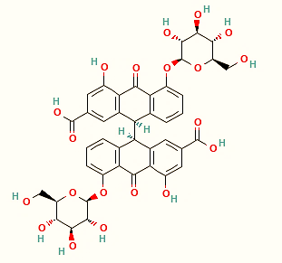
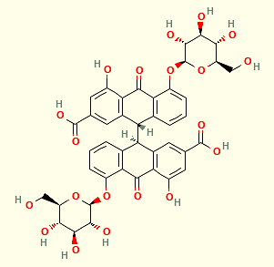
This linkage makes C10 and C10′ stereochemically relevant positions. Sennosides A and B have the same basic composition but differ in the relative configurations at C10/C10′ and are therefore diastereomers. In the corresponding sennidins, the A form generally has the 10R,10′R configuration, whereas the B form is the meso 10R,10′S isomer [2].
This structural feature shows that dimerization does more than increase molecular weight. Formation of a C–C bond between two anthrones simultaneously introduces new stereochemical and conformational features into the molecule.
Compared with highly fused naphthodianthrones, the two aromatic portions of a dianthrone remain connected mainly through the central C–C bond and retain greater conformational freedom relative to one another. Theoretical studies of sennidins have also shown that the C10–C10′ linkage differs markedly in its properties from an ordinary C–C bond between monomeric anthrones, consistent with the ability of dianthrone systems to undergo subsequent reductive cleavage and metabolic transformation [2].
2.2 The Development of Sennoside Activity Depends on Intestinal Metabolism
Sennosides A and B contain two glucose residues. They are not readily hydrolyzed by human digestive enzymes or absorbed in the upper gastrointestinal tract, allowing a substantial proportion to reach the large intestine [3,4].
After they enter the colon, β-glucosidases and reductases produced by intestinal microorganisms jointly participate in their transformation. Current evidence supports at least two reaction sequences. In one pathway, the sugar residues are first removed to form sennidins, followed by reductive cleavage of the C10–C10′ bond. In the other, the sennoside is first reductively cleaved to form 8-glucosylrhein anthrone, which is subsequently hydrolyzed to rhein anthrone. Both pathways can converge on rhein anthrone, the metabolite responsible for the laxative activity [3,12].
In 2025, Xu et al. used a photoaffinity probe to identify an NfrA-type sennoside A reductase in Bifidobacterium pseudocatenulatum. Purified recombinant NfrA was able to catalyze the reduction of sennoside A in the presence of flavin mononucleotide (FMN) and NADPH. The experiments detected 8-glucosylrhein anthrone and its readily oxidized product, rhein 8-O-β-D-glucoside, supporting a metabolic pathway in which sennoside A first undergoes reductive cleavage of the C10–C10′ bond and is then deglycosylated [12].
Rhein anthrone acts in the colon, where it can affect colonic motility and the transport of water and electrolytes. The pharmacological activity of sennosides is therefore directly associated with structural transformation of the parent compounds in the intestine rather than with extensive absorption of unchanged sennosides from the upper gastrointestinal tract [3].
This process reflects an important property of dianthrone structures: the central dimeric structure retains considerable potential for chemical and biological transformation.
3 Oxidation and Dehydrogenation Alter the Electronic State of Dimeric Anthracene Structures
3.1 Bianthraquinones: Two Anthracene-Derived Units in Higher Oxidation States
Bianthraquinones consist of two anthraquinone units connected by a carbon–carbon bond. Compared with dianthrones, their two units are, overall, in higher oxidation states; however, the two classes are not necessarily direct oxidation counterparts that retain the same linkage positions.
Skyrin is a representative naturally occurring bianthraquinone. Recent metabolic studies of Hypericum species have drawn increased attention to this compound. Researchers have detected skyrin and related glycosides in multiple Hypericum species and observed distributional associations between them and hypericin and related metabolites. Matrix-assisted laser desorption/ionization high-resolution mass spectrometry imaging (MALDI-HRMS imaging) has also shown that skyrin and certain hypericin-related metabolites can be spatially colocalized in specific tissues [6].
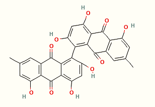
These findings have made skyrin an important candidate intermediate in studies of hypericin biosynthesis. However, spatial colocalization and metabolic correlation alone cannot establish a complete precursor–product relationship. Current reviews continue to identify the subsequent steps of hypericin biosynthesis in plants as questions requiring further investigation [5].
3.2 Dehydrodianthrones: Increased Unsaturation in the Central Linkage Region
Further dehydrogenation of a dianthrone can produce a dehydrodianthrone-type structure.
Relative to a dianthrone, dehydrogenation increases the degree of unsaturation in the central linkage region and changes the π-electron interactions between the two anthracene-derived units. Its effects are not limited to the loss of two hydrogen atoms from the molecular formula but also involve readjustments in bond order, conformation, and electron distribution.
The effects of oxidation or dehydrogenation can be divided into two levels:
① Changes in oxidation state alter the carbonyl groups and associated redox properties;
② Changes in the degree of unsaturation alter the electronic coupling between the two anthracene-derived portions.
The actual extent of conjugation still depends on the linkage position, molecular torsion angle, and substituents. The strength of conjugation cannot be inferred directly from the oxidation level alone.
4 Further Cyclization: Helianthrones and Naphthodianthrones
4.1 Additional Transannular C–C Bonds Restrict Molecular Conformation
When a new transannular C–C bond forms between two anthracene-derived units, the molecular connectivity changes substantially.
Helianthrone-type compounds exhibit this structural feature. Compared with dianthrones, which are formed mainly through a central linkage, a helianthrone skeleton contains an additional covalent linkage spanning the two aromatic portions, further fixing the two units that previously possessed greater freedom of relative motion.
This change primarily affects conformational freedom and skeletal rigidity. Formation of the new ring system reduces the number of relative orientations available to the aromatic rings and simultaneously changes the spatial relationship between their π orbitals.
Protohypericin is a substituted helianthrone-type compound and is an important example for studying this structural change. Chemical synthesis studies have demonstrated that protohypericin can undergo further cyclization under visible-light irradiation to generate hypericin [7].
4.2 Photocyclization of Protohypericin to Form Hypericin
A well-established photochemical transformation exists between protohypericin and hypericin:
Protohypericin ──hν (visible light)──→ Hypericin
Here, hν represents photon energy.
In a 2018 synthetic study, protohypericin was prepared from emodin anthrone and subsequently irradiated with monochromatic visible light having a peak wavelength of approximately 575 nm, resulting in efficient photocyclization of protohypericin to hypericin [7].
This step introduces a new transannular linkage and produces a more fully developed polycyclic naphthodianthrone system. The two anthracene-derived units are thereby fixed by multiple covalent bonds and no longer display the relatively distinct independent conformational characteristics found in dianthrones.
It is important to distinguish the photocyclization of protohypericin to hypericin, a specific transformation supported by evidence from experimental and natural systems, from the sequence “dianthrone → bianthraquinone → dehydrodianthrone → helianthrone → naphthodianthrone,” which is not an established universal biosynthetic pathway in plants. The exact precursors and associated enzyme systems involved in hypericin biosynthesis in plants remain under investigation [5,6].
5 Why Hypericin Exhibits Pronounced Photochemical Properties
5.1 Extensive Conjugation Alters the Electronic Energy-Level Structure
Hypericin is a naturally occurring naphthodianthrone pigment with the molecular formula C₃₀H₁₆O₈. Its structure contains a highly fused polycyclic quinone system and multiple phenolic hydroxy groups.
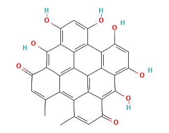
Compared with the dianthrone structure of sennosides, hypericin has a larger continuous π-electron system and a substantially more rigid skeleton. Extensive π-electron delocalization alters its electronic energy levels, resulting in strong absorption in the visible region. The absorption maxima of hypericin vary with solvent and aggregation state, with a common long-wavelength absorption band located near 590 nm [10].
Hypericin is not a completely planar rigid molecule. Steric crowding at the molecular periphery can produce a degree of conformational distortion. Proton transfer between phenolic hydroxy and carbonyl groups, together with the molecular aggregation state, also affects its spectroscopic and excited-state behavior [8].
The optical properties of hypericin arise from the combined effects of its fused polycyclic skeleton, extended π conjugation, quinonoid electronic structure, and hydroxy/carbonyl system.
5.2 Photoexcitation Produces the Triplet State and Contributes to Reactive Oxygen Species Generation
After absorbing light of an appropriate wavelength, hypericin is promoted from the ground state to an excited singlet state. A proportion of the excited molecules subsequently undergo intersystem crossing (ISC) to enter a longer-lived triplet state.
The process can be represented in simplified form as follows:
Hyp + hν → ¹Hyp* → ³Hyp*
Here, Hyp represents hypericin, while ¹Hyp* and ³Hyp* represent singlet- and triplet-state excited hypericin, respectively.
Triplet-state hypericin can subsequently participate in reactions through two types of photosensitization pathway.
5.2.1 Type I Photochemical Reaction
Excited-state hypericin undergoes electron or hydrogen transfer with surrounding substrates, forming radicals or radical ions that can subsequently react with oxygen to generate reactive oxygen species such as the superoxide radical anion.
5.2.2 Type II Photochemical Reaction
Triplet-state hypericin transfers energy to ground-state triplet oxygen, converting it into singlet oxygen:
³Hyp* + ³O₂ → Hyp + ¹O₂
Electron paramagnetic resonance and spin-trapping studies have detected signals consistent with the formation of semiquinone radicals, singlet oxygen, and superoxide radical anions [9]. Modern photophysical studies also support triplet-state formation by hypericin and its participation in singlet-oxygen generation [8].
The relative contributions of Type I and Type II reactions vary with the solvent, oxygen concentration, substrate environment, irradiation conditions, hypericin concentration, and aggregation state.
These properties provide the chemical basis for research into the use of hypericin in photodynamic therapy (PDT) [10].
6 Structural and Property Differences Between Sennosides and Hypericin
Sennosides and hypericin represent two different states of dimeric anthracene structures.
Comparison item | Sennosides A and B | Hypericin |
Structural class | Dianthrone diglucosides | Naphthodianthrone |
Linkage between the anthracene-derived units | Centered on a C10–C10′ linkage | Multiple C–C bonds form a fused ring system |
Conformational characteristics | Retain greater relative conformational freedom | Substantially increased skeletal rigidity |
Electronic structure | The two anthracene-derived portions retain pronounced localized structural characteristics | A more extensive continuous π-conjugated system is formed |
Key structural reactions | Glycoside hydrolysis, reduction, and transformation of the dimeric system | Photoexcitation, electron transfer, and energy transfer |
Principal external factors | Intestinal microbial enzyme systems | Light, oxygen, solvent, and aggregation state |
Typical activity process | Laxative effect after formation of rhein anthrone | Photosensitization and reactive oxygen species generation |
Representative research areas | Gut microbiota and natural-product metabolism | Photophysics, photochemistry, and photodynamic research |
The differences between these two classes illustrate a central principle governing structural changes in dimeric anthracene derivatives:
Dianthrones retain a distinct transformable dimeric structure, whereas naphthodianthrones undergo further cyclization and π-electron delocalization to form conjugated systems with distinctive photophysical properties.
Sennosides retain dianthrone glycoside structures that can be transformed by intestinal microorganisms. Hypericin, by contrast, is based on a rigid fused skeleton and extended π conjugation and exhibits photosensitizing properties that include visible-light absorption, triplet-state formation, and reactive oxygen species generation.
7 Testable Research Questions Concerning Dimeric Anthracene Natural Products
7.1 How Can a Candidate Biosynthetic Intermediate Be Demonstrated Rather Than Merely Associated with Other Metabolites?
Skyrin and hypericin show correlations in abundance and tissue colocalization in Hypericum species, but this evidence is insufficient to establish a definitive precursor–product relationship.
Further validation can combine:
① Stable-isotope tracing: Determine whether carbon atoms from a labeled precursor are incorporated into the hypericin skeleton;
② Functional validation of candidate enzymes: Recombinantly express relevant oxidases or coupling enzymes and determine whether they catalyze the predicted bond-forming steps;
③ In vitro reconstruction of enzyme cascades: Verify the dimerization, oxidation, and cyclization processes step by step;
④ Integration of gene-function perturbation with metabolomics: Compare changes in candidate-gene expression with the accumulation of intermediates.
These studies can advance “colocalization and correlation” evidence toward causal evidence for specific chemical transformations.
7.2 Can Differences in Sennoside Metabolism Be Traced to Specific Intestinal Bacteria and Reductases?
The development of sennoside activity depends on intestinal microorganisms participating in reductive cleavage of the C10–C10′ bond and subsequent metabolism. Differences in microbial community composition may therefore affect the efficiency with which active metabolites are generated.
Variables warranting further investigation include:
① The ability of different intestinal bacterial strains to reduce sennosides A and B;
② Differences in the substrate selectivity and kinetics of NfrA-type reductases;
③ The relationship between microbial community composition and the rate of rhein anthrone formation;
④ The effects of antibiotic use, diet, or other drugs on sennoside metabolism through changes in the gut microbiota.
This research direction connects the reactivity of a specific bond within a dimeric anthracene structure with the function of the gut microbiota and may help explain interindividual differences in natural-product metabolism.
7.3 How Do Photosensitive Precursors Affect the Quantitative Analysis of Natural Products?
Protohypericin can be converted to hypericin upon exposure to light, and protopseudohypericin undergoes a corresponding phototransformation. Sample-handling conditions can therefore directly affect the measured proportions of these components [11].
Analytical methods should focus on controlling:
① Light exposure during sample collection and extraction;
② Storage time and temperature;
③ Exposure time in the autosampler;
④ The mass balance between precursor and product at different light doses.
Samples protected from light can be analyzed in parallel with samples exposed to controlled irradiation. Protohypericin, hypericin, protopseudohypericin, and pseudohypericin can be determined simultaneously to establish whether observed changes in composition originate in the plant itself or arise during sample handling.
8 Classification Tables of Representative Chemicals for Research on Sennoside–Hypericin Dimeric Anthracene Structures, Metabolic Transformation, and Conjugated Properties
Table 1. Dianthrone Glycosides and Their Deglycosylated Aglycones
Classification | CAS No. | Aladdin Product No. | Name | Specification or purity | Product features and applications |
Rhein anthrone homodimeric diglucoside, type A | 81-27-6 | Sennoside A | Analytical standard, ≥96% | Used to investigate the stereochemical configurations of sennosides A/B, carbon–carbon linkages in dianthrones, and deglycosylation and reductive transformation by the gut microbiota; also used for compositional analysis of senna leaf- and rhubarb-related samples. | |
Rhein anthrone homodimeric diglucoside, type B | 128-57-4 | Sennoside B | Analytical standard, ≥97% (HPLC) | Used in comparison with sennoside A to investigate dianthrone linkage configurations, chromatographic behavior, and differences in intestinal metabolism, and to study the formation of rhein anthrone. | |
Heterodimeric diglucoside of rhein anthrone and aloe-emodin anthrone, type C | 37271-16-2 | Sennoside C | ≥98% | Used to investigate heterodianthrone structures, glycosylation, and intestinal metabolism, and to evaluate effects associated with rhein anthrone and aloe-emodin anthrone. | |
Heterodimeric diglucoside of rhein anthrone and aloe-emodin anthrone, type D | 37271-17-3 | Sennoside D | Moligand™, ≥98% | Used in comparison with sennoside C to investigate heterodianthrone stereochemistry, chromatographic behavior, and metabolic transformation, and for compositional analysis of dianthrone glycosides in senna leaves and rhubarb. | |
Deglycosylated dianthrone aglycone of sennoside A | 641-12-3 | Sennidin A | Moligand™, ≥98% | Used to investigate sennoside A deglycosylation, stereochemical differences between sennidins A and B, and subsequent reduction to rhein anthrone. | |
Deglycosylated dianthrone aglycone of sennoside B | 517-44-2 | Sennidin B | Moligand™, ≥98% | Used to investigate sennoside B deglycosylation, the stereochemistry of dianthrone aglycones, and their intestinal metabolic conversion to rhein anthrone. |
Table 2. Monomeric Anthracene Metabolites and Anthrone–Anthraquinone Oxidation-State Reference Compounds
Classification | CAS No. | Aladdin Product No. | Name | Specification or purity | Product features and applications |
Rhein anthrone-type active intestinal metabolite | 480-09-1 | Rhein-9-anthrone | ≥95% | Used to investigate the active metabolite formed through transformation of sennosides by the gut microbiota, and to evaluate anthrone–anthraquinone redox chemistry, colonic propulsion, and water transport. | |
Rhein anthraquinone-type oxidation-state reference compound | 478-43-3 | Rhein | Moligand™, ≥98% | Used with rhein anthrone to establish an anthraquinone–anthrone oxidation-state comparison, investigate the oxidative transformation of sennoside metabolites, and analyze rhein-related constituents. | |
Aloe-emodin anthraquinone-type reference compound for a heterodimeric unit | 481-72-1 | Aloe-emodin | ≥95% | Used to investigate the constituent units of sennoside C/D heterodianthrones, hydroxymethyl substitution effects, and structural differences from rhein-related compounds. | |
Emodin anthrone-type reduced-state reference compound | 491-60-1 | Emodin anthrone | Moligand™, ≥98% | Used to investigate the reduced state of anthrones, keto–enol tautomerism, oxidation to emodin, and the chemical reactivity of anthrone structures. | |
Emodin anthraquinone-type oxidized-state reference compound | 518-82-1 | Emodin | Analytical standard, ≥96% (HPLC) | Used in comparison with emodin anthrone to investigate differences between anthraquinone and anthrone oxidation states, and the effects of hydroxy and methyl substituents on electronic and redox properties. |
Table 3. Reference Compounds for Dimeric, Dehydrogenated, and Fused Conjugated Structures
Classification | CAS No. | Aladdin Product No. | Name | Specification or purity | Product features and applications |
Dianthrone structural reference compound linked by a carbon–carbon single bond | 434-84-4 | Bianthronyl | ≥97% | Used to investigate the bond length, twisted conformation, and conformational freedom resulting from dimerization of two anthrone units through a central carbon–carbon single bond, and for comparison with dehydrodianthrones and fused skeletons. | |
Dehydrodianthrone conjugated-structure reference compound | 434-85-5 | Bianthrone | ≥95% (HPLC) | Used to investigate the conjugated structure formed after dehydrogenation of a dianthrone, changes in skeletal conformation, and photochromic behavior, and as a reference for the transition from dimeric to fused conjugated structures. | |
Nonfused natural bianthraquinone with a carbon–carbon linkage | 602-06-2 | Skyrin, Glucagon Receptor Antagonist | ≥97% | Used to investigate carbon–carbon linkages in nonfused bianthraquinones, phenolic hydroxy substitution, and the biosynthesis of dimeric skeletons, and to evaluate glucagon receptor antagonism and hepatocyte metabolism. | |
Photoconversion precursor of hypericin | 548-03-8 | Protohypericin | ≥95% | Used to investigate light-induced cyclization and dehydrogenative transformation during hypericin formation, and to compare visible-light absorption and photosensitizing properties before and after conversion. | |
Fused naphthodianthrone photosensitizer | 548-04-9 | Hypericin | Moligand™, ≥96% | Used to investigate highly fused naphthodianthrone conjugated skeletons, visible-light absorption, fluorescence, reactive oxygen species generation, and photosensitization. |
Note: The products listed above are representative research-related products from Aladdin. Their specific uses should be determined with reference to product specifications, lot-specific certificates of analysis (COAs), and the intended reaction or evaluation system. Additional specifications, grades, and COA information can be retrieved from the Aladdin website using the product name, CAS number, or product number.
References
[1] Hua H, Lou H. Natural Medicinal Chemistry. 8th ed. Beijing: People’s Medical Publishing House; 2022.
[2] Szymanski S, Majerz I. In Silico Studies on Sennidines—Natural Dianthrones from Senna. Biology. 2021;10(6):468. doi:10.3390/biology10060468.
[3] Le J, Ji H, Zhou X, et al. Pharmacology, Toxicology, and Metabolism of Sennoside A, A Medicinal Plant-Derived Natural Compound. Frontiers in Pharmacology. 2021;12:714586. doi:10.3389/fphar.2021.714586.
[4] European Medicines Agency. European Union Herbal Monograph on Senna alexandrina Mill. (Cassia senna L.; Cassia angustifolia Vahl), folium—Revision 1. EMA/HMPC/625849/2015. 25 September 2018.
[5] Rizzo P, Altschmied L, Ravindran BM, Rutten T, D’Auria JC. The Biochemical and Genetic Basis for the Biosynthesis of Bioactive Compounds in Hypericum perforatum L., One of the Largest Medicinal Crops in Europe. Genes. 2020;11(10):1210. doi:10.3390/genes11101210.
[6] Revuru B, Bálintová M, Henzelyová J, et al. MALDI-HRMS Imaging Maps the Localization of Skyrin, the Precursor of Hypericin, and Pathway Intermediates in Leaves of Hypericum Species. Molecules. 2020;25(17):3964. doi:10.3390/molecules25173964.
[7] Zhang Y, Shang K, Wu X, et al. Highly Efficient Green Synthesis and Photodynamic Therapeutic Study of Hypericin and Its Derivatives. RSC Advances. 2018;8:21786–21792. doi:10.1039/C8RA03732A.
[8] Gattuso H, Marazzi M, Dehez F, Monari A. Deciphering the Photosensitization Mechanisms of Hypericin towards Biological Membranes. Physical Chemistry Chemical Physics. 2017;19:23187–23193. doi:10.1039/C7CP03723F.
[9] Diwu Z, Lown JW. EPR Studies of Photodynamic Action of Hypericin: Formation of Semiquinone Radical and Activated Oxygen Species on Illumination. Free Radical Biology and Medicine. 1993;14(2):209–215. doi:10.1016/0891-5849(93)90012-J.
[10] de Andrade GP, de Souza TFM, Cerchiaro G, et al. Hypericin in Photobiological Assays: An Overview. Photodiagnosis and Photodynamic Therapy. 2021;35:102343. doi:10.1016/j.pdpdt.2021.102343.
[11] Baugh SF. Simultaneous Determination of Protopseudohypericin, Pseudohypericin, Protohypericin, and Hypericin Without Light Exposure. Journal of AOAC International. 2005;88(6):1607–1612. doi:10.1093/jaoac/88.6.1607.
[12] Xu Y, Lv S, Li X, et al. Photoaffinity Probe-Enabled Discovery of Sennoside A Reductase in Bifidobacterium pseudocatenulatum. Journal of Pharmaceutical Analysis. 2025;15:101108. doi:10.1016/j.jpha.2024.101108.
For more related articles, see below:
Extraction, isolation and characterization of anthraquinones in Rheum palmatum (Rheum palmatum)
Tetramethyl-1,4-benzoquinone(DQ)
Extraction and isolation of free hydroxyanthraquinone components from Cynodon dactyliflorus
Chloranil, Tetrachloro-1,4-benzoquinone
Tetramethyl-1,4-benzoquinone(DQ)
Glucosidases: Classification Framework, Catalytic Mechanisms, and Application Essentials
Biological Characteristics and Application Value of the Aminoglycoside Antibiotic Hygromycin B
n-Dodecyl-β-D-maltoside (DDM): Structure, Properties, and Applications as a Non-ionic Surfactant
Molecular Basis of Glycosylation Modification and Functional Release in Natural Products
Will glycosylation affect the biological activity of recombinant protein?
Plant proteomics and glycosylation experiments


 危险品化学品经营许可证(带存储)