甘油(Glycerol),化学名称丙三醇,分子式 C3H8O3,是一种无色无臭的黏稠液体。作为甘油三酯的核心组成成分,甘油是脂肪分解的重要代谢产物,因此血液甘油含量是评价机体脂解水平的关键指标,在代谢研究与药物研发中具有重要应用价值。 此外,甘油因具有良好的保湿性与稳定性,被广泛应用于食品、饮料、医药及化妆品等领域,其精准定量在产品开发与质量控制中同样至关重要。 本试剂盒采用高灵敏度的 Amplex Red 荧光探针检测甘油含量。在 ATP 存在下,甘油经甘油激酶(GK)磷酸化为甘油 - 3 - 磷酸,后者在甘油磷酸氧化酶(GPO)催化下生成过氧化氢( H2O2);在辣根过氧化物酶(HRP)作用下,Amplex Red 与 H2O2以 1:1 定量反应生成强荧光产物试卤灵(Resorufin)。通过检测试卤灵荧光强度(560 nm 激发、590 nm 发射)或吸光度(A570),即可实现甘油的高灵敏定量检测,信号强度与甘油浓度成正比。
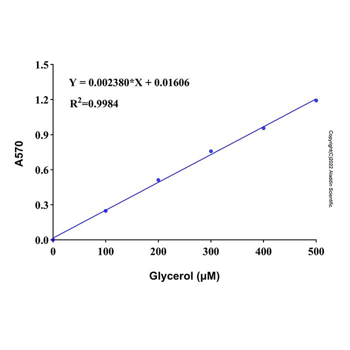
本试剂盒可以用吸光度和荧光两种方法来进行检测,检测灵敏度高,线性范围宽,样品用量少。在样品体积为20 µL时,采用吸光度法检测在10-500μM浓度范围内有良好的线性关系;采用荧光法检测在1-100μM浓度范围内有良好的线性关系。
使用方法
1. 样本制备
(1)血液样品的准备:对于血清样品,将全血在常温下静置30分钟-2小时,4ºC约1000-2000×g离心10分钟,取上清;对于血浆样品,将全血用肝素或者EDTA进行抗凝,4ºC约1000-2000×g离心10分钟,取上清。
(2)细胞样本的准备:对于贴壁细胞,弃掉培养基,用胰酶消化或适当拍打使细胞脱离培养器皿并转移至离心管中,用PBS 重悬洗涤一次,离心 (400-500×g, 5分钟)收集细胞到离心管内,弃上清并吸净残留液体。对于培养的悬浮细胞,离心 (400-500×g, 5分钟)收集细胞到离心管内,PBS 重悬洗涤一次,离心 (400-500×g, 5分钟)收集细胞,弃上清并吸净残留液体。每100万个细胞加入100-200μl异丙醇,适当吹打,然后在4℃或冰浴等低温条件下进行匀浆或者超声破碎细胞。4ºC, 12,000×g离心5分钟,取上清用于后续检测。
(3)对于组织样品,每10 mg组织加入100 -200μL 异丙醇,在4℃或冰浴等低温条件下进行匀浆或者超声破碎。4ºC约12,000×g离心5分钟,取上清用于后续检测。
(4)细胞培养上清样品的准备:对于贴壁细胞,直接取培养液;对于悬浮细胞,离心取培养液。
注意:以上所有操作均需在4ºC或冰上操作。制备好的细胞或组织样品如果不能立即检测,可以-20ºC或-80ºC冻存。
2. 甘油显色工作液的配制
首先将检测缓冲液平衡至室温,然后按照每个检测反应80 μL的体积配制显色工作液。根据待检测样品(包括标准品)的数量,配制适量的显色工作液。显色工作液的各个组份的具体配制方法参考下表。由于Amplex Red室温容易见光分解,建议尽量现配现用,如果不能及时检测,请置于4℃或者冰浴避光保存且不要过夜,以免影响检测效果。
样本数 | 1 | 10 | 20 | 50 | 甘油激酶 | 5 μL | 50 μL | 100 μL | 250 μL | 酶混合物 | 5 μL | 50 μL | 100 μL | 250 μL | 辅因子 | 4 μL | 40 μL | 80 μL | 200 μL | Amplex Red | 2 μL | 20 μL | 40 μL | 100 μL | 检测缓冲液 | 64 μL | 640 μL | 1280 μL | 3200 μL |
|
3. 样本测定
标准曲线绘制:
(1) 吸光度检测法:取10μl甘油标准溶液(10mM),加入190μl检测缓冲液,配置成浓度为500uM的甘油标准溶液。分别取500μM的甘油标准溶液0μL,2μL,4μL,8μL,12μL,16μL,20μL加入96孔板的标准品孔中,然后用检测缓冲液或者异丙醇(如果样品是用异丙醇制备,建议用异丙醇稀释标品绘制标准曲线)补足至20μL,此时,标准溶液的浓度分别为0、50、100、200、300、400、500μM。
(2) 荧光检测法:取2μl甘油标准溶液(10mM),加入398μl检测缓冲液,配置成浓度为50uM的甘油标准溶液。分别取50μM的甘油标准溶液0μL,2μL,4μL,8μL,12μL,16μL,20μL加入96孔板的标准品孔中,然后用检测缓冲液或者异丙醇(如果样品是用异丙醇制备,建议用异丙醇稀释标品绘制标准曲线)补足至20μL,此时,标准溶液的浓度分别为0、5、10、20、30、40、50μM。
样品测定
(3) 取1-20 μL样品或稀释后的样品至96孔板样品孔中,并相应地加入检测缓冲液或者异丙醇补足至20 µL。同时设置仅含检测缓冲液或异丙醇的孔为空白对照孔。每孔加入甘油检测工作液80μl,混匀,37ºC避光反应10分钟。
(4) 反应结束后,如果是吸光度法检测,读取A570;如果是荧光法检测,设置激发波长560nm,发射波长590nm进行荧光强度检测。
4. 结果计算
建立标准曲线时,需要减去空白孔对照。标准曲线建立完毕后,可根据标准曲线计算出样品中的甘油浓度(C),计算公示如下:
C(μM)=A×n
注:A为根据标准曲线计算出的甘油浓度(μM);n为样品稀释倍数。也可以根据甘油的分子量92.09计算出质量浓度(μg/ml) = C × 0.09209
注意事项
1. 本试剂盒如果用于细胞或组织样品的检测,需用户自备异丙醇。
2. 如果样品为异丙醇制备,在绘制标准曲线时,建议用和样品相同含量的异丙醇稀释标准品并绘制标准曲线。
3. 显色工作液建议尽量现配现用。
4. Amplex Red容易室温见光分解,开启后应尽快使用,且在使用过程中一定要注意适当避光。
5. 建议反应体系的pH值维持在7-8之间,否则可能会影响Amplex Red的稳定性,从而影响检测信号。
6. 在样品检测时,需合理设置样品背景对照孔,例如甘油-3-磷酸和或者 H2O2的存在都会使得检测信号偏高,从而干扰检测结果。因此,如果样品自身含有甘油-3-磷酸和或者 H2O2时,在设置样品背景对照孔时,需要将甘油显色工作液中的甘油激酶组份用等体积的检测缓冲液替换掉,配置成不含甘油激酶的“显色工作液”
Glycerol (chemical name: Glycerol), with the molecular formula C3H8O3, is a colorless, odorless, viscous liquid. As the core component of triglycerides, glycerol is an important metabolic product of fat decomposition. Therefore, the blood glycerol content is a key indicator for evaluating the level of body lipolysis, and has important application value in metabolic research and drug development. In addition, due to its good moisturizing properties and stability, glycerol is widely used in food, beverages, medicine, cosmetics and other fields, and its accurate quantification is also crucial in product development and quality control. This kit uses a high-sensitivity Amplex Red fluorescent probe to detect glycerol content. In the presence of ATP, glycerol is phosphorylated to glycerol-3-phosphate by glycerol kinase (GK); the latter is catalyzed by glycerol phosphate oxidase (GPO) to generate hydrogen peroxide (H2O2); under the action of horseradish peroxidase (HRP), Amplex Red reacts quantitatively with H2O2 at a ratio of 1:1 to generate a strong fluorescent product, resorufin. High-sensitivity quantitative detection of glycerol can be achieved by detecting the fluorescence intensity (560 nm excitation, 590 nm emission) or absorbance (A570) of resorufin, and the signal intensity is proportional to the glycerol concentration.
This kit can be detected by two methods: absorbance and fluorescence, with high detection sensitivity, wide linear range and small sample volume. When the sample volume is 20 µL, the absorbance method has a good linear relationship in the concentration range of 10-500 μM; the fluorescence method has a good linear relationship in the concentration range of 1-100 μM.
Usage Method
1. Sample Preparation
(1) Preparation of blood samples: For serum samples, let the whole blood stand at room temperature for 30 minutes to 2 hours, centrifuge at 4ºC at about 1000-2000×g for 10 minutes, and take the supernatant; for plasma samples, anticoagulate the whole blood with heparin or EDTA, centrifuge at 4ºC at about 1000-2000×g for 10 minutes, and take the supernatant.
(2) Preparation of cell samples: For adherent cells, discard the culture medium, digest with trypsin or tap appropriately to detach the cells from the culture vessel and transfer to a centrifuge tube, resuspend and wash once with PBS, centrifuge (400-500×g, 5 minutes) to collect the cells into the centrifuge tube, discard the supernatant and aspirate the residual liquid. For cultured suspension cells, centrifuge (400-500×g, 5 minutes) to collect the cells into a centrifuge tube, resuspend and wash once with PBS, centrifuge (400-500×g, 5 minutes) to collect the cells, discard the supernatant and aspirate the residual liquid. Add 100-200 μl of isopropanol per 1 million cells, pipette properly, then homogenize or sonicate the cells at low temperature such as 4℃ or ice bath. Centrifuge at 4ºC, 12,000×g for 5 minutes, and take the supernatant for subsequent detection.
(3) For tissue samples, add 100-200 μL of isopropanol per 10 mg of tissue, and homogenize or sonicate at low temperature such as 4℃ or ice bath. Centrifuge at 4ºC at about 12,000×g for 5 minutes, and take the supernatant for subsequent detection.
(4) Preparation of cell culture supernatant samples: For adherent cells, directly take the culture medium; for suspension cells, centrifuge and take the culture medium.
Note: All the above operations must be performed at 4ºC or on ice. If the prepared cell or tissue samples cannot be detected immediately, they can be stored at -20ºC or -80ºC.
2. Preparation of Glycerol Chromogenic Working Solution
First, equilibrate the Assay Buffer to room temperature, then prepare the chromogenic working solution according to the volume of 80 μL per detection reaction. Prepare an appropriate amount of chromogenic working solution according to the number of samples to be detected (including standards). For the specific preparation method of each component of the chromogenic working solution, refer to the following table. Since Amplex Red is easily decomposed by light at room temperature, it is recommended to prepare it fresh as much as possible. If it cannot be detected in time, please store it at 4℃ or ice bath protected from light and do not leave it overnight to avoid affecting the detection effect.
Number of sample | 1 | 10 | 20 | 50 | Glycerol Kinase | 5 μL | 50 μL | 100 μL | 250 μL | Enzyme Mix | 5 μL | 50 μL | 100 μL | 250 μL | Cofactor | 4 μL | 40 μL | 80 μL | 200 μL | Amplex Red | 2 μL | 20 μL | 40 μL | 100 μL | Assay Buffer | 64 μL | 640 μL | 1280 μL | 3200 μL |
|
3. Sample Determination
Standard Curve Drawing:
(1) Absorbance detection method: Take 10 μl of glycerol standard solution (10mM), add 190 μl of Assay Buffer to prepare a glycerol standard solution with a concentration of 500 μM. Take 0 μL, 2 μL, 4 μL, 8 μL, 12 μL, 16 μL and 20 μL of 500 μM glycerol standard solution into the standard wells of a 96-well plate, then make up to 20 μL with Assay Buffer or isopropanol (if the sample is prepared with isopropanol, it is recommended to dilute the standard with isopropanol to draw the standard curve). The concentrations of the standard solutions are 0, 50, 100, 200, 300, 400 and 500 μM respectively.
(2) Fluorescence detection method: Take 2 μl of glycerol standard solution (10mM), add 398 μl of Assay Buffer to prepare a glycerol standard solution with a concentration of 50 μM. Take 0 μL, 2 μL, 4 μL, 8 μL, 12 μL, 16 μL and 20 μL of 100 μM glycerol standard solution into the standard wells of a 96-well plate, then make up to 20 μL with Assay Buffer or isopropanol (if the sample is prepared with isopropanol, it is recommended to dilute the standard with isopropanol to draw the standard curve). The concentrations of the standard solutions are 0, 5, 10, 20, 30, 40 and 50 μM respectively.
Sample Determination
(3) Take 1-20 μL of sample or diluted sample into the sample well of a 96-well plate, and add Assay Buffer or isopropanol to make up to 20 µL accordingly. At the same time, set a well containing only Assay Buffer or isopropanol as a blank control well. Add 80 μl of glycerol detection working solution to each well, mix well, and incubate at 37ºC protected from light for 10 minutes.
(4) After the reaction, if using the absorbance method, read A570; if using the fluorescence method, set the excitation wavelength to 560 nm and the emission wavelength to 590 nm for fluorescence intensity detection.
4. Result Calculation
When establishing the standard curve, the blank well control needs to be subtracted. After the standard curve is established, the glycerol concentration (C) in the sample can be calculated according to the standard curve, and the calculation formula is as follows:
C(μM) = A × n
Note: A is the glycerol concentration (μM) calculated according to the standard curve; n is the sample dilution factor. The mass concentration (μg/ml) can also be calculated according to the molecular weight of glycerol (92.09) as follows: mass concentration (μg/ml) = C × 0.09209
Precautions
1. If this kit is used for the detection of cell or tissue samples, the user must prepare isopropanol by themselves.
2. If the sample is prepared with isopropanol, it is recommended to dilute the standard with isopropanol at the same concentration as the sample when drawing the standard curve.
3. It is recommended to prepare the chromogenic working solution fresh as much as possible.
4. Amplex Red is prone to photodegradation at room temperature. It should be used as soon as possible after opening, and proper protection from light must be ensured during use.
5. It is recommended that the pH value of the reaction system be maintained between 7 and 8; otherwise, the stability of Amplex Red may be affected, thereby interfering with the detection signal.
6. During sample detection, it is necessary to reasonably set up sample background control wells. For example, the presence of glycerol-3-phosphate or H₂O₂ will increase the detection signal, thus interfering with the detection results. Therefore, if the sample itself contains glycerol-3-phosphate or H₂O₂, when setting up the sample background control wells, the glycerol kinase component in the glycerol chromogenic working solution should be replaced with an equal volume of Assay Buffer to prepare a "chromogenic working solution" without glycerol kinase.

 ZJ26F0434697


 危险品化学品经营许可证(带存储)