G6P (Glucose-6-phosphate,葡萄糖-6-磷酸,又称6-磷酸葡萄糖)是葡萄糖的第6位碳上的羟基在己糖激酶催化下发生磷酸化后生成的小分子,是生物体葡萄糖代谢的首个关键中间产物,也是连接多条核心代谢通路的关键枢纽,可分别进入糖酵解途径参与能量供给、磷酸戊糖途径生成NADPH与核糖,同时参与糖原合成、糖异生及脂质合成等生化过程,直接调控细胞能量代谢、氧化稳态与物质合成代谢平衡。G6P的胞内含量是反映细胞代谢活性、糖代谢紊乱程度的核心指标,其异常波动与多种生理病理过程密切相关,包括葡萄糖-6-磷酸脱氢酶缺乏症、糖代谢异常、氧化应激损伤及代谢类疾病等,是基础代谢研究、疾病机制探索与药物筛选的重要检测靶点。
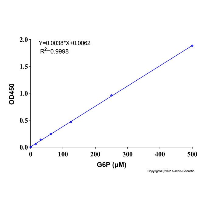
本试剂盒检测原理:G6P在葡萄糖-6-磷酸脱氢酶(G6PD)的作用下氧化生成6-磷酸葡萄糖酸内酯(6-phosphogluconate, 6-PG),在这一反应过程中NADP⁺被还原成NADPH,生成的NADPH在电子耦合剂作用下将WST- 8还原生成橙黄色的formazan,在450 nm左右有最大吸收峰。反应体系中生成的formazan与样品中的G6P的量呈正比关系。 该方法检测灵敏度高,线性范围宽,样品用量少。在样品体积为50 µL时,采用吸光度法检测在15 μM-500 μM浓度范围内有良好的线性关系。
G1511290 | 组件 | 物料外观 | 100 T | 储存条件 | 单次测试使用量 |
G1511290A | 葡萄糖-6-磷酸脱氢酶 | 无色液体 | 200 µL | -20℃.避光. | 2 µL |
G1511290B | G6P标准品(2mM) | 无色液体 | 1 mL | -20℃.避光. | 按需 |
G1511290C | NADP⁺ | 无色液体 | 200 µL | -20℃.避光. | 2 µL |
G1511290D | 反应缓冲液 | 无色液体 | 9.2 mL | -20℃. | 92 µL |
G1511290E | 提取液 | 无色液体 | 20 mL | -20℃. | 按需 |
G1511290F | 显色液 | 红褐色液体 | 400 µL | -20℃.避光. | 4 µL |
使用方法
1. 样本制备
(1) 细胞样本的准备:对于贴壁细胞,弃掉培养基,用胰酶消化或适当拍打使细胞脱 离培养器皿并转移至离心管中,用PBS 重悬洗涤一次,离心 (400-500×g, 5分钟)收集细胞到离心管内,弃上清并吸净残留液体。对于培养的悬浮细胞,离心 (400-500×g, 5分钟)收集细胞到离心管内,PBS 重悬洗涤一次,离心 (400-500×g, 5分钟)收集细胞,弃上清并吸净残留液体。每100万个细胞加入100-200μL提取液,适当吹打,然后在4℃或冰浴等低温条件下进行匀浆或者超声破碎细胞。4ºC, 12,000×g离心5分钟,取上清用于后续检测。
(2) 对于组织样品,每10 mg组织加入100 -200μL 提取液,在4℃或冰浴等低温条件下进行匀浆或者超声破碎。4ºC约12,000×g离心5分钟,取上清用于后续检测。
(3) 对于液体样品,直接检测或者用提取液稀释合适倍数后检测。
注意:以上所有操作均需在4ºC或冰上操作。制备好的细胞或组织样品如果不能立即检测,可以-20ºC或-80ºC冻存。
2. 显色工作液的配制
首先将反应缓冲液平衡至室温,然后按照每个检测反应100 μL的体积配制显色工作液。根据待检测样品(包括标准品)的数量,配制适量的显色工作液。显色工作液的各个组份的具体配制方法参考下表。由于显色液室温容易见光分解,建议尽量现配现用,如果不能及时检测,请置于4℃或者冰浴避光保存且不要过夜,以免影响检测效果。
样本数 | 1 | 10 | 50 | 100 |
反应缓冲液 | 92 μL | 920 μL | 4600 μL | 9200 μL |
G6PD | 2 μL | 20 μL | 100 μL | 200 μL |
NADP⁺ | 2 μL | 20 μL | 100 μL | 200 μL |
显色液 | 4 μL | 40 μL | 200 μL | 400 μL |
3. 样本测定
(1)标准曲线绘制:提前将2mM的G6P标准品拿出,冰上解冻,取100 μL G6P标准品,加300 μL超纯水稀释至500 μM,然后用提取液分别将500μM的G6P梯度稀释至500 μM,250 μM,125 μM,62.5 μM,31.25 μM,15.625 μM和0 μM。其中0 μM为空白孔,仅含提取液。取50 μL不同浓度的标品溶液至96孔板孔中,然后每孔添加100 μL显色工作液,混匀后,于37℃反应10min后读取 A₄₅₀ 。
(2)样品测定:取50 μL样品或稀释后的样品至96孔板样品孔中,然后每孔加入添加100μL显色工作液,混匀后,于37℃反应10min后读取 A₄₅₀ 。
4. 结果计算
建立标准曲线时,需要减去空白孔对照。标准曲线建立完毕后,可根据标准曲线计算出样品中的G6P浓度(C),计算公示如下:
C(μM)=A×n
注:A为根据标准曲线计算出的G6P浓度(μM);n为样品稀释倍数。
注意事项
1. NADP⁺,显色液等组分不稳定,建议标品现用现配,且注意避光。
2. 显色工作液同样建议尽量现配现用。
3. β巯基乙醇、二硫苏糖醇等会干扰本检测实验,请样品中尽量避免添加以上物质。
G6P (Glucose-6-phosphate) is a small molecule produced when the hydroxyl group on the 6th carbon of glucose undergoes phosphorylation catalyzed by hexokinase. As the first key intermediate in glucose metabolism in living organisms, it also acts as a critical hub linking multiple core metabolic pathways.G6P participates in diverse biochemical processes: it enters the glycolysis pathway to supply energy, proceeds through the pentose phosphate pathway to generate NADPH and ribose, and is involved in glycogen synthesis, gluconeogenesis and lipid synthesis. It directly regulates cellular energy metabolism, redox homeostasis and the balance of anabolic metabolism. The intracellular level of G6P serves as a core indicator reflecting cellular metabolic activity and the severity of glucose metabolism disorders. Its abnormal fluctuations are closely associated with a wide range of physiological and pathological conditions, including glucose-6-phosphate dehydrogenase deficiency, impaired glucose metabolism, oxidative stress damage and other metabolic diseases. Therefore, G6P is an important detection target for basic metabolism research, disease mechanism investigation and drug screening.
Detection Principle of This Kit : G6P is oxidized to 6-phosphogluconate (6-PG) catalyzed by glucose-6-phosphate dehydrogenase (G6PD). During this reaction, NADP⁺ is reduced to NADPH. The resulting NADPH reduces WST-8 to orange-yellow formazan in the presence of an electron coupling agent, which exhibits a maximum absorbance peak at approximately 450 nm. The amount of formazan produced in the reaction system is proportional to the concentration of G6P in the sample. This assay features high detection sensitivity, a wide linear range and low sample consumption. When the sample volume is 50 µL, the absorbance measurement shows an excellent linear relationship within the concentration range of 15 μM to 500 μM.
G1511290 | Components | Appearance | 100T | Storage | Usage volume per test |
G1511290A | G6PD | Colorless liquid | 200 µL | -20℃.Store in the dark. | 2µL |
G1511290B | G6P Standard (2 mM) | Colorless liquid | 1 mL | -20℃.Store in the dark. | As need |
G1511290C | NADP⁺ | Colorless liquid | 200 µL | -20℃.Store in the dark. | 2µL |
G1511290D | Reaction Buffer | Colorless liquid | 9.2 mL | -20℃. | 92 µL |
G1511290E | Extraction Buffer | Colorless liquid | 20 mL | -20℃. | As need |
G1511290F | Chromogenic solution | Reddish-brown liquid | 400 µL | -20℃..Store in the dark | 4µL |
Usage Method
1. Sample Preparation
(1) Cell Sample Preparation:For adherent cells: Discard the culture medium. Detach cells from the culture vessel using trypsin digestion or gentle tapping, then transfer the cell suspension into a centrifuge tube. Resuspend and wash cells with PBS, then centrifuge at 400–500 × g for 5 minutes. Discard the supernatant and thoroughly remove residual liquid; For suspension cells: Transfer the cell culture into a centrifuge tube and centrifuge at 400–500 × g for 5 minutes. Resuspend and wash the cell pellet with PBS, followed by another centrifugation at 400–500 × g for 5 minutes. Discard the supernatant and remove all residual liquid completely. Add 100–200 μL of extraction buffer per 1×10⁶ cells, and mix well by gentle pipetting. Lyse the cells via homogenization or ultrasonication under low temperature (4°C or ice bath). Centrifuge the lysate at 12,000 × g for 5 minutes at 4°C, and collect the supernatant for subsequent assays
(2) Tissue Sample Preparation:Add 100–200 μL of extraction buffer per 10 mg tissue. Perform tissue homogenization or ultrasonication on ice or at 4°C. Centrifuge the homogenate at 12,000 × g for 5 minutes at 4°C, and collect the supernatant for subsequent assays.
(3) Liquid Sample Preparation: Test liquid samples directly, or dilute them to an appropriate concentration with extraction buffer prior to testing.
Note: All operations above must be performed at 4°C or on ice. If the prepared cell or tissue samples cannot be assayed immediately, they can be stored frozen at −20°C or −80°C.
2. Preparation of Chromogenic Working Solution
First equilibrate the Reaction Buffer to room temperature. Prepare the chromogenic working solution at a volume of 100 μL per reaction. Prepare an appropriate amount according to the number of test samples (including standards). Refer to the table below for the detailed formulation of each component in the chromogenic working solution.
Number of sample | 1 | 10 | 50 | 100 |
Reaction Buffer | 92 μL | 920 μL | 4600 μL | 9200 μL |
G6PD | 2 μL | 20 μL | 100 μL | 200 μL |
NADP⁺ | 2 μL | 20 μL | 100 μL | 200 μL |
Chromogenic solution | 4 μL | 40 μL | 200 μL | 400 μL |
The chromogenic solution is prone to photodegradation at room temperature; it is recommended to prepare it immediately before use. If assay cannot be performed promptly, store the solution at 4 °C or in an ice bath under dark conditions, and do not store overnight to avoid affecting detection performance.
3. Sample Determination
(1) Standard Curve Construction: Take out the 2 mM G6P standard solution in advance and thaw it on ice. Pipette 100 μL of the 2 mM G6P standard, add 300 μL of ultrapure water to dilute it to 500 μM. Subsequently, use the extraction solution to prepare serial dilutions of the 500 μM G6P solution to obtain concentrations of 500 μM, 250 μM, 125 μM, 62.5 μM, 31.25 μM, 15.625 μM and 0 μM. The 0 μM group serves as the blank control, containing extraction solution only. Add 50 μL of each diluted standard solution into corresponding wells of a 96-well plate, then dispense 100 μL of chromogenic working solution into each well. Mix thoroughly, incubate the plate at 37°C for 10 minutes, and finally measure the absorbance at 450 nm.
(2) Sample Determination: Pipette 50 μL of sample or diluted sample into respective sample wells of the 96-well plate. Add 100 μL of chromogenic working solution to each well and mix well. Incubate at 37°C for 10 minutes, then measure the absorbance at 450 nm .
4. Result Calculation
When establishing the standard curve, the blank well control needs to be subtracted. After the standard curve is established, the glycerol concentration (C) in the sample can be calculated according to the standard curve, and the calculation formula is as follows:
C(μM) = A × n
Note: A represents the G6P concentration (μM) calculated from the standard curve; n is the sample dilution factor.
Precautions
1. Components including NADP⁺ and chromogenic solution are unstable. It is recommended to prepare the standard solution freshly before use and protect it from light.
2. The chromogenic working solution is also recommended to be prepared immediately prior to use.
3. β-Mercaptoethanol, dithiothreitol (DTT) and similar substances will interfere with this assay. Avoid adding the above reagents to samples.

 ZJ26F0737241


 危险品化学品经营许可证(带存储)