葡萄糖被认为是自然界中分布最广且最为重要的一种单糖,可以被直接吸收并进入血液,是人体能量代谢和维持生命所必需的营养物质之一,对维持机体代谢与内环境稳态有非常重要的作用。葡萄糖是生物体重要的能量来源和代谢中间产物,血糖水平已经成为多种代谢疾病的重要诊断指标。
本试剂盒中的Amplex Red是一种对H₂O₂高度敏感的荧光探针。在辣根过氧化物酶存在的情况下,Amplex Red能与H₂O₂反应,产生强烈的红色荧光物质试卤灵。试卤灵的最大激发波长为571 nm,最大发射波长为585 nm,并且在激发波长处有很强的可见光吸收。因此本试剂盒可以用吸光度和荧光两种方法来进行检测。
本试剂盒的检测原理如下:葡萄糖在葡萄糖氧化酶的催化作用下和氧气发生氧化反应生成D-葡萄糖醛酮和H₂O₂。再通过检测H₂O₂与Amplex Red的反应产物试卤灵的荧光强度或吸光度来计算样品中葡萄糖的含量。试卤灵的荧光强度或吸光度与样品中葡萄糖的含量成正比。
产品组分
G1522838 | 组件 | 物理外观 | 100 T | 500 T | 储存 | 单次测试使用量 |
G1522838A | 裂解液 | 液体 | 20 mL | 100 mL | -20℃.避光 | 按需 |
G1522838B | 检测缓冲液 | 液体 | 20 mL | 100 mL | -20℃ | 按需 |
G1522838C | Amplex Red | 液体 | 200 μL | 1 mL | -20℃.避光 | 2 μL |
G1522838D | 葡萄糖氧化酶 | 液体 | 200 μL | 1 mL | -20℃.避光 | 2 μL |
G1522838E | 辣根过氧化物酶 | 液体 | 200 μL | 1 mL | -20℃.避光 | 2 μL |
G1522838F | 葡萄糖标准品 (5 mM) | 液体 | 200 μL | 1 mL | -20℃.避光 | 按需 |
使用方法
1. 样品准备
(1)血液样品的准备。对于血清样品,将全血在常温下放置30分钟-2小时,不要剧烈摇晃以免溶血,待全血自然凝固并析出血清后,4ºC约1000 g离心10分钟,取黄色上清即得血清;对于血浆样品,将全血用肝素或者EDTA进行抗凝,4ºC约1000 g离心10分钟,取黄色或淡黄色上清即得血浆。
(2)细胞或组织样品的准备。对于培养的贴壁细胞,PBS洗涤一次。对于培养的悬浮细胞,离心收集细胞,弃上清并吸净残留液体。按照每100万细胞加入100-200 μL裂解液的比例加入裂解液,适当吹打,冰浴5-10分钟以充分裂解细胞。4ºC约12,000×g离心5分钟,取上清用于后续检测。
(3)组织样品的制备。按照每10 mg组织加入100 μL 裂解液的比例,冰浴等低温条件下进行匀浆。4ºC约12,000×g离心3-5分钟,取上清用于后续检测。
(4)细胞培养上清样品的准备。对于贴壁细胞,直接取培养液;对于悬浮细胞,离心取培养液。
以上所有操作均需在4ºC或冰上操作。制备好的细胞或组织样品如果不能立即检测,可以-20ºC或-80ºC冻存。
2、葡萄糖标准曲线设置
吸光度或荧光检测,可选取其中的一种,对于样品量较少或浓度较低的情况,优先推荐采用荧光检测。吸光度检测时建议使用透明96孔板,荧光检测时建议使用96孔黑板。
在稀释标准品时,如果检测的是裂解液制备的细胞或组织样品,使用裂解液稀释标准品;如果检测血液、上清等无需处理的样品,使用检测缓冲液稀释标准品,这样结果更准确。
(1)吸光度检测:取4 μL葡萄糖标准品 (5 mM),加入196 μL裂解液或者检测缓冲液,混匀,配制成浓度为100 µM的葡萄糖标准溶液。分别取100 µM的葡萄糖标准溶液0、2.5、5、10、20、30、40、50 μL加入透明96孔板中,并相应地用裂解液或者检测缓冲液补足至50 μL,此时,标准曲线的浓度分别为0、5、10、20、40、60、80、100 µM。
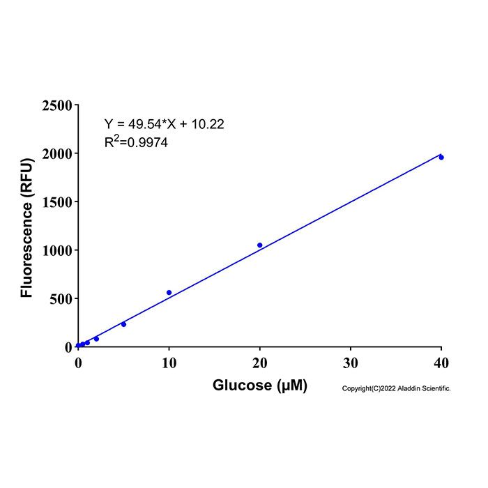
(2)荧光检测:取2 μL葡萄糖标准品 (5 mM),加入198 μL裂解液或者检测缓冲液,混匀,配制成浓度为50 µM的葡萄糖标准溶液。分别取50 µM的葡萄糖标准溶液0、0.5、1、2、5、10、20、40 μL加入黑色96孔板中,并相应地用裂解液或者检测缓冲液补足至50 μL,此时,标准曲线的浓度分别为0、0.5、1、2、5、10、20、40 µM。
3、反应工作液的配制
按照每个反应50 µL的体积配制适量的Amplex Red反应工作液。均匀混合44 µL检测缓冲液、2 µL Amplex Red、2 µL葡萄糖氧化酶、2 µL辣根过氧化物酶,即可配制成50 µL Amplex Red反应工作液。根据待检测样品(包括标准品)的数量,配制适量的反应工作液,建议尽量现配现用。具体配制方法参考下表:
样本数 | 1 | 10 | 20 | 50 |
检测缓冲液 | 44 µL | 440 µL | 880 µL | 2200 µL |
Amplex Red | 2 µL | 20 µL | 40 µL | 100 µL |
葡萄糖氧化酶 | 2 µL | 20 µL | 40 µL | 100 µL |
辣根过氧化物酶 | 2 µL | 20 µL | 40 µL | 100 µL |
总体积 | 50 µL | 500 µL | 1000 µL | 2500 µL |
注:H₂O₂的存在会对葡萄糖的检测产生干扰。如果样品含有H₂O₂,须同时设置背景对照孔,加入不含葡萄糖氧化酶的反应工作液,即配制反应工作液时2 µL葡萄糖氧化酶用检测缓冲液替代。
4. 样品测定
(1)取50 μL样品加入样品孔中,同时设置仅含裂解液或检测缓冲液的孔为空白对照。
(2)向标准品孔和样品孔中加入Amplex Red反应工作液50 µL,混匀,37ºC避光反应30分钟。
(3)如果使用吸光度检测,测定A570;如果使用荧光检测,设置激发波长为560 nm,发射波长为590 nm进行荧光强度检测。
5. 结果计算
建立标准曲线,并计算样品中葡萄糖的浓度(A),如果样品的背景对照信号比较高,样品的信号值应减去样品背景对照的信号值。葡萄糖浓度的计算公式如下: C (µM)=A×n
注:A为根据标准曲线确定的葡萄糖浓度(µM);n为样品总稀释倍数。可根据葡萄糖的分子量180计算出样品中葡萄糖的质量浓度(µg/ml)=C×0.18。
6. 结果展示
标准曲线


注意事项
1. Amplex Red在空气中不太稳定,开启后应尽快使用,且在使用过程中需注意适当避光。
2. Amplex Red须要完全解冻并平衡至室温后再使用,否则会影响检测结果。
3. 葡萄糖氧化酶冰上解冻,使用前后避免室温放置。
4 .为减少稀释液产生的荧光背景带来的误差,样品和标准品的稀释液应该根据样品的种类来定。当样品为裂解液制备的细胞或组织的裂解样品时,应使用裂解液稀释,当样品为血液等其它样品时,宜使用检测缓冲液稀释。
5. 吸光度检测时建议使用透明96孔板,荧光检测时建议使用96孔黑板。
6. 为了您的安全和健康,请穿实验服并戴一次性手套操作。
Glucose is considered one of the most widely distributed and important monosaccharides in nature. It can be directly absorbed into the bloodstream and is an essential nutrient for human energy metabolism and life maintenance, playing a critical role in sustaining metabolic homeostasis and the internal environment. Glucose is a major energy source and metabolic intermediate in living organisms, and blood glucose level has become an important diagnostic indicator for various metabolic diseases.
In this kit, Amplex Red is a fluorescent probe highly sensitive to H₂O₂. In the presence of horseradish peroxidase (HRP), Amplex Red reacts with H₂O₂ to produce the strongly red-fluorescent compound resorufin. Resorufin exhibits a maximum excitation wavelength of 571 nm and a maximum emission wavelength of 585 nm, and also shows strong visible light absorption at its excitation wavelength. Therefore, this kit can be used for both absorbance and fluorescence detection. The detection principle of this kit is as follows: Glucose is oxidized by glucose oxidase (GOD) in the presence of oxygen to produce D-glucono-δ-lactone and H₂O₂. The glucose concentration in the sample is then calculated by measuring the fluorescence intensity or absorbance of resorufin, the reaction product between H₂O₂ and Amplex Red. The fluorescence intensity or absorbance of resorufin is directly proportional to the glucose concentration in the sample.
Components
G1522838 | Components | Appearance | 100 T | 500 T | Storage | Quantity Per Test |
G1522838A | Lysis Buffer | Liquid | 20 mL | 100 mL | -20°C, dark | As needed |
G1522838B | Assay Buffer | Liquid | 20 mL | 100 mL | -20°C | As needed |
G1522838C | Amplex Red | Liquid | 200 μL | 1 mL | -20°C, dark | 2 μL |
G1522838D | Glucose Oxidase | Liquid | 200 μL | 1 mL | -20°C, dark | 2 μL |
G1522838E | Horseradish Peroxidase | Liquid | 200 μL | 1 mL | -20°C, dark | 2 μL |
G1522838F | Glucose Standard (5 mM) | Liquid | 200 μL | 1 mL | -20°C, dark | As needed |
Usage Protocol
1. Sample Preparation
1) Preparation of blood samples. For serum samples, place whole blood at room temperature for 30 minutes to 2 hours without vigorous shaking to avoid hemolysis. After the blood has naturally clotted and the serum has separated, centrifuge at approximately 1000 × g for 10 minutes at 4 °C, and collect the yellow supernatant as serum. For plasma samples, anticoagulate whole blood with heparin or EDTA, centrifuge at approximately 1000 × g for 10 minutes at 4 °C, and collect the yellow or light yellow supernatant as plasma.
2) Preparation of cell or tissue samples. For cultured adherent cells, wash once with PBS. For cultured suspension cells, collect cells by centrifugation, discard the supernatant, and completely remove any residual liquid. Add lysis buffer at a ratio of 100–200 μL per 1 × 10⁶ cells, pipette gently to mix, and lyse on ice for 5–10 minutes to ensure complete cell lysis. Centrifuge at approximately 12,000 × g for 5 minutes at 4 °C, and collect the supernatant for subsequent analysis.
3) Preparation of tissue samples. Add lysis buffer at a ratio of 100 μL per 10 mg of tissue, and homogenize under low‑temperature conditions such as on ice. Centrifuge at approximately 12,000 × g for 3–5 minutes at 4 °C, and collect the supernatant for subsequent analysis.
4) Preparation of cell culture supernatant samples. For adherent cells, directly collect the culture medium. For suspension cells, centrifuge and collect the culture medium.
All of the above operations should be performed at 4 °C or on ice. If the prepared cell or tissue samples cannot be analyzed immediately, they may be stored at –20 °C or –80 °C..
2. Glucose Standard Curve Setup
For absorbance or fluorescence detection, either method can be selected. For samples with low volume or low concentration, fluorescence detection is recommended. For absorbance detection, a clear 96‑well plate is recommended; for fluorescence detection, a black 96‑well plate is recommended.
When diluting the standard, use lysis buffer if detecting cell or tissue samples prepared with lysis buffer; use assay buffer if detecting samples such as blood or supernatant that do not require processing. This yields more accurate results.
1) Absorbance detection: Pipette 4 μL of Glucose Standard (5 mM) and add to 196 μL of lysis buffer or assay buffer, mix well, to prepare a 100 μM glucose standard solution. Add 0, 2.5, 5, 10, 20, 30, 40, and 50 μL of the 100 μM glucose standard solution into a clear 96‑well plate, and then bring the volume to 50 μL in each well with the corresponding lysis buffer or assay buffer. The resulting standard curve concentrations are 0, 5, 10, 20, 40, 60, 80, and 100 μM, respectively.
2) Fluorescence detection: Pipette 2 μL of Glucose Standard (5 mM) and add to 198 μL of lysis buffer or assay buffer, mix well, to prepare a 50 μM glucose standard solution. Add 0, 0.5, 1, 2, 5, 10, 20, and 40 μL of the 50 μM glucose standard solution into a black 96‑well plate, and then bring the volume to 50 μL in each well with the corresponding lysis buffer or assay buffer. The resulting standard curve concentrations are 0, 0.5, 1, 2, 5, 10, 20, and 40 μM, respectively.
3. Preparation of Reaction Working Solution
Prepare an appropriate amount of Amplex Red Reaction Working Solution based on a volume of 50 μL per reaction. Mix 44 μL of Assay Buffer, 2 μL of Amplex Red, 2 μL of Glucose Oxidase, and 2 μL of Horseradish Peroxidase to prepare 50 μL of Amplex Red Reaction Working Solution. Prepare an appropriate amount according to the number of samples to be tested (including standards). It is recommended to prepare the working solution fresh immediately before use. Refer to the table below for specific preparation methods:
Number of samples | 1 | 10 | 20 | 50 |
Assay Buffer | 44 µL | 440 µL | 880 µL | 2200 µL |
Amplex Red | 2 µL | 20 µL | 40 µL | 100 µL |
Glucose Oxidase | 2 µL | 20 µL | 40 µL | 100 µL |
Horseradish Peroxidase | 2 µL | 20 µL | 40 µL | 100 µL |
Total volume | 50 µL | 500 µL | 1000 µL | 2500 µL |
Note: The presence of H₂O₂ may interfere with glucose detection. If the sample contains H₂O₂, set up a background control well in parallel, and add the Reaction Working Solution without Glucose Oxidase (replace the 2 μL of Glucose Oxidase with Assay Buffer when preparing the Reaction Working Solution).
4. Sample Detection
1) Add 50 μL of sample to the sample wells, and set up wells containing only lysis buffer or assay buffer as blank controls.
2) Add 50 μL of Amplex Red Reaction Working Solution to each standard well and sample well, mix well, and incubate at 37 °C in the dark for 30 minutes.
3) For absorbance detection, measure A₅₇₀; for fluorescence detection, set the excitation wavelength to 560 nm and the emission wavelength to 590 nm for fluorescence intensity measurement.
5. Result Calculation
Establish a standard curve and calculate the glucose concentration in the sample (A). If the background signal of the sample is relatively high, subtract the signal value of the sample background control from that of the sample. The formula for calculating glucose concentration is as follows: C (µM) = A × n
Note: A is the glucose concentration (µM) determined from the standard curve; n is the total dilution factor of the sample. The mass concentration of glucose in the sample (µg/mL) can be calculated using the molecular weight of glucose (180): mass concentration = C × 0.18.
6. Results Presentation
Standard curve
Precautions
1. Amplex Red is relatively unstable in the air. Use it as soon as possible after opening, and take care to protect it from light during use.
2. Amplex Red must be completely thawed and equilibrated to room temperature before use; otherwise, the detection results may be affected.
3. Thaw Glucose Oxidase on ice and avoid leaving it at room temperature before or after use.
4. To reduce errors caused by fluorescence background from the diluent, the diluent for samples and standards should be selected according to the sample type. When the sample is a cell or tissue lysate prepared with lysis buffer, lysis buffer should be used for dilution; for other samples such as blood, assay buffer should be used for dilution.
5. For absorbance detection, a clear 96‑well plate is recommended; for fluorescence detection, a black 96‑well plate is recommended.
6. For your safety and health, please wear a lab coat and disposable gloves during operation.

 ZJ26F0737244


 危险品化学品经营许可证(带存储)