二磷酸腺苷(Adenosine diphosphate, ADP),即腺苷 5′‑焦磷酸酯,是由一分子腺苷与两个相连磷酸基团构成的化合物,分子式 C₁₀H₁₅N₅O₁₀P₂ 相对分子质量 427.2。ADP 属于核苷酸,在能量转移反应中发挥关键作用。生物体内,ADP一般由三磷酸腺苷(Adenosine triphosphate, ATP)水解脱去一分子磷酸基团、断裂一条高能磷酸键并释放能量生成;ADP 自身含有一条高能磷酸键,可在腺苷酸激酶催化下可逆转化为 ATP。ATP 与 ADP 的相互转化过程有多种磷酸(酯) 酶、磷酸化酶(激酶)参与。
本试剂盒中的ADHP底物含有对过氧化氢 (H₂O₂) 高度敏感的探针组份。在辣根过氧化物酶 (Horseradish peroxidase, HRP) 存在条件下,ADHP可与 H₂O₂以 1:1 摩尔比反应,生成具有红色荧光的产物试卤灵(Resorufin)。试卤灵最大激发波长 571 nm,最大发射波长 585 nm,同时在可见光区具有特征吸收。反应产物信号强度与样品中 ADP 含量成正比,可在 570 nm 处进行分光光度测定。因此,本试剂盒支持荧光检测法与分光光度法 (吸光度) 检测法两种读数方式。
该试剂盒仅用于科研领域,不适用于临床诊断或其他用途。
注意事项:
1. ADHP底物在空气中不太稳定,开启后应尽快使用,且在使用过程中需注意适当避光。
2. 请确保反应体系的pH值在7-8之间,否则会影响ADHP 底物的稳定性和荧光值。
3. ADHP与H₂O₂反应生成的氧化产物试卤灵,在还原剂存在条件下易分解,实验体系应避免引入还原剂,体系内还原剂浓度建议低于10 µM。
4. ADHP底物、ADP检测缓冲液需充分解冻并平衡至室温后方可使用,否则将干扰检测结果;其余试剂建议置于冰上操作。
5. 为减少稀释液产生的荧光背景带来的误差,样品和标准品的稀释液应该根据样品的种类来定。细胞或组织的裂解样品用代谢裂解缓冲液来稀释。
6. 使用荧光酶标仪检测时,请选用黑色不透光荧光酶标板。
7. 操作时请穿戴实验服与一次性手套,做好个人防护。
8. 所有试剂避免反复冻融,多次冻融会导致试剂活性下降,建议分装保存。
H1511292 | 组件 | 物理外观 | 100T | 储存 | 单次测试使用量 |
H1511292A | 代谢裂解缓冲液 | 无色透明液体 | 20 mL | -20°C | 按需 |
H1511292B | ADP检测缓冲液 | 无色透明液体 | 20 mL | -20°C | 72 µL |
H1511292C | ADHP 底物 | 淡粉色透明液体 | 0.2 mL | -20°C,Protect from light | 2 µL |
H1511292D | 酶溶液 A | 无色透明液体 | 0.2 mL | -20°C | 2 µL |
H1511292E | 酶溶液 B | 淡粉黄色透明液体 | 0.2 mL | -20°C | 2 µL |
H1511292F | 辅因子混合液 | 黄色透明溶液 | 0.2 mL | -20°C,Protect from light | 2 µL |
H1511292G | ADP 标准品(10 mM) | 无色透明液体 | 0.2 mL | -20°C | 按需 |
使用说明:
1. 试剂准备
额外所需材料:
(1) 实验材料:细胞或组织。
(2) 所需仪器:酶标仪、离心机、水浴锅。
(3) 其它:透明96孔板、96孔黑板、离心管。
2. 准备样品
细胞或组织样品的准备:
(1)对于培养的贴壁细胞,用预冷PBS洗涤一次并吸净残留液体。对于培养的悬浮细胞,先适当离心(如100-500×g,5分钟)收集细胞到离心管内,弃上清并吸净残留液体。按照每100万细胞加入100-200 µL 代谢裂解缓冲液的比例加入裂解液,适当吹打,冰浴5-10分钟以充分裂解细胞。4ºC约12,000×g离心5-10分钟,取上清用于后续检测。
(2)对于组织样品,按照每10 mg组织加入100 µL代谢裂解缓冲液的比例,使用高通量组织研磨仪、手持式组织研磨或玻璃匀浆器在约4ºC或冰浴等低温条件下进行匀浆。4ºC约12,000×g离心5-10分钟,取上清用于后续检测。
注:以上裂解、匀浆全过程均需在 4℃或冰上操作。制备好的细胞或组织样品如果不能立即检测,建议分装储存于-80ºC,避免反复冻融。
3. ADHP反应工作液的配制:
配制前先将 ADHP 底物、ADP检测缓冲液充分解冻并平衡至室温;酶溶液 A、酶溶液 B、辅因子混合液置于冰上备用。工作液配制全程避光操作。
配制建议:优先混合代谢检测缓冲液、辅因子混合液、酶溶液 A、酶溶液 B,最后加入 ADHP 底物,减少 ADHP 氧化。
单个反应体系总体积为 80 µL。各组分配比:代谢检测缓冲液 72 µL、ADHP 底物 2 µL、酶溶液 A 2 µL、酶溶液 B 2 µL、辅因子混合液 2 µL。将上述组分充分混匀,即得到 ADHP 反应工作液。
配制完成的 ADHP 反应工作液可在 4℃避光或冰浴避光条件下当日使用,强烈建议现配现用。试剂使用结束后,请依据试剂盒储存条件及时存放。具体配制参考下表:
样品(µL) | 1 份 | 10 份 | 20 份 | 50 份 |
ADP 检测缓冲液 | 72 | 720 | 1440 | 3600 |
ADHP 底物 | 2 | 20 | 40 | 100 |
酶溶液 A | 2 | 20 | 40 | 100 |
酶溶液 B | 2 | 20 | 40 | 100 |
辅因子混合液 | 2 | 20 | 40 | 100 |
ADHP 反应工作液 | 80 | 800 | 1600 | 4000 |
4. 样品测定:
ADP 标准曲线设置:可选择分光光度检测或荧光检测其中一种模式;样品量较少、ADP 浓度偏低时,优先推荐荧光检测。
(1) 分光光度检测:
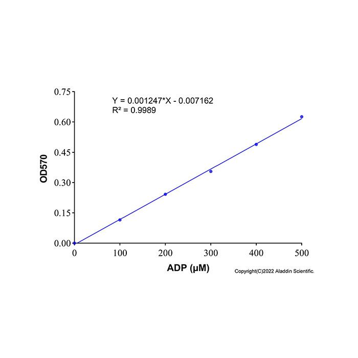
取5 μL ADP 标准品 (10 mM),加入95 μL代谢裂解缓冲液,混匀,配制成浓度为500 μM的ADP标准溶液。分别取500 μM的ADP标准溶液0、4、8、12、16、20 μL加入96孔板的标准品孔中,并相应地用代谢裂解缓冲液补足至20 µL, 此时,标准曲线的浓度分别为0、100、200、300、400、500 μM。
注:分光光度检测时建议使用透明96孔板。
(2) 荧光检测:
取2 μL ADP 标准品 (10mM),加入98 μL代谢裂解缓冲液,混匀,配制成浓度为 200 µM的ADP标准溶液。分别取200μM的ADP标准溶液0、1、2、5、10、20μl加入96孔板的标准品孔中,并相应地用代谢裂解缓冲液补足至20 μL,此时,标准曲线的浓度分别为0、10、 20、50、100、200 μM。
注:荧光检测时建议使用96孔黑板。
(3) 加样:
取20 μL样品或稀释后的样品至96孔板样品孔中,标准品、待测样品建议设置复孔。并相应地加入代谢裂解缓冲液至样品孔中,补足至20 µL。同步设置空白对照孔(仅加入 20 μL代谢裂解缓冲液);如需扣除样品基质背景,需设置不含反应工作液的样品背景孔。
注:建议先开展预实验,设置多个样品稀释梯度,确保检测信号落在标准曲线线性区间;若信号超出线性范围,请调整样品稀释倍数后重新测定。样品总稀释倍数记为 n。例:细胞裂解步骤稀释 10 倍,本步骤取10 µL 稀释后样品+10 µL 裂解缓冲液补足至 20 µL,则总稀释倍数 n =10×(20/10)= 20。
(4) 启动反应:
各孔加入ADPH反应工作液80 µL,轻柔振荡混匀,酶标板加盖,37ºC避光反应 30分钟。
注1:荧光反应于室温条件下孵育 5 min 即可进行读数;若荧光信号偏弱,可适当延长反应时间。
注2:如果吸光度偏低,可适当延长反应时间。例如反应45或60分钟。
(5) 信号读取:
如果使用分光光度检测,测定A₅₇₀;如果使用荧光检测,设置激发波长为560 nm,发射波长为590 nm进行荧光强度检测。
注:试卤灵理论最大激发波长 571 nm、最大发射波长 585 nm;本参数为通用酶标仪滤光片推荐条件,可根据仪器硬件适当微调。
(6) 数据计算:
根据标准曲线拟合方程,求出 20 µL 反应体系内稀释样品的 ADP 浓度。原始样品中 ADP浓度=计算得到的浓度×总稀释倍数n,如果样品的背景对照信号比较高,样品的信号值应减去样品背景对照的信号值。
Adenosine diphosphate (ADP), also known as adenosine 5′‑pyrophosphate, is composed of one adenosine molecule linked to two consecutive phosphate groups, with the molecular formula C₁₀H₁₅N₅O₁₀P₂ and a relative molecular mass of 427.2. As a nucleotide, ADP plays an essential role in energy transfer reactions. In vivo, ADP is generally generated when adenosine triphosphate (ATP) is hydrolyzed to lose one phosphate group, breaking one high-energy phosphate bond and releasing energy. ADP itself contains one high-energy phosphate bond and can be reversibly converted to ATP under the catalysis of adenylate kinase. A variety of phosphate (ester) enzymes and phosphorylases (kinases) participate in the interconversion between ATP and ADP.
ADHP Substrate included in this kit contains a probe component highly sensitive to hydrogen peroxide (H₂O₂). In the presence of Horseradish peroxidase (HRP), ADHP reacts with(H₂O₂) at a 1:1 molar ratio to produce resorufin, a red fluorescent product. Resorufin exhibits a maximum excitation wavelength of 571 nm and a maximum emission wavelength of 585 nm, and shows characteristic absorbance in the visible region. The signal intensity of the reaction product is proportional to the ADP content in samples, and spectrophotometry measurement can be performed at 570 nm. Therefore, both fluorescence detection and spectrophotometry (absorbance) detection are supported by this kit.
This kit is intended for research purposes only and is not suitable for clinical diagnosis or other applications.
Precautions
1. ADHP substrate is unstable upon exposure to air. Use it promptly after opening and protect it from light during operation.
2. Ensure the pH of the reaction system is maintained between 7 and 8; deviation from this range will compromise ADHP stability and fluorescence signals.
3. Resorufin, the oxidized product formed from the reaction between ADHP and H₂O₂,
decomposes readily in the presence of reducing agents. Avoid introducing reducing agents into the reaction system; the concentration of reducing agents should preferably be lower than 10 µM.
4. Fully thaw ADHP Substrate and Metabolic Assay Buffer and equilibrate them to room temperature before use; otherwise, detection results will be affected. Keep all other reagents on ice.
5. To minimize errors caused by fluorescence background from diluents, select appropriate diluents according to sample types. Use Metabolic Lysis Buffer for diluting cell or tissue lysate samples.
6. Use black opaque 96-well plates for fluorescence measurement with a fluorescence microplate reader.
7. Wear a lab coat and disposable gloves during operation for personal safety and health protection.
8. Avoid repeated freeze-thaw cycles for all reagents, as repeated freezing and thawing reduces reagent activity. Aliquot reagents for storage as recommended.
H1511292 | Components | Appearance | 100T | Storage | Quantity Per Test |
H1511292A | Metabolic Assay Lysis Buffer | Colorless transparent liquid | 20 mL | -20°C | As needed |
H1511292B | ADP Assay Buffer | Colorless transparent liquid | 20 mL | -20°C | 72 µL |
H1511292C | ADHP Substrate | Light pink transparent liquid | 0.2 mL | -20°C,Protect from light | 2 µL |
H1511292D | Enzyme Solution A | Colorless transparent liquid | 0.2 mL | -20°C | 2 µL |
H1511292E | Enzyme Solution B | pinkish-yellow transparent liquid | 0.2 mL | -20°C | 2 µL |
H1511292F | Cofactor Mix | Yellow transparent solution | 0.2 mL | -20°C,Protect from light | 2 µL |
H1511292G | ADP Standard (10mM) | Colorless transparent liquid | 0.2 mL | -20°C | As needed |
Instructions for Use
1. Reagent Preparation
Additional materials required:
(1) Samples: cultured cells or tissues。
(2) Equipment: microplate reader, centrifuge, water bath。
(3) Consumables: transparent 96-well plates, black 96-well plates, centrifuge tubes。
2. Sample Preparation
Preparation of cell and tissue samples:
(1) For adherent cultured cells: Wash cells once with pre-cooled PBS and thoroughly aspirate residual liquid. For suspension cultured cells: Harvest cells into centrifuge tubes by centrifugation (e.g.,100-500×g, 5 min), discard the supernatant and remove residual liquid completely. Add Metabolic Lysis Buffer at a ratio of 100-200 µL per 1× 10⁶ cells, pipette gently, and lyse cells on ice for 5-10 min. Centrifuge at 12,000×g,
4°C for 5-10 min, and collect the supernatant for subsequent detection.
(2) For tissue samples: Add Metabolic Lysis Buffer at a ratio of 100 µL per 10 mg tissue. Homogenize samples on ice or at 4 °C using a high-throughput tissue homogenizer, handheld tissue homogenizer or glass homogenizer. Centrifuge at 12,000×g, 4°C for 5-10 min, and collect the supernatant for subsequent detection.
Note: All lysis and homogenization procedures must be carried out on ice or at 4°C. If prepared cell or tissue supernatants cannot be tested immediately, aliquot and store at -80°C to avoid repeated freeze-thaw cycles.
3. Preparation of ADHP Working Solution
(1) Fully thaw ADHP Substrate and Metabolic Assay Buffer and equilibrate them to room temperature before preparation; keep Enzyme Solution A, Enzyme Solution B and Cofactor Mix on ice. Protect all operations from light during working solution preparation.
(2) Preparation suggestion: Mix Metabolic Assay Buffer, Cofactor Mix, Enzyme Solution A and Enzyme Solution B first, then add ADHP Substrate last to reduce ADHP oxidation.
(3) The total volume for a single reaction is 80 µL. Formulation:
Metabolic Assay Buffer 72 µL, ADHP Substrate 2 µL, Enzyme Solution A 2 µL, Enzyme Solution B 2 µL, Cofactor Mix 2 µL. Thoroughly mix all components to obtain ADHP Working Solution.
(4) The prepared ADHP Working Solution may be used on the same day when stored at 4°C protected from light or kept on ice with light shielding. Fresh preparation is strongly recommended. After use, store all reagents according to the storage requirements specified in the kit. See the table below for detailed preparation volumes.
Item (µL) | 1 reaction | 10 reactions | 20 reactions | 50 reactions |
ADP Assay Buffer | 72 | 720 | 1440 | 3600 |
ADHP Substrate | 2 | 20 | 40 | 100 |
Enzyme Solution A | 2 | 20 | 40 | 100 |
Enzyme Solution B | 2 | 20 | 40 | 100 |
Cofactor Mix | 2 | 20 | 40 | 100 |
ADHP Working Solution | 80 | 800 | 1600 | 4000 |
4. Sample Measurement
Setup of ADP Standard Curve:Either colorimetric detection or fluorometric detection can be selected. Fluorometric detection is recommended when sample volume is limited or ADP concentration is low.
(1) Spectrophotometry Detection
Mix 5 μL ADP Standard (10 mM) with 95 μL Metabolic Lysis Buffer to prepare 500 μM ADP standard stock solution. Transfer 0, 4, 8, 12, 16, 20 μL of 500 μM ADP standard stock solution into designated standard wells of a 96-well plate. Adjust the final volume to 20 µL with Metabolic Lysis Buffer. The resulting standard concentrations are 0, 100, 200, 300, 400, 500 μM.
Note: Use transparent 96-well plates for colorimetric measurement.
(2) Fluorometric Detection
Mix 2 μL ADP Standard (10 mM) with 98 μL Metabolic Lysis Buffer to prepare 200 μM ADP standard stock solution. Transfer 0, 1, 2, 5, 10, 20 μL of 200 μM ADP standard stock solution into designated standard wells of a 96-well plate. Adjust the final volume to 20 μL with Metabolic Lysis Buffer. The resulting standard concentrations are 0, 10, 20, 50, 100, 200 μM.
Note: Use black 96-well plates for fluorometric measurement.
(3) Sample Loading
Transfer 20 µL undiluted or diluted sample into sample wells. Stan dards and unknown samples are recommended to be tested in replicates. If sample volume is less than 20 µL, top up to 20 µL using Metabolic Assay Lysis Buffer .
Set blank control wells containing only 20 µL Metabolic Assay Lysis Buffer . If matrix background subtraction is required, prepare sample background wells without ADHP Working Solution.
Note: A preliminary experiment with serial sample dilutions is suggested to ensure signals fall within the linear range of the standard curve. Adjust sample dilution factor and re-test if signals exceed the linear range. Let n denote the total dilution factor of the sample. e.g.: Samples are diluted 10-fold during cell lysis; 10 µL diluted sample plus 10 µL lysis buffer is supplemented to reach 20 µL in this step. Total dilution factor n=10× (20/10)= 20.
(4) Reaction Initiation
Add 80 µL ADHP Working Solution into each well. Mix gently by shaking, cover the microplate, and incubate at 37°C for 30 min protected from light.
Note 1: For the fluorescence reaction, incubate at room temperature for 5 min before reading. If the fluorescence signal is weak, the reaction time may be appropriately extended.
Note 2: If s pectrophotometry signal is weak, incubation time can be extended to 45 or 60 min accordingly. Maintain identical incubation duration across all wells.
(5) Signal Reading
1) For spectrophotometry detection: Measure absorbance at A₅₇₀.
2) For fluorometric detection: Set excitation wavelength at 560 nm and emission wavelength at 590 nm to record fluorescence intensity.
Note: Resorufin has theoretical maximum excitation at 571 nm and maximum emission at 585 nm. The above parameters are recommended filter settings for common microplate readers and may be fine-tuned according to instrument configuration.
(6) Data Calculation
Calculate the diluted ADP concentration within the 20 µL reaction system based on the fitted standard curve equation.
ADP concentration in original sample=calculated concentration×total dilution factor n.
Note: If sample matrix background is high, subtract the signal value of corresponding sample background control from the sample signal.

 ZJ26F0838768


 危险品化学品经营许可证(带存储)