产品介绍
泛素(Ub)介导的蛋白质翻译后修饰是核心调控通路,几乎参与全部细胞生理活动。除经典的介导蛋白降解功能外,现有研究证实泛素还参与细胞信号传导、DNA 损伤应答、蛋白胞内转运、细胞周期调控、炎症反应、免疫应答及细胞凋亡调控。 泛素修饰依靠异肽键完成,泛素C端与靶蛋白赖氨酸残基的ε-氨基共价结合,该过程称为泛素化修饰。泛素自身含7个赖氨酸位点(K6、K11、K27、K29、K33、K48、K63),任意赖氨酸均可连接新的泛素分子,形成多聚泛素链。不同赖氨酸连接型泛素链赋予泛素系统多样化细胞调控功能。研究最透彻的两类泛素链为 K48、K63 连接型:K48多聚泛素链主要介导靶蛋白经蛋白酶体降解;K63多聚泛素链调控蛋白功能、亚细胞定位与蛋白互作。大量研究证明 K11型泛素链参与细胞有丝分裂调控,未来针对该链型的研究规模将逐步比肩K48、K63。随着更多链型与疾病、特异性细胞通路的关联被报道,其余稀有泛素链的研究热度将快速上升。泛素修饰是动态可逆过程。E1激活酶、E2结合酶、E3连接酶协同催化蛋白发生单泛素化或多聚泛素化;而去泛素化酶(DUBs)可移除蛋白上单/多聚泛素链、水解泛素链。
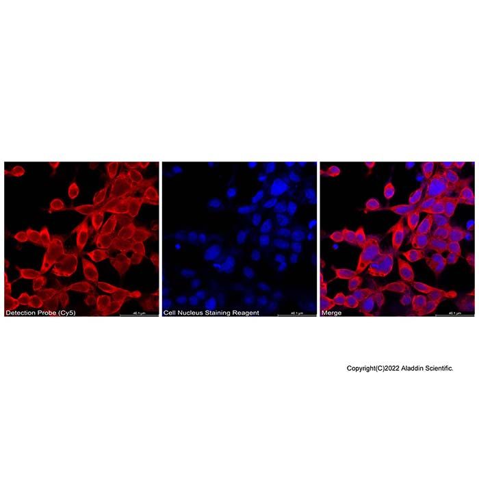
本试剂盒专为细胞或组织样本中泛素化蛋白的分布与水平检测而开发,采用高特异性泛素识别探针,结合荧光成像技术,可直观、灵敏地反映样本中泛素化蛋白的定位与相对丰度。
产品特点
1. 高灵敏度与特异性:采用高亲和力特异性靶向泛素化蛋白,即使在低丰度样本中也能准确捕捉泛素化信号,非特异性结合背景极低。
2. 操作简便:提供即用型工作液,减少配制步骤,简化实验流程。
3. 稳定性强:经严格质量控制,批次间性能一致,数据重复性好。
4. 广泛适用性:兼容细胞爬片、组织切片等多种样本类型,适配荧光显微镜及共聚焦显微镜。
检测原理
本试剂盒基于泛素识别探针与样本中泛素化蛋白的特异性结合。探针共价偶联近红外荧光染料Cy5(激发/发射波长:647 nm/665 nm),其优势在于背景荧光低、光稳定性良好,尤其适合细胞与组织成像。当探针结合靶标后,在647 nm波长激发下发射665 nm荧光信号,从而直观显示泛素化蛋白的亚细胞定位与相对丰度。试剂盒提供的细胞核染色试剂(激发/发射波长:350 nm/460 nm)可透过细胞膜,选择性结合DNA双链,呈现明亮蓝色荧光,用于辅助定位细胞核及评估细胞状态。染色后样本可于荧光显微镜或共聚焦显微镜下观察。
产品组分
| L1375166 | 组件 | 物理外观 | 10 T | 储存 | 单次测试使用量 |
| L1375166A | 固定液 | 无色液体
| 10 mL | 2-8℃ | 6孔板,1 mL |
| L1375166B | 通透液 | 无色液体 | 10 mL | 2-8℃ | 6孔板,1 mL |
| L1375166C | 洗涤液 | 无色液体
| 150 mL | 2-8℃ | 6孔板,1mL*12-14次 |
| L1375166D | 封闭液 | 浅黄色或黄色液体
| 30 mL | 2-8℃ | 6孔板,1 mL |
| L1375166E | Cy5标记的检测探针 | 蓝色液体 | 20 μL | -20℃. Store in the dark. | 6孔板,1-2 μL |
| L1375166F | 细胞核染色试剂
| 黄色液体 | 20 μL | -20℃. Store in the dark. | 6孔板,1-2 μL |
| L1375166G | 抗荧光淬灭封片液 | 浅黄色或黄色液体 | 2 mL | -20℃. Store in the dark. | 按需 |
使用方法
注:固定液、通透液、洗涤液、封闭液、 抗荧光淬灭封片液,在使用前需完全恢复至室温后方可加样;所有含荧光探针、细胞核染色试剂、封片液的孵育步骤全程避光操作,建议使用锡箔包裹湿盒避光孵育。
1. 对于贴壁细胞:
1.1 细胞培养生长至约50%-70%融合度。吸弃培养液,用PBS洗涤1-2次。
1.2 加入足量固定液完全覆盖细胞表面,室温固定 10-30 min;参考用量:6 孔板 1 mL/孔,96 孔板 100 μL/孔。
1.3 小心吸除固定液,用洗涤液洗涤3次,每次3-5分钟。每次洗涤时须尽量吸尽残余液体,同时要保持样品表面有些湿润,不能干掉,最后一次洗涤完时吸尽洗涤液。
1.4 加入足量通透液(6 孔板1 mL/孔),室温通透15分钟。最佳通透时间需根据细胞类型进行优化。
1.5 小心吸除通透液,用洗涤液洗涤3次,每次3-5分钟。每次洗涤时须尽量吸尽残余液体,同时要保持样品表面有些湿润,不能干掉,最后一次洗涤完时吸尽洗涤液。
1.6 加入足量封闭液 (6 孔板1 mL/孔),室温封闭1小时。
1.7 无需弃去封闭液,按体系终稀释比例 1:500-1:1000 加入Cy5标记的检测探针,4 ℃避光湿盒孵育过夜。提示:若追求更低背景、更强特异性信号,可弃去封闭液并洗涤 1 次后再加探针。
1.8 小心吸出检测探针,用洗涤液洗涤3-5次,每次5分钟。每次洗涤时须尽量吸尽残余液体,同时要保持样品表面有些湿润,不能干掉,最后一次洗涤完时吸尽洗涤液。
1.9 加入细胞核染色试剂,室温避光染色5-10分钟(6 孔板1 mL/孔)。
1.10 吸除细胞核染色液,用洗涤液洗涤3次,每次3-5分钟。每次洗涤时须尽量吸尽残余液体,同时要保持样品表面有些湿润,不能干掉,最后一次洗涤完时吸尽洗涤液。
1.11 成像处理:6 孔板 / 细胞爬片:滴加适量抗荧光淬灭封片液,盖玻片封片(避免气泡),避光静置后荧光显微镜下镜检;96 孔板:可保留少量洗涤液直接上机成像,需快速完成拍摄避免荧光衰减。
2. 对于悬浮细胞:
2.1 收集细胞悬液于离心管中,500×g离心5分钟,小心吸弃上清,用PBS洗涤1-2次。
2.2 加入固定液重悬,室温固定10-30分钟。500×g离心5分钟,小心吸弃固定液,洗涤液洗涤3次,每次3-5分钟。
2.3 加入通透液重悬,室温通透15分钟。500×g离心5分钟,小心吸弃通透液,洗涤液洗涤3次,每次3-5分钟。最佳通透时间需根据细胞类型进行优化。
2.4 加入封闭液重悬,室温封闭1小时。
2.5 无需弃去封闭液,按体系终稀释比例 1:500-1:1000 加入Cy5标记的检测探针,4 ℃避光湿盒孵育过夜。提示:若追求更低背景、更强特异性信号,可弃去封闭液并洗涤1次后再加探针。 500×g离心5分钟,小心吸弃探针液,洗涤液洗涤3~5次,每次3-5分钟。
2.6 加入细胞核染色液重悬,室温避光染色5-10分钟。500×g离心5分钟,小心吸弃细胞核染色液,洗涤液洗涤3次,每次3-5分钟。
2.7 将细胞重悬于适量PBS中,滴加至载玻片上,轻柔吹匀,自然微干后滴加适量的抗荧光淬灭封片液,封片(避免气泡)后避光镜检。
3. 对于组织切片:
3.1 对于石蜡切片先进行常规的二甲苯脱蜡和梯度乙醇水化处理后,需进行抗原修复(推荐微波热修复法,于柠檬酸钠缓冲液或EDTA缓冲液中加热至95-100°C,维持10-20分钟),冷却至室温后再从固定步骤开始操作。
3.2 对于冷冻切片取出恢复至室温后,PBS充分漂洗去除OCT包埋剂后,直接进行后续固定步骤。
3.3 后续固定、通透、封闭、探针孵育、核染、洗涤、封片流程同贴壁细胞操作规范。
注意事项
1. 试剂回温:固定液、通透液、洗涤液、封闭液、 抗荧光淬灭封片液,在使用前需完全恢复至室温后再进行加样操作,低温试剂直接接触样本易造成细胞皱缩、背景升高。
2. 避光操作:所有涉及荧光探针、细胞核染色液及封片液的步骤均需避光操作,荧光染料见光易淬灭,建议使用锡箔纸包裹或在不透光的湿盒中进行孵育。
3. 对照设置:每次实验需设置阴性对照和阳性对照,以排除假阳性、试剂自发荧光干扰。
4. 安全操作:固定液含多聚甲醛,具有毒性,请在通风橱中操作并佩戴防护用品。细胞核染色液有腐蚀和对人体有潜在危害,操作时请戴手套。
5. 防止干片:洗涤和孵育过程中,务必保持样品湿润,避免干片导致高背景或脱片。
Product Introduction
Ubiquitin (Ub)-mediated post‑translational modification is a core regulatory pathway that participates in virtually all cellular physiological activities. Beyond its canonical function in proteasomal degradation, ubiquitin is now known to be involved in signal transduction, DNA damage response, intracellular protein trafficking, cell‑cycle regulation, inflammatory responses, immune responses, and apoptosis regulation. Ubiquitin modification is achieved through an isopeptide bond: the C‑terminus of ubiquitin is covalently conjugated to the ε‑amino group of a lysine residue on the target protein, a process termed ubiquitination. Ubiquitin itself contains seven lysine residues (K6, K11, K27, K29, K33, K48, and K63), each of which can link to another ubiquitin molecule, forming polyubiquitin chains. Different lysine‑linked chain types confer diverse regulatory functions upon the ubiquitin system. The two best‑characterised polyubiquitin chain types are K48‑ and K63‑linked chains: K48‑linked chains primarily target proteins for proteasomal degradation, whereas K63‑linked chains regulate protein function, subcellular localisation, and protein–protein interactions. Substantial evidence demonstrates that K11‑linked chains are involved in mitotic regulation, and research on this chain type is expected to expand to a scale comparable to that of K48 and K63 in the future. As more associations between other chain types and diseases or specific signalling pathways are reported, interest in the less common ubiquitin chains will rapidly increase. Ubiquitination is a dynamic and reversible process. E1 activating enzymes, E2 conjugating enzymes, and E3 ligases cooperate to catalyse mono‑ or polyubiquitination, whereas deubiquitinases (DUBs) can remove mono‑/polyubiquitin moieties from proteins and hydrolyse ubiquitin chains.
This kit is specifically developed for detecting the distribution and levels of ubiquitinated proteins in cell or tissue samples. It employs a highly specific ubiquitin recognition probe combined with fluorescence imaging technology to provide intuitive and sensitive detection of ubiquitinated protein localization and relative abundance.
Product Features
1. High Sensitivity and Specificity: High-affinity probes specifically target ubiquitinated proteins, enabling precise detection of ubiquitination signals even in low-abundance samples, with minimal nonspecific binding and ultra‑low background.
2. Easy to Use: Ready-to-use working solutions minimize preparation steps and simplify the experimental workflow.
3. High Stability: Strict quality control ensures consistent performance across batches and excellent data reproducibility.
4. Broad Compatibility: Compatible with various sample types including cell smears and tissue sections; adaptable to fluorescence microscopes and confocal microscopes.
Detection Principle
This kit is based on the specific binding of a ubiquitin recognition probe to ubiquitinated proteins in the sample. The probe is covalently conjugated to the Near-red fluorescent dye Cy5 (Ex/Em: 647 nm/665 nm), which offers low background fluorescence and good photostability, making it particularly suitable for cell and tissue imaging. Upon probe binding to the target, excitation at 647 nm generates an emission signal at 665 nm, allowing direct visualization of the subcellular localization and relative abundance of ubiquitinated proteins.
The provided nuclear staining reagent (Ex/Em: 350 nm/460 nm) is cell-permeable and selectively binds to double-stranded DNA, emitting bright blue fluorescence. This reagent assists in nuclear localization and assessment of cell status. Stained samples can be observed under a fluorescence microscope or confocal microscope.
Components
| L1375166 | Components | Appearance | 10 T | Storage | Quantity Per Test |
| L1375166A | Fixation Buffer | Colourless liquid | 10 mL | 2-8℃ | 6 wells,1 mL |
| L1375166B | Permeabilization Buffer | Colourless liquid | 10 mL | 2-8℃ | 6 wells,1 mL |
| L1375166C | Washing Buffer | Colourless liquid | 150 mL | 2-8℃ | 6 wells,1 mL*12-14 times |
| L1375166D | Blocking Buffer | Light yellow to yellow liquid | 30 mL | 2-8℃ | 6 wells,1 mL |
| L1375166E | Detection Probe (Cy5) | Blue liquid | 20 μL | -20℃. Store in the dark. | 6 wells, 1-2 μL |
| L1375166F | Cell Nucleus Staining Reagent
| Yellow liquid
| 20 μL | -20℃. Store in the dark. | 6 wells, 1-2 μL |
| L1375166G | Antifade Mounting Medium | Light yellow to yellow liquid | 2 mL | -20℃. Store in the dark. | As needed |
Usage Protocol
Note: Fixation Buffer, Permeabilization Buffer, Washing Buffer, Blocking Buffer, and Anti-fade Mounting Medium must be fully equilibrated to room temperature before use. All incubation steps involving the fluorescent probe, nuclear staining reagent, or mounting medium must be performed under light‑protected conditions; it is recommended to wrap the humid chamber with aluminum foil during incubation.
1. For Adherent Cells
1.1 Culture cells to approximately 50%-70% confluence. Aspirate the culture medium and wash 1-2 times with PBS.
1.2 Add sufficient fixative to completely cover the cell surface and fix at room temperature for 10–30 min. Recommended volumes: 1 mL/well for a 6‑well plate; 100 μL/well for a 96‑well plate.
1.3 Carefully aspirate the fixative and wash 3 times with wash solution, 3-5 min each. During each wash, remove as much residual liquid as possible while keeping the sample surface slightly moist; do not allow the sample to dry out. After the last wash, aspirate the wash solution completely.
1.4 Add sufficient permeabilization solution (1 mL/well for a 6‑well plate) and permeabilize at room temperature for 15 min. Optimal permeabilization time should be determined empirically for different cell types.
1.5 Carefully aspirate the permeabilization solution and wash 3 times with wash solution, 3-5 min each. During each wash, remove as much residual liquid as possible while keeping the sample surface slightly moist; do not allow the sample to dry out. After the last wash, aspirate the wash solution completely.
1.6 Add sufficient blocking solution (1 mL/well for a 6‑well plate) and block at room temperature for 1 h.
1.7 Without removing the blocking solution, add the Cy5‑labeled detection probe to a final dilution of 1:500-1:1000 (based on the total system volume). Incubate overnight at 4 °C in a light‑protected humid chamber. Tip: If lower background and stronger specific signal are desired, the blocking solution may be discarded and the samples washed once before adding the probe.
1.8 Carefully aspirate the probe solution and wash 3-5 times with wash solution, 5 min each. During each wash, remove as much residual liquid as possible while keeping the sample surface slightly moist; do not allow the sample to dry out. After the last wash, aspirate the wash solution completely.
1.9 Add nuclear staining reagent (1 mL/well for a 6‑well plate) and stain at room temperature in the dark for 5-10 min.
1.10 Aspirate the nuclear staining solution and wash 3 times with wash solution, 3-5 min each. During each wash, remove as much residual liquid as possible while keeping the sample surface slightly moist; do not allow the sample to dry out. After the last wash, aspirate the wash solution completely.
1.11 Imaging: For 6‑well plates or cells grown on coverslips: apply a suitable amount of antifade mounting medium, mount with a coverslip (avoid air bubbles), allow to stand in the dark, and then examine under a fluorescence microscope. For 96‑well plates: a small volume of wash solution may be left in the wells and the plate directly imaged; complete the acquisition quickly to minimize fluorescence decay.
2. For Suspension Cells
2.1 Collect the cell suspension in a centrifuge tube, centrifuge at 500×g for 5 min, carefully aspirate the supernatant, and wash 1-2 times with PBS.
2.2 Resuspend the cells in fixative and fix at room temperature for 10-30 min. Centrifuge at 500×g for 5 min, carefully aspirate the fixative, and wash 3 times with wash solution, 3–5 min each.
2.3 Resuspend in permeabilization solution and permeabilize at room temperature for 15 min. Centrifuge at 500×g for 5 min, carefully aspirate the permeabilization solution, and wash 3 times with wash solution, 3-5 min each. Optimal permeabilization time should be determined empirically for different cell types.
2.4 Resuspend in blocking solution and block at room temperature for 1 h.
2.5 Without removing the blocking solution, add the Cy5‑labeled detection probe to a final dilution of 1:500-1:1000. Incubate overnight at 4 °C in a light‑protected humid chamber. Tip: For lower background and stronger specific signal, the blocking solution may be discarded and the cells washed once before adding the probe. Then centrifuge at 500×g for 5 min, carefully aspirate the probe solution, and wash 3-5 times with wash solution, 3-5 min each.
2.6 Resuspend in nuclear staining reagent and stain at room temperature in the dark for 5-10 min. Centrifuge at 500×g for 5 min, carefully aspirate the nuclear staining reagent, and wash 3 times with wash solution, 3-5 min each.
2.7 Resuspend the cells in an appropriate volume of PBS, pipette onto a glass slide, gently spread the suspension, allow to air‑dry slightly, then add a suitable amount of antifade mounting medium, mount with a coverslip (avoid air bubbles), and examine under light‑protected conditions.
3. For Tissue Sections
3.1 Paraffin sections: After routine deparaffinization with xylene and rehydration through graded ethanol, perform antigen retrieval (microwave heat‑induced retrieval is recommended: heat sections in sodium citrate buffer or EDTA buffer to 95–100 °C and maintain for 10–20 min). Allow to cool to room temperature, then proceed from the fixation step.
3.2 Frozen sections: Remove sections, equilibrate to room temperature, wash thoroughly with PBS to remove OCT embedding medium, and then directly proceed with the subsequent fixation step.
3.3 All subsequent steps (fixation, permeabilization, blocking, probe incubation, nuclear staining, washing, and mounting) are the same as those described for adherent cells.
Precautions
1. Reagent temperature equilibration: Fixative, permeabilization solution, wash solution, blocking solution, and antifade mounting medium must be fully equilibrated to room temperature before use. Addition of cold reagents directly to samples may cause cell shrinkage and elevated background.
2. Light protection: All steps involving the fluorescent probe, nuclear staining reagent, or mounting medium must be performed under light‑protected conditions, as fluorescent dyes are readily quenched by light. It is recommended to wrap the humid chamber with aluminum foil or incubate in a light‑proof container.
3. Control setup: Negative and positive controls should be included in each experiment to exclude false‑positive signals and interference from reagent autofluorescence.
4. Safety precautions: The fixative contains paraformaldehyde, which is toxic. Please operate in a fume hood and wear appropriate personal protective equipment. The nuclear staining reagent is corrosive and potentially hazardous; wear gloves during handling.
5. Prevent drying of samples: During washing and incubation steps, ensure that samples remain moist at all times. Drying may cause high background staining or detachment of sections.

 ZJ26F0838244


 危险品化学品经营许可证(带存储)