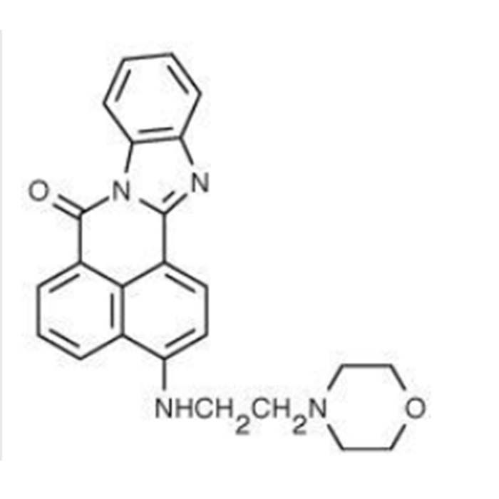
本品具有较低的 pKa 值,仅在酸性区室内才具有荧光。本品以溶于无水 DMSO 的 1 mM 储存液形式提供。原料均采用美国进口原料。该探针是主要对活细胞中的酸性区室 (如溶酶体、顶体精子) 动态合成和功能进行研究的一类荧光探针,该类探针的染色具有向酸性,可通过质子化作用在酸性细胞器内累积。该类探针的这种质子化作用还可解除染料的荧光淬灭性,使其所发射的荧光强度更强。该探针随着细胞器的酸化程度其荧光强度呈 pH 依赖型 (向酸)增强。
使用方法(仅供参考)
使用前,先将本品取出回温至室温,并对其进行简短离心使 DMSO 溶液集中于管底。最佳工作浓度需根据不同的实验要求、细胞类型、细胞或组织的膜通透性等进行优化。
1.工作液的配制
利用培养基或合适的缓冲液将 1 mM 储存液稀释至工作浓度,推荐工作液浓度至少为 1 µM;
注意
(1)为了降低探针加载过度可能引起的假阳性,建议在不影响染色效果的情况下尽量使用低浓度。
(2)若细胞在染色后于不含染料的培养基中孵育,会观察到荧光信号的衰减和细胞的空泡化现象。
(3)工作液现配现用。
2. 染色
2.1 对于贴壁细胞
(1) 将细胞置于培养皿中的盖玻片上,加入合适培养基,使其爬片生长。
(2) 待细胞生长到合适丰度,吸除培养液,加入适量 37℃预热的含探针工作液。于生长状态下孵育 30 min~2 h(具体孵育时间需根据细胞类型而定)。
(3) 利用新鲜培养基替换上述染色液并在荧光显微镜(含合适滤片)下观察。若染色不够充分,建议增加染料浓度或延长染色时间。
2.2 对于悬浮细胞
(1) 离心,吸除上清。
(2) 利用 37℃预热的探针工作液重悬细胞,于生长状态下孵育 30 min~2 h(具体时间需根据细胞类型而定)。
(3) 离心,吸除染色液,加入新鲜培养液重悬细胞。
(4) 置于荧光镜下观察。若染色不够充分,建议增加染料浓度或加长染色时间。
注:对于悬浮细胞,也可将细胞贴附于经 BD Cell-Tak 处理过的盖玻片上,然后使用类似于贴壁细胞的方法进行染色。
注意事项
1.我司产品为非无菌包装,若用于细胞培养,请提前做预处理,除去热原细菌,否则会导致染菌。
2.部分产品我司仅能提供部分信息,我司不保证所提供信息的权威性,以上数据仅供参考交流研究之用。
This product has a low pKa value and only emits fluorescence within acidic compartments. It is supplied as a 1 mM stock solution dissolved in anhydrous DMSO, with all raw materials imported from the United States. As a fluorescent probe specifically targeting acidic compartments in live cells (such as lysosomes and sperm acrosomes), it enables research on their dynamic biogenesis and functions. The probe exhibits acidotropic staining and accumulates in acidic organelles via protonation. This protonation relieves fluorescence quenching of the dye, resulting in significantly stronger fluorescence signals. Its fluorescence intensity increases in a pH-dependent manner with the acidification of organelles.
Protocol (For Reference Only)
Before use, take the product out and equilibrate it to room temperature, then perform a brief centrifugation to collect all DMSO solution at the bottom of the tube. The optimal working concentration must be optimized according to experimental requirements, cell type, and membrane permeability of cells or tissues.
1. Preparation of Working Solution
Dilute the 1 mM stock solution with culture medium or appropriate buffer to prepare the working solution; the recommended minimum working concentration is 1 µM.
Notes:
(1) To reduce false positive signals caused by overloading the probe, use the lowest feasible concentration without compromising staining quality.
(2) If stained cells are incubated in dye-free medium after labeling, attenuation of fluorescence signals and cellular vacuolation may be observed.
(3) Prepare the working solution immediately before use.
2. Staining Procedure
2.1 Adherent Cells
1.Seed cells onto coverslips placed in culture dishes with adequate medium for adherent growth.
2.When cells reach optimal confluency, discard the medium and add pre-warmed probe working solution at 37 °C. Incubate under normal culture conditions for 30 min to 2 h (incubation time varies by cell line).
3.Replace the staining solution with fresh medium and observe under a fluorescence microscope equipped with appropriate filter sets. Increase dye concentration or extend incubation time if staining intensity is insufficient.
2.2 Suspension Cells
1.Centrifuge the cell suspension and discard the supernatant.
2.Resuspend the cell pellet in pre-warmed probe working solution at 37 °C, then incubate under normal culture conditions for 30 min to 2 h (incubation time varies by cell line).
3.Centrifuge again, remove the staining solution, and resuspend cells in fresh culture medium.
4.Observe under a fluorescence microscope. Increase dye concentration or extend incubation time if staining intensity is insufficient.
Note: Suspension cells may also be immobilized on BD Cell-Tak-coated coverslips, followed by the staining protocol for adherent cells.
Precautions
1.This product is not sterilely packaged. If used for cell culture, pretreat the solution to remove pyrogens and bacteria in advance to avoid microbial contamination.
2.Only partial product information is available for some items. The company does not warrant the authority of the provided data, which are for reference and academic research only.



 危险品化学品经营许可证(带存储)