脂质过氧化是由氧化损伤造成的脂质降解,是氧化应激的重要标志。多不饱和脂质通常易受活性氧的氧化攻击,引起明确的链反应并生成最终产物,如丙二醛(MDA)。脂质过氧化影响许多疾病的病理,包括动脉粥样硬化、糖尿病和阿尔茨海默氏病。脂质氧化(MDA)检测试剂盒(Lipid Peroxidation MDA Assay Kit)采用一种基于MDA和硫代巴比妥酸(thiobarbituric acid,TBA)反应产生红色产物的显色反应,随后通过比色法用于对血浆、血清、尿液、动植物组织或细胞裂解液中MDA进行定量检测,广泛用于脂质氧化(lipid peroxidation)水平检测的试剂盒。
丙二醛(Malondialdehyde,MDA)是一种生物体脂质氧化的天然产物。动物或植物细胞发生氧化应激(oxidative stress)时,会发生脂质氧化。一些脂肪酸氧化后逐渐分解为一系列复杂的化合物,其中包括MDA。此时通过检测MDA的水平即可检测脂质氧化的水平,因此MDA的测定被广泛用作脂质氧化的指标。生物体内的一些其它生化反应也会产生MDA,例如thromboxane synthase也可以催化产生,但只要在测定时设置适当对照即可观察到脂质氧化水平的变化。丙二醛在较高温度及酸性环境中可与TBA发生反应,形成红色的MDA-TBA加合物,相应的反应原理图如下:

MDA-TBA加合物在535 nm处有最大吸收,据此可以通过比色法进行检测。另外,MDA-TBA加合物也可以在530 nm被激发产生发射波长560 nm,据此也可以进行荧光检测。特点:本试剂盒中采用了特殊的抗氧化剂,可以有效地抑制样品在检测过程中产生新的MDA,使检测更加准确。同时本检测试剂盒在检测过程中可以把部分MDA天然形成的聚丙二醛分解成MDA,使对脂质氧化的测定更加准确。
本试剂盒可以检测低达0.5 μM的MDA,也可检测高达200 μM的MDA。低浓度样本可用荧光法进行检测更灵敏。血浆、血清样品中的MDA含量通常在约2-4 μM,尿液中的MDA含量通常在约5-30 μM,在本试剂盒的检测范围内,可以直接用本试剂盒检测血浆、血清、尿液样品等。


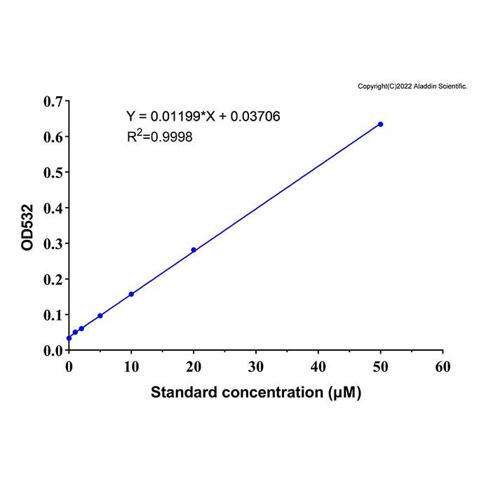
不同浓度标准品使用本试剂盒的检测效果图。实测数据会因检测仪器等的不同而存在差异,图中数据仅供参考。
产品组分
M1524526
| Component | 外观 | 100 T | 500 T | 储存 | 单次使用量 |
| M1524526A | TBA | 白色至黄色粉末 | 25 mg | 125 mg | -20℃避光 | 按需 |
| M1524526B | TBA配制液 | 无色液体 | 7 mL | 35 mL | 2-8℃.
| 按需 |
| M1524526C | TBA稀释液 | 无色液体 | 15 mL | 75 mL | 2-8℃.
| 按需 |
| M1524526D | 抗氧化剂 | 无色液体 | 0.3 mL | 1.5 mL | -20℃避光 | 按需 |
| M1524526E | 标准品 (1mM) | 无色液体 | 0.2 mL | 1 mL | -20℃避光 | 按需 |
注意事项
1) 醛、较高浓度的可溶性糖(例如250 mM蔗糖)对反应有干扰,可溶性糖与TBA显色反应的产物在532nm也有吸收(最大吸收在450 nm)。如果可溶性糖对测定有干扰,可以通过测定450 nm作为参考波长进行双波长测定,消除其干扰。
2) 本产品仅限于专业人员的科学研究用,不得用于临床诊断或治疗,不得用于食品或药品,不得存放于普通住宅内。
3) 为了您的安全和健康,请穿实验服并戴一次性手套操作。
使用方法
1. 样品的准备:
1) 血浆、血清或尿液样品制备后可以直接用于MDA测定。
2)组织或细胞可以使用PBS或裂解液进行匀浆或裂解。对于组织,组织重量占匀浆液或裂解液的比例为10%;对于细胞,每100万细胞使用0.1ml裂解液或匀浆液。匀浆或裂解后,10,000 g-12,000 g离心10分钟取上清用于后续测定。对于一些特殊样品,离心不能获得澄清的上清溶液的,可以使用0.2微米孔径的过滤器过滤以获得澄清的样品溶液。匀浆或裂解等样品制备步骤宜在冰浴或4℃进行操作。
3) 对于组织或细胞样品,样品准备完毕后用BCA蛋白浓度测定试剂盒测定蛋白浓度,便于后续计算单位蛋白重量组织或细胞内的MDA含量。
4) 本试剂盒对于样品中的常见化学成分的兼容性参考下表:
试剂类别 | 化学成分 | 是否干扰 |
缓冲试剂 | Borate (50 mM) | 否 |
缓冲试剂 | HEPES (10 0mM) | 否 |
缓冲试剂 | Phosphate (100 mM) | 否 |
缓冲试剂 | Tris (25 mM) | 否 |
去垢剂 | CHAPS (≤1%) | 否 |
去垢剂 | Triton X-100 (≤1%) | 否 |
去垢剂 | Tween 20 (≤1%) | 否 |
抑制剂 / 螯合剂 | Antipain (≤100 μg/mL) | 否 |
抑制剂 / 螯合剂 | Chymostatin (≤10 μg/mL) | 否 |
抑制剂 / 螯合剂 | Leupeptin (≤10 μg/mL) | 否 |
抑制剂 / 螯合剂 | PMSF (≤200 μM) | 否 |
抑制剂 / 螯合剂 | Trypsin (≤10 μg/mL) | 否 |
抑制剂 / 螯合剂 | EDTA (≤1 mM) | 否 |
抑制剂 / 螯合剂 | EGTA (≤1 mM) | 否 |
其它试剂 | Sucrose (250 mM) | 是 |
其它试剂 | Glycerol (≤10%) | 否 |
2. 试剂盒的准备工作:
1) TBA储存液的配制:称取适量TBA,用TBA配制液配制成浓度为0.37%的TBA储存液。例如18.5 mg TBA用5mL TBA配制液配制,或者25 mg TBA用6.76 mL TBA配制液配制,最终浓度即为0.37%。TBA配制液需完全溶解后再使用,可以加热到70℃以促进溶解。TBA储存液较难溶解,需加热到70℃,并通过剧烈Vortex以促进溶解。配制好的TBA储存液室温避光保存,至少3个月内有效。
2) MDA检测工作液的配制: 根据待测定的样品数(含对照),参考下表在临检测前新鲜配制适量的MDA检测工作液
试剂名称 | 1 次检测用量 | 10 次检测用量 | 20 次检测用量 | 50 次检测用量 |
TBA 稀释液 | 150 μL | 1500 μL | 3000 μL | 7500 μL |
TBA 储存液 | 50 μL | 500 μL | 1000 μL | 2500 μL |
抗氧化剂 | 3 μL | 30 μL | 60 μL | 150 μL |
注意: MDA检测工作液较难溶解,可以70℃加热,并剧烈Vortex以促进溶解。也可以通过超声处理以促进溶解。配制好的MDA检测工作液必须当天使用。
3) 标准品的稀释:取适量标准品用蒸馏水稀释至0、0.5、1、2、5、10、20、50 μM,用于后续制作标准曲线。如果样品中MDA的浓度很高,可以增加100、150和200 μM的标准品浓度。
3. 样品测定:
1) 在离心管或其它适当容器内加入0.1 mL匀浆液、裂解液或PBS等适当溶液作为空白对照,加入0.1 mL上述不同浓度标准品用于制作标准曲线,加入0.1 mL样品用于测定;随后加入0.2 mL MDA检测工作液。可参考下表设置检测反应体系:
2) 混匀后,100 ℃或沸水浴加热15分钟。加热时务必注意避免液体暴沸溅出。如果使用加热块(Heat block)进行加热注意用重物压紧离心管盖;如果使用沸水浴,则需使用可把盖子锁死的离心管或螺旋盖离心管,或用Parafilm封住离心管口,用针头刺一小孔。最方便和准确的加热方法是使用带有热盖并可以加热0.5 ml PCR管的PCR仪。
3) 水浴冷却至室温,1000 g室温离心10分钟。取200 μL上清加入到96孔板中,随后用酶标仪在532 nm测定吸光度。如果不方便测定532 nm的吸光度,也可以测定530-540 nm之间的吸光度。可以设定450 nm为参考波长进行双波长测定。如测荧光可取200 μL上清加入到96孔黑色不透光板中,随后用酶标仪在530 nm激发产生发射波长560 nm测定吸光度。
4) MDA含量的计算:对于血浆、血清或尿液等样品可以直接根据标准曲线计算获得MDA的摩尔浓度,对于细胞、或组织样品,计算出样品溶液中的MDA含量后,可以通过单位重量的蛋白含量或组织重量等来表示最初样品中的MDA含量,例如μmol/mg蛋白或μmol/mg组织。
常见问题
没有检测到MDA。可能样品中MDA浓度过低。在检测组织或细胞的MDA时,请使用更多的组织或细胞。不要稀释样品。
应用
脂质过氧化(MDA)检测试剂盒已用于确定丙二醛(MDA)的水平。
适用性
适于测定组织、细胞和血浆等各类样品中的丙二醛(MDA)
Lipid peroxidation is the oxidative damage-induced degradation of lipids and serves as an important marker of oxidative stress. Polyunsaturated lipids are generally susceptible to oxidative attack by reactive oxygen species (ROS), triggering a well-defined chain reaction that generates end products such as malondialdehyde (MDA). Lipid peroxidation is implicated in the pathology of numerous diseases, including atherosclerosis, diabetes, and Alzheimer’s disease.
The Lipid Peroxidation MDA Assay Kit utilizes a colorimetric reaction based on the formation of a red product from the reaction between MDA and thiobarbituric acid (TBA), followed by spectrophotometric quantification of MDA in plasma, serum, urine, animal/plant tissues, or cell lysates. This kit is widely used for the detection of lipid peroxidation levels.
Malondialdehyde (MDA) is a natural byproduct of lipid peroxidation in organisms. When animal or plant cells undergo oxidative stress, lipid peroxidation occurs. Oxidation of certain fatty acids leads to their gradual decomposition into a series of complex compounds, including MDA. Thus, the level of lipid peroxidation can be assessed by measuring MDA, and MDA determination is widely used as an indicator of lipid peroxidation. MDA can also be produced by other biochemical reactions in vivo, such as those catalyzed by thromboxane synthase; however, changes in lipid peroxidation levels can be observed by setting appropriate controls during the assay. Under high-temperature and acidic conditions, MDA reacts with TBA to form a red MDA-TBA adduct. The corresponding reaction scheme is shown below:
The MDA-TBA adduct exhibits maximum absorbance at 535 nm, enabling its quantification by colorimetric assay. Additionally, the MDA-TBA adduct can also be excited at 530 nm to generate an emission wavelength of 560 nm, allowing for detection via fluorometric assay.
Features: This kit incorporates a proprietary antioxidant system that effectively inhibits de novo MDA formation during the assay, ensuring more accurate measurements. Furthermore, the kit is designed to decompose naturally occurring poly-MDA oligomers into monomeric MDA during the reaction, thereby improving the accuracy of lipid peroxidation quantification.
The kit can detect MDA concentrations as low as 0.5 μM and as high as 200 μM. The fluorescence method offers higher sensitivity when detecting samples at low concentrations. MDA levels in plasma and serum samples are typically approximately 2-4 μM, while those in urine samples range from approximately 5-30 μM, all of which fall well within the detection range of this kit, enabling direct analysis of these sample types.
This figure shows the detection results of standard solutions with different concentrations using this kit. The actual measured values may vary depending on factors such as the detection instrument used; the data in the figure is for reference only.
Component
M1524526
| Component | Appearance | 100 T | 500 T | Storage conditions | Quantity Per Test | | M1524526A | TBA | White to Yellow Powder
| 25 mg | 125 mg | -20℃. Store in the dark. | As needed | | M1524526B | TBA preparation solution | Colourless liquid | 7 mL | 35 mL | 2-8℃.
| As needed
| | M1524526C | TBA diluent | Colourless liquid | 15 mL | 75 mL | 2-8℃.
| As needed
| | M1524526D | Antioxidant | Colourless liquid | 0.3 mL | 1.5 mL | -20℃. Store in the dark. | As needed
| | M1524526E | Standard(1 mM) | Colourless liquid | 0.2 mL | 1 mL | -20℃. Store in the dark. | As needed
|
|
Notes
1. Aldehydes and high concentrations of soluble sugars (e.g., 250 mM sucrose) interfere with the reaction. The colored product formed by the reaction of soluble sugars with TBA also absorbs at 532 nm (with maximum absorbance at 450 nm). If soluble sugar interference is present, it can be eliminated by performing a dual-wavelength assay using 450 nm as the reference wavelength.
2. This product is for research use only by qualified personnel. It is not intended for clinical diagnosis or treatment, nor for use in food or pharmaceutical applications. Do not store in residential areas.
3. For your personal safety and health, please wear a lab coat and disposable gloves during handling.
Protocol
1. Sample Preparation
1) Prepared plasma, serum or urine samples can be directly used for MDA detection.
2) Tissues or cells can be homogenized or lysed with PBS or lysis buffer. For tissue samples, the mass ratio of tissue to homogenate/lysis solution is 10%. For cell samples, add 0.1 mL of homogenate/lysis solution per 1 million cells. After homogenization or lysis, centrifuge the sample at 10,000-12,000 g for 10 minutes, and collect the supernatant for subsequent assays. For certain special samples that cannot yield a clear supernatant after centrifugation, filter the solution through a 0.2 μm filter to obtain a clear sample. All procedures including homogenization and lysis should be performed on ice or at 4 °C.
3) After preparation of tissue or cell samples, determine the protein concentration using a BCA Protein Assay Kit. The result is used to calculate MDA content per unit mass of protein in tissues or cells.
4) Refer to the table below for the compatibility of this kit with common chemical components in samples:
Reagent Category | Chemical Component | Interference |
Buffers | Borate (50 mM) | No |
Buffers | HEPES (100 mM) | No |
Buffers | Phosphate (100 mM) | No |
Buffers | Tris (25 mM) | No |
Detergents | CHAPS (≤1%) | No |
Detergents | Triton X-100 (≤1%) | No |
Detergents | Tween 20 (≤1%) | No |
Inhibitors / Chelators | Antipain (≤100 μg/mL) | No |
Inhibitors / Chelators | Chymostatin (≤10 μg/mL) | No |
Inhibitors / Chelators | Leupeptin (≤10 μg/mL) | No |
Inhibitors / Chelators | PMSF (≤200 μM) | No |
Inhibitors / Chelators | Trypsin (≤10 μg/mL) | No |
Inhibitors / Chelators | EDTA (≤1 mM) | No |
Inhibitors / Chelators | EGTA (≤1 mM) | No |
Other Reagents | Sucrose (250 mM) | Yes |
Other Reagents | Glycerol (≤10%) | No |
2. Kit Preparation
1) Preparation of TBA Stock Solution
Weigh an appropriate amount of TBA and dissolve it with TBA Diluent to prepare a 0.37% TBA stock solution. For example, dissolve 18.5 mg of TBA in 5 mL of TBA Diluent, or dissolve 25 mg of TBA in 6.76 mL of TBA Diluent to obtain a final concentration of 0.37%. Ensure TBA is fully dissolved before use; heating to 70 °C can facilitate dissolution. Since TBA is poorly soluble, heat the mixture to 70 °C and vortex vigorously to achieve complete dissolution. The prepared TBA stock solution can be stored at room temperature away from light and remains stable for at least 3 months.
2) Preparation of MDA Working Solution
Prior to assay, prepare an appropriate volume of fresh MDA working solution according to the number of samples to be tested (including controls) and refer to the table below.
Reagent Name
Reagent Name | Volume for 1 test | Volume for 10 tests | Volume for 20 tests | Volume for 50 tests |
TBA Diluent | 150 μL | 1500 μL | 3000 μL | 7500 μL |
TBA Stock Solution | 50 μL | 500 μL | 1000 μL | 2500 μL |
Antioxidant | 3 μL | 30 μL | 60 μL | 150 μL |
Note: The MDA working solution may be difficult to dissolve. Heat the solution to 70 °C and vortex vigorously to facilitate dissolution. Sonication is also an alternative method. The prepared MDA working solution must be used on the day of preparation.
3) Dilution of Standard Solution
Dilute the stock standard with distilled water to final concentrations of 0, 0.5, 1, 2, 5, 10, 20 and 50 μM for standard curve generation. If the MDA concentration in test samples is relatively high, additional standard points at 100, 150 and 200 μM can be included.
3. Sample Assay
1) Add 0.1 mL of homogenate, lysis buffer or PBS into centrifuge tubes or other suitable vessels as the blank control. Add 0.1 mL of serially diluted standard solutions for standard curve generation, and 0.1 mL of test samples for detection. Then add 0.2 mL of MDA working solution. Refer to the table below for reaction system setup.
Component Blank Control Standard Test Sample
Component | Blank Control | Standard | Test Sample |
Homogenate, Lysis Buffer or PBS | 0.1 mL | - | - |
Standard Solution | - | 0.1 mL | - |
Test Sample | - | - | 0.1 mL |
MDA Working Solution | 0.2 mL | 0.2 mL | 0.2 mL |
2) Mix thoroughly, then heat the samples in a boiling water bath or at 100 °C for 15 minutes. Take precautions to prevent liquid bumping and splashing during heating.
If using a heat block, secure the centrifuge tube caps with weights.If using a boiling water bath, use lock-lid or screw-cap centrifuge tubes. Alternatively, seal the tube openings with Parafilm and poke a small hole with a needle.A PCR instrument with a heated lid (compatible with 0.5 mL PCR tubes) is recommended for the most convenient and accurate heating.
3) Cool the samples to room temperature in water, then centrifuge at 1000 g for 10 minutes at room temperature. Transfer 200 μL of the supernatant into a 96-well plate, and measure the absorbance at 532 nm using a microplate reader. Absorbance can also be detected within the range of 530–540 nm if 532 nm is unavailable. A dual-wavelength assay can be performed with 450 nm set as the reference wavelength. For fluorescence measurement, transfer 200 μL supernatant into a black opaque 96-well plate. Measure the fluorescence intensity using a microplate reader with excitation wavelength at 530 nm and emission wavelength at 560 nm.
4) Calculation of MDA Content
For plasma, serum, urine and similar samples, the molar concentration of MDA can be directly calculated from the standard curve. For cell and tissue samples, calculate the MDA concentration in the supernatant first. The final MDA level in the original sample is expressed relative to total protein or tissue weight, such as μmol/mg protein or μmol/mg tissue.
Frequently Asked Questions
No MDA signal detected. This may result from an excessively low MDA concentration in the sample. For MDA detection in tissues or cells, increase the sample dosage and avoid sample dilution.
Applications
This Lipid Peroxidation MDA Assay Kit is used for the quantification of malondialdehyde (MDA) levels.
Suitability
It is applicable for the determination of malondialdehyde (MDA) in various samples including tissues, cells and plasma.

 J2614474


 危险品化学品经营许可证(带存储)