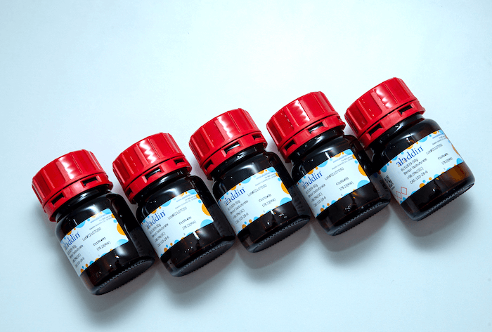
NA-Red (EB升级换代产品)

  • 2000X
有货

库存信息

关闭

库存信息

关闭
货号 (SKU) 包装规格 是否现货 价格 数量
N748354-1ml
1ml 期货 Stock Image
N748354-5ml
5ml 期货 Stock Image
查看相关系列
电泳染料 (7)

基本描述

规格或纯度 2000X
稳定性与储存 室温保存,至少两年有效。
英文名称 NA-Red
储存温度 室温
运输条件 常规运输
产品介绍

阿拉丁的NA-Red (Nucleic Acid Red,核酸红)是一种EB (Ethidium bromide,溴化乙锭)的升级换代产品,用于凝胶中DNA、RNA等核酸的染色。NA-Red具有安全(致突变性极低且检测不到显著的细胞毒性)、灵敏度高、稳定性好等优点,凝胶中的核酸在使用本产品染色后用适当紫外灯(300nm左右波长)检测呈现红色荧光,适用于原先使用EB为染料的凝胶成像系统。NA-Red比EB和SYBR Green更安全。NA-Red在远高于其工作浓度范围时均没有细胞毒性及诱变性。艾姆斯氏试验(Ames test)结果表明,NA-Red的诱变性远小于EB。EB在多种致突变测试中呈现很强的诱变性,而NA-Red仅仅在浓度高达20微克/毫升时,并在S9代谢活化时有非常微弱的诱变性。通常NA-Red在凝胶中的工作浓度约为2微克/毫升,大大低于可导致诱变的浓度。NA-Red和阿拉丁的另外一种安全型核酸染料NA-Green,由于它们特殊的化学结构使其难以进入细胞,从而大大降低甚至避免了染料的致突变性和细胞毒性。而SYBR Green染料可以穿透细胞膜,进入活细胞并染色DNA,并且有报道SYBR Green可以强烈增强紫外线诱导的基因突变。NA-Red检测灵敏度高,对于小分子量核酸的染色效果好。NA-Red的检测灵敏度比EB高8-10倍,在检测低浓度、微量DNA或RNA方面比EB更佳,尤其对小分子量的DNA检测非常灵敏。在使用浸泡染色法时,NA-Red和SYBR Gold的灵敏度相近甚至更高;与SYBR Gold不同的是,NA-Red预先配制在凝胶中也有很高的灵敏度。EB对于小分子量核酸的染色效果差,而NA-Red对于小分子量核酸的染色效果很好,便于观察酶切或PCR获得的小分子量核酸片段。推荐使用的NA-Red浓度,其检测效果略优于EB。如果希望获得更高的染色灵敏度,可以适当提高NA-Red的工作浓度。NA-Red的稳定性好,染色重复性高。含SYBR Green的凝胶核酸染色的重复性比较差,这通常是由于SYBR系列染料的稳定性差导致的。而NA-Red的稳定性很好,可以室温长时间保存及使用微波炉加热。NA-Red的光稳定性良好,可以在室内正常光线下操作而无需避光。由于其热稳定性和光稳定性,含NA-Red的凝胶在核酸染色时的重复性非常好。NA-Red可以使用和EB相同的检测体系。NA-Red和EB的激发光和发射光都非常接近,可以直接用NA-Red替换EB,而不必更换已有的凝胶观察、拍照或成像系统(约300nm激发)。NA-Red的激发光谱和发射光谱请参考图1。NA-Red的使用方法和EB一致。NA-Red可以按适当比例直接加入琼脂糖中配制成凝胶,也可以在电泳完毕后对凝胶进行染色。前者更加方便,而后者灵敏度要更高一些。但由于NA-Red本身已经非常灵敏,通常采用把NA-Red直接配制在凝胶中就可以了。对于一些特殊情况,如核酸样品量特别少的情况等,则可以考虑电泳后再对凝胶进行染色。NA-Red对于核酸的迁移率影响非常小,小于SYBR Green对于核酸迁移率的影响。通过凝胶回收试剂盒(如阿拉丁或Qiagen的凝胶回收试剂盒)或酚氯仿抽提,可以有效去除与DNA或RNA结合的NA-Red,从而确保不会影响后续的连接、酶切、PCR、测序等常规的分子生物学用途。


注意事项

为确保使用的不是假冒的NA-Red,可以用细胞培养液把NA-Red稀释至1X,然后对培养的活细胞进行染色。随后在荧光显微镜下尝试用各种激发光观察,如果发现活细胞细胞核出现明显的荧光,则可以判定为假冒产品。如果各种激发光下活细胞均无荧光,则说明该核酸染料是不能进入活细胞的高度安全的染料。具有强致突变性的吖啶橙染色核酸后呈现荧光,但其可以染色活细胞,而NA-Red不会染色活细胞。制备好的NA-Red琼脂糖凝胶,在4℃避光条件下通常可以保存3-5天。NA-Red琼脂糖凝胶再次熔化使用时,为取得更好的观察效果,需要添加适量NA-Red。电泳之后的凝胶不建议重复使用。电泳后再使用NA-Red染色的凝胶一般不需要脱色。如果发现背景太高,可以使用不含核酸酶的水进行脱色处理。NA-Red和NA-Green除了可以染色双链DNA外,也可染色单链DNA和RNA。NA-Red对单链核酸的染色灵敏度约为对双链DNA染色灵敏度的一半。NA-Red对单链核酸的染色灵敏度约为NA-Green的5倍。如果使用聚丙烯酰胺凝胶,请使用浸泡染色法染胶,并延长染色时间至30分钟-1小时。如果观察到条带弥散或者分离不理想,建议使用浸泡染色法染色以确认是否与染料有关。如果浸泡染色法染色后仍然出现类似的问题,说明与染料无关,请尝试以下方法:使用新鲜配制的电泳缓冲液、降低核酸的上样量、降低染料的浓度、降低琼脂糖浓度、选用更长的凝胶、降低电泳电压一倍以上并延长凝胶电泳时间以改善电泳效果、使用更薄的梳齿等。本产品兼容常用的电泳缓冲液,例如TAE和TBE。NA-Red不属于有毒有害物质,并通过了环境安全相关测试,相关废弃物无需特殊处理,可以参考常规化学试剂进行处理。本产品仅限于专业人员的科学研究用,不得用于临床诊断或治疗,不得用于食品或药品,不得存放于普通住宅内。为了您的安全和健康,请穿实验服并戴一次性手套操作。


使用说明

NA-Red和EB(溴化乙锭)一样可以根据使用者的偏好或实验目的采用以下方法中的一种: 1. 琼脂糖凝胶中添加NA-Red。 根据需要配制适当浓度(例如1-3%)的琼脂糖胶液。在琼脂糖完全融解后,适当冷却但又不会使琼脂糖凝固时,按照每100毫升胶液加入50微升NA-Red的比例(2000:1)加入NA-Red。混匀后即可把琼脂糖胶液倒入制备凝胶的模具中。适量的DNA或RNA在该胶中电泳后,在紫外灯下可以观察到明亮的核酸条带。说明:NA-Red非常稳定,所以NA-Red可以像EB一样在琼脂糖凝胶液加热融解后但未凝固前加入并混匀,也可以在琼脂糖融解前加入,然后再微波炉加热融解并混匀。2. 电泳完毕后对琼脂糖凝胶染色。按照每100毫升100mM NaCl溶液或水中加入100-200微升NA-Red的比例(500-1000:1)加入NA-Red,配制成NA-Red染色液。把电泳完毕的琼脂糖凝胶放到适当的容器中,加入适量上述配制好的NA-Red染色液,确保至少盖住凝胶。在摇床上缓慢摇动(约30-50rpm)染色20-30分钟。染色时间根据胶的厚度而定,胶厚则染色时间需要长一些,胶薄则染色时间可以短一些。染色完毕后,在紫外灯下即可观察核酸条带。要观察到更为清晰的条带,可以在染色后用水漂洗1-2次,每次3-5分钟,以消除背景,然后在适当紫外灯下或用凝胶成像系统观察。NA-Red染色液可以重复使用3次左右。NA-Red染色液也可以一次大量制备,在室温下避光保存,直至用完。对于核酸需要回收的情况,操作过程中需要注意避免核酸酶污染。附录:如何快速鉴别EB(溴化乙锭)和NA-Red由于EB和NA-Red溶液颜色相似,在核酸染色后又采用相同的检测系统进行检测,所以除了使用液相色谱-质谱(LC-MS)技术鉴定分子量外,特别需要有一个快速区别EB和NA-Red的简单方法。我们研究发现通过固定发射波长(Emission wavelength)、扫描激发波长(Excitation wavelength)的方式,可以快速鉴别EB和NA-Red。如图2,固定发射波长为600nm,240-360nm扫描激发波长。可以发现在没有核酸的情况下,不同浓度的EB本身可以在300nm处有激发最高峰,整体扫描图谱非常清晰(图A),与在有核酸(如质粒)的情况下也基本一致(图B);而NA-Red在没有核酸的情况下,本身很难被激发,即荧光背景非常弱,虽然在250-300nm有一定的激发峰,但不是很明显(图A),与在有核酸(如质粒)的情况下形成鲜明的对比(图B)。最为明显的是,在没有核酸的情况下,EB本身的荧光强度大大高于NA-Red。这样就可以通过简单的荧光检测来区分EB和NA-Red了。图2. 对于EB、NA-Red及各自与质粒的混合物,固定发射波长为600nm,在240-360nm范围内进行激发波长扫描。

Aladdin's NA-Red is a new generation fluorescent nucleic acid dye designed to replace the highly toxic and mutagenic ethidium bromide (EB).NA-Red is superior to EB in noncytotoxicity, sensitivity, and stability. The nucleic acid stained by NA-Red exhibits red fluorescence when excited by UV light at approximately 300nm, which can be examined by the gel imaging system originally used for the observance of EB.NA-Red is safer than EB or SYBR Green. NA-Red is not cytotoxic or mutagenic even at concentrations far above its working range. The Ames test also proves that the mutagenicity of NA-Red is far less than that of EB. NA-Red and NA-Green have a special chemical structure that makes it difficult to enter cells, thus greatly reducing or even avoiding the mutagenicity and cytotoxicity of the dye. SYBR Green, on the other hand, can penetrate cell membranes, enter live cells and stain DNA, and has been reported to strongly enhance UV-induced mutagenesis.NA-Red is highly sensitive and effective for staining nucleic acids with small molecular weight. NA-Red is 8-10 times more sensitive than EB for detecting trace amounts of DNA or RNA, and is especially sensitive for small molecular weight DNA. When staining gel after electrophoresis, NA-Red has similar or even higher sensitivity than SYBR Gold. Different from SYBR Gold, NA-Red is also highly sensitive when precast in a gel. The staining effect of NA-Red of concentration recommended in this manual is slightly better than EB, and the working concentration of NA-Red can be increased if higher sensitivity is desired.NA-Red has good stability and high reproducibility of staining. The reproducibility of SYBR Green nucleic acid staining method is poor, usually due to the low stability of the SYBR dyes. In contrast, NA-Red is thermostable and has strong light resistance, and thus produces nucleic acid staining results with good reproducibility.NA-Red can be examined with the same detection system as EB. NA-Red has almost the same excitation and emission spectra as EB, so it can be used as a direct substitute for EB. Please refer to Figure 1 for the excitation and emission spectra of NA-Red.Figure 1. The excitation and emission spectra of NA-Red NA-Red can be used in the same way as EB. NA-Red can be added directly to agarose gel after melting in an appropriate proportion, or the gel can be stained after electrophoresis is completed. The former method is more convenient, while the latter is a bit more sensitive. However, since NA-Red itself is already very sensitive, it is usually sufficient to precast it in the gel. For some special cases, such as samples with particularly small amounts of nucleic acid, gel staining after electrophoresis is recommended.The effect of NA-Red on the mobility of nucleic acids is very small and less than that of SYBR Green.NA-Red bound to DNA or RNA can be effectively removed by gel recovery kits or phenol/chloroform extraction, thus enabling the subsequent ligation, digestion, PCR, sequencing, and other routine molecular biology applications.


Precautions

To check the quality of NA-Red, dilute the stock to 1X with cell culture medium which is then used to stain cultured cells, followed by examination by fluorescence microscopy. High quality NA-Red does not stain live cells, while the counterfeit does.The prepared agarose gel containing NA-Red can be stored at 4ºC in the dark for 3-5 days for future use.If the NA-Red agarose gel will be used after melting, an appropriate amount of NA-Red needs to be added for better observation.Reuse of the gel after sample loading and electrophoresis is not recommended.Gels that are stained with NA-Red after electrophoresis generally do not need to be decolorized prior to examination. If the background is too high, decolorization can be performed using nuclease-free water.NA-Red, like NA-Green, can stain dsDNA, ssDNA and RNA.For polyacrylamide gels, stain after electrophoresis and extend the staining time to 30 minutes - 1 hour.If DNA smear is observed or DNA could not be separated well, we recommend gel staining after electrophoresis to determine whether the problem is due to the dye. If the problem still exists other attempts can be tried, such as using freshly prepared buffer, reducing the amount of nucleic acid loaded, reducing the concentration of dye or agarose, using a longer gel, reducing the voltage for electrophoresis and extending the electrophoresis time to improve electrophoresis.NA-Red is compatible with commonly used electrophoresis buffer solutions, such as TAE and TBE.NA-Red is not a toxic or hazardous substance and has passed environmental safety related tests. The related waste does not require special treatment and can be disposed of as conventional chemical reagents.This product is for R&D only. Not for drug, household, or other uses.For your safety and health, please wear a lab coat and disposable gloves during the operation.


Instructions for Use

Similar to EB, NA-Red can be used by the following two methods. 1. Add NA-Red to the gel Prepare agarose gel of the desired concentration (e.g., 1-3%). After the agarose is melted and properly cooled, add NA-Red at a ratio of 1:10000 (e.g., add 10µl of NA-Red per 100ml of gel). Mix well, and pour the gel. After electrophoresis of DNA or RNA samples, bright nucleic acid bands in the gel should be visible when excited by UV light.Note: NA-Red has good thermal stability, so it can be added directly to the hot agarose solution without cooling. It also can be mixed together with the agarose powder and the electrophoresis buffer prior to melting.2. Stain gel after electrophoresisPrepare the NA-Red staining solution by diluting NA-Red with 100 mM NaCl solution or water at a ratio of 1:2500-1:5000 (e.g., add 20-40µl NA-Red per 100 ml of water). After electrophoresis, immerse the agarose gel in an appropriate amount of NA-Red staining solution, and stain gel for 20-30 minutes with slow shaking (30-50rpm) on a shaker. The staining time depends on the thickness of the gel. The thicker the gels, the longer the staining time. After staining, the nucleic acid bands can be examined by blue light. To obtain clearer bands, the stained gel can be rinsed 1-2 times with water for 3-5 minutes each time to eliminate the background and then examined by blue light with excitation wavelength at approximately 500 nm or by other appropriate gel imaging systems. The NA-Red staining solution can be reused about 3 times or prepared in large quantities at one time and stored at room temperature in the dark. For cases where nucleic acids need to be recovered, care needs to be taken during operation to avoid nuclease contamination.Appendix: How to Quickly Distinguish EB (Ethidium Bromide) from NA-RedSince EB and NA-Red solutions are similar in color and are examined by the same detection system after nucleic acid staining, there is a particular need for a simple method to quickly differentiate between EB and NA-Red, besides the liquid chromatography-mass spectrometry (LC-MS) approach determining their molecular weight. We found that EB and NA-Red can be distinguished quickly by measuring the fluorescence intensity at a fixed emission wavelength when scanned within a range of excitation wavelength. As shown in Figure 2, the emission wavelength was fixed at 600 nm, and the excitation wavelength was ranged from 240 to 360 nm. It was found that different concentrations of free EB had an excitation maximum at 300 nm, and the overall scanning spectra were very clear (Figure 2A), which was basically consistent with the plasmid DNA-bound EB (Figure 2B). However, free NA-Red was hardly excited (Figure 2A), while the plasmid DNA-bound NA-Red had the maximum fluorescence intensity when excited at 300nm (Figure 2B). Therefore, EB and NA-Red in their free state can be differentiated by fluorescence examination.Figure 2. The fluorescence intensity of EB, NA-Red and their respective bound forms with plasmid DNA, when excited in the range of 240-360nm. A. The fluorescence intensity of free EB and NA-red. B. The florescence intensity of EB and NA-red binding to plasmid DNA.

AI解读

技术规格说明书

质检证书(CoA,COO,BSE/TSE 和分析图谱)

C of A & Other Certificates(BSE/TSE, COO):
输入批号以搜索分析图谱:

溶液计算器