泛素 (Ub) 介导的蛋白质翻译后修饰是核心调控通路,几乎参与全部细胞生理活动。除经典的介导蛋白降解功能外,现有研究证实泛素还参与细胞信号传导、DNA损伤应答、蛋白胞内转运、细胞周期调控、炎症反应、免疫应答及细胞凋亡调控。泛素修饰依靠异肽键完成,泛素C端与靶蛋白赖氨酸残基的氨基共价结合,该过程称为泛素化修饰。泛素自身含7个赖氨酸位点 (K6、K11、K27、K29、K33、K48、K63),任意赖氨酸均可连接新的泛素分子,形成多聚泛素链。不同赖氨酸连接型泛素链赋予泛素系统多样化细胞调控功能。研究最透彻的两类泛素链为K48、K63连接型:K48多聚泛素链主要介导靶蛋白经蛋白酶体降解;K63多聚泛素链调控蛋白功能、亚细胞定位与蛋白互作。大量研究证明K11型泛素链参与细胞有丝分裂调控,未来针对该链型的研究规模将逐步比肩K48、K63。随着更多链型与疾病、特异性细胞通路的关联被报道,其余稀有泛素链的研究热度将快速上升。泛素修饰是动态可逆过程。E1激活酶、E2结合酶、E3连接酶协同催化蛋白发生单泛素化或多聚泛素化;而去泛素化酶(DUBs)可移除蛋白上单 / 多聚泛素链、水解泛素链。精准富集并检测细胞内泛素化修饰蛋白,是研究泛素调控机制、筛选泛素化靶蛋白、验证蛋白修饰功能的核心实验手段。
泛素化蛋白富集洗脱试剂盒采用His标签泛素结合蛋白,能够体外特异性识别并结合泛素化蛋白,与泛素链稳定结合,可在裂解、孵育富集阶段减缓泛素链被内源去泛素化酶降解,不添加抑制剂的情况下也是如此,最大程度保留样本原始修饰状态;再利用His标签与Ni树脂亲和作用捕获复合物;洗涤去除非特异性杂蛋白,高浓度咪唑竞争性洗脱获得洗脱产物。洗脱产物可直接用于Western Blot验证靶蛋白泛素化水平,或脱盐后开展质谱蛋白质组学。试剂盒配套完整裂解、结合、洗涤、洗脱缓冲液体系,内置蛋白酶抑制剂混合物与去泛素化酶抑制剂,可全程抑制蛋白降解与泛素链去除,最大程度保留样本中泛素化修饰的完整性。适配哺乳动物细胞、组织样本。
PR-619,是一种细胞渗透性广谱去泛素化酶 (DBU) 可逆抑制剂,已被证明可以保护多泛素化蛋白免于降解。在裂解缓冲液中加入 PR-619 可以提高细胞和组织提取物制备过程中多泛素化蛋白的产量。
1,10-菲咯啉 (o-PA),是一种强效的金属蛋白酶抑制剂,有助于防止多聚泛素链的降解。
产品组分
| 储运单元货号 | 储运单元名称 | 组分货号 | 组分名称 | 物理外观 | 10T | 储存 | 单次测试使用量 |
| O1375167NT | 泛素化蛋白富集洗脱试剂盒 (常温组分) | O1375167A | DMSO | 无色透明液体 | 1.2 mL | 室温 | 按需 |
| | | O1375167B | 1,10-菲罗啉 | 白色至白粉色粉末 | 1 mg
| 室温. 避光 | 按需 |
| O1375167WT | 泛素化蛋白富集洗脱试剂盒 (2-8℃组分) | O1375167C | Ni-TED树脂 | 蓝色混悬液体 | 0.2 mL | 2-8℃. 禁止冷冻 | 按需 |
| | | O1375167D | 裂解液 | 无色透明液体 | 10 mL | 2-8℃. | 按需 |
| | | O1375167E | 洗涤液 | 无色透明液体 | 20 mL | 2-8℃. | 按需 |
| | | O1375167F | 500 mM咪唑洗脱液 | 无色透明液体 | 2 mL | 2-8℃. | 按需 |
| O1375167DT | 泛素化蛋白富集洗脱试剂盒 (-20℃组分) | O1375167G | His标签泛素结合蛋白 | 无色透明液体 | 1 mg | -20℃. 避免反复冻融. | 按需 |
| | | O1375167H | 蛋白酶抑制剂混合物 (100×) | 无色透明液体 | 0.1 mL | -20℃. | 按需 |
| | | O1375167I | PR-619 | 黄色固体 | 1 mg
| -20℃. | 按需 |
注:发货时各组分依据储存温度 (RT/2‑8℃/-20℃) 独立包装,差异化温控运输以保障产品性能。同一订单含多温区物料可能分批次送达,请以实际收货为准。收货后按组分表标注温度分类存放,有效期自生产之日起 12 个月。
试剂盒未提供需自备材料
1. SDS-PAGE系统和缓冲液。
2. PVDF膜,蛋白印迹转移系统和缓冲液。
3. 封闭试剂。
4. 目标蛋白的一抗。
5. HRP标记的二抗。
6. 化学发光检测仪器。
使用说明
注意:全程冰上 / 4℃低温操作,抑制剂现配现加。用户可依据样本蛋白丰度优化孵育时长、试剂用量。
1. 试剂配制
1.1. PR-619 (200×) 储液:试剂盒提供的PR-619试剂,在使用前和打开样品瓶之前,置于室温下回温至少1小时,通过离心使试剂到管底部,用448 μL DMSO(试剂盒提供)溶解,得到200×储液。分装为10 × 50 μL等份,有助于减少反复冻融,冻融会降低抑制剂效力。
1.2. 1,10-菲罗啉 (200×) 储液:试剂盒提供的1,10-菲罗啉试剂,在使用前和打开样品瓶之前,通过离心使试剂到管底部,用555 μL DMSO(试剂盒提供)溶解,得到200×储液。分装为10 × 50 μL等份,有助于减少反复冻融,冻融会降低抑制剂效力。
2. 样本预处理 (细胞裂解)
2.1. 按细胞培养要求培养细胞。
2.2. 准备裂解液、离心管预冷至4℃;向裂解液中分别添加蛋白酶抑制剂混合物 (100×)、PR-619 (200×) 储液与1,10-菲罗啉 (200×) 储液,使三种试剂的最终浓度均为1×;即蛋白酶抑制剂混合物 (1× 终浓度),PR-619 (1× 终浓度),1,10-菲罗啉 (1× 终浓度),现配现用。
2.3. 将His标签泛素结合蛋白加入含抑制剂的裂解液中,至终浓度100-200 μg/mL,冰上保存。加入的His标签泛素结合蛋白能有效保护多泛素化蛋白免于降解,同时具有较好的富集效率。
2.4. 细胞常规清洗。移除培养基,PBS洗涤2次,弃尽上清。
2.5. 细胞刮收集贴壁细胞或离心收集悬浮细胞,加入1.3步骤的含有His标签泛素结合蛋白的裂解液重悬细胞,转移至预冷的离心管中。参考起始用量:10 cm 培养皿 (约1×10⁷个细胞,总蛋白约1 mg) 加入0.5 mL裂解液。最优细胞用量根据细胞株、靶蛋白丰度调整。
2.6. 冰上孵育15-30 min。
2.7. 4℃ 14000×g 离心10 min,澄清细胞裂解液,收集上清。吸取部分上清作为Input样本用于WB检测 (示例:20 μL细胞裂解液加入5μL 5×Protein Loading Buffer,100℃加热5-10min;10000×g 离心1 min,上清进行SDS-PAGE与Western Blot检测)。
3. Ni-TED树脂平衡
3.1. 充分颠倒混匀Ni-TED树脂混悬液;吸取所需Ni-TED树脂混悬液 (按照1mg总蛋白量取20μL Ni-TED树脂混悬液) 至离心管,500×g、4°C离心5 min,弃上清。注意不要扰动树脂,以避免树脂损失。
3.2. 使用10倍树脂体积的洗涤液重悬树脂,500×g、4°C离心5 min,弃上清。重复洗涤至少2次。
4. 亲和捕获
4.1. 将步骤2.2平衡好的Ni-TED树脂加入至步骤1.7收集的细胞裂解液上清中,4℃摇床孵育2 h以上,使His标签复合物充分结合树脂。
4.2. 500×g、4°C离心5 min收集树脂;保留上清,标记为未结合组分 (Unbound Fraction),吸取部分液体按照Input样本同等方式制备用于后续分析。
5. 洗脱
5.1. 使用10倍树脂体积的洗涤液轻柔重悬树脂,混匀5 min,500×g、4°C离心5 min弃上清,避免扰动树脂,重复洗涤3次;或选择5-10倍低浓度咪唑洗涤液 (如10、20、50 mM咪唑,用洗涤液稀释500 mM咪唑洗脱液配制) 轻柔重悬树脂洗涤,每次5 min,充分去除弱吸附杂蛋白。
5.2. 尽量吸净全部洗涤液,避免扰动树脂,树脂中加入50-100 μL 500 mM咪唑洗脱液,轻柔重悬,室温孵育10 min;缩小洗脱体积可提升检测灵敏度,可重复洗脱2次,提升回收率。
5.3. 500×g、4°C离心5 min,收集洗脱上清,保留洗脱后的Ni-TED树脂。按照Input样本同等方式制备用于后续分析。
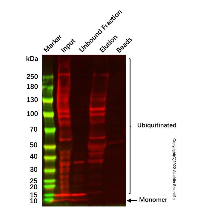
5.4. 洗脱样本可直接用于Western Blot,与Input、未结合组分 (Unbound Fraction)、洗脱后的Beads做归一化对比。若用于质谱分析,必须脱盐处理。
注意事项
1. His标签泛素结合蛋白避免反复冻融,开封后建议按需分装。
2. Ni-TED树脂严禁冷冻,不可完全风干,风干造成介质失活;禁止长时间高速离心压实树脂,建议不超过1000×g。
3. 全程冰上低温操作,抑制剂必须裂解前即时添加,减少泛素链降解丢失。
4. 建议同步保留Input、未结合组分 (Unbound Fraction)、洗脱产物、洗脱后的树脂,避免实验结果误判。
5. 梯度咪唑浓度根据实验需求可优化洗脱浓度和体积。
6. 本产品仅用于科研领域,不得用于临床诊断或其他用途。
7. 为了您的安全和健康,请穿实验服并戴一次性手套操作。
Ubiquitin (Ub)-mediated post-translational modification is a core regulatory pathway that participates in virtually all cellular physiological activities. In addition to its classical role in mediating protein degradation, ubiquitin is also involved in cell signaling, DNA damage response, intracellular protein trafficking, cell cycle regulation, inflammatory responses, immune responses, and apoptosis regulation. Ubiquitin modification is accomplished via an isopeptide bond, formed between the C-terminus of ubiquitin and the ε-amino group of a lysine residue on the target protein—a process termed ubiquitination. Ubiquitin itself contains seven lysine residues (K6, K11, K27, K29, K33, K48, and K63), each of which can serve as a conjugation site for additional ubiquitin molecules, generating polyubiquitin chains. Different lysine-linked ubiquitin chains confer diverse regulatory functions to the ubiquitin system. The two most extensively characterized linkage types are K48- and K63-linked chains: K48-linked polyubiquitin chains primarily mediate proteasomal degradation of target proteins, whereas K63-linked chains regulate protein function, subcellular localization, and protein–protein interactions. Accumulating evidence indicates that K11-linked chains are involved in cell cycle regulation, and research on this linkage type is expected to expand substantially, approaching the level of K48 and K63 in the future. As more linkages are associated with diseases and specific cellular pathways, interest in other rare ubiquitin chains is also increasing rapidly. Ubiquitination is a dynamic and reversible process. E1 activating enzymes, E2 conjugating enzymes, and E3 ligases coordinately catalyze mono- or polyubiquitination of proteins; conversely, deubiquitinating enzymes (DUBs) remove mono- or polyubiquitin chains from proteins and hydrolyze ubiquitin chains. Precise enrichment and detection of ubiquitinated proteins within cells are essential experimental approaches for studying ubiquitin regulatory mechanisms, screening ubiquitination target proteins, and validating protein modification functions.
The Ubiquitinated Protein Enrichment and Elution Kit utilizes a His-tagged ubiquitin-binding protein that specifically recognizes and binds ubiquitinated proteins in vitro, with stable association to ubiquitin chains. This binding protects ubiquitin chains from endogenous DUB-mediated degradation during lysis and enrichment phases, even in the absence of exogenous inhibitors, thereby preserving the original modification state of the sample to the greatest extent possible. The His-tagged complexes are subsequently captured via affinity binding to Ni resin. After washing to remove non‑specifically bound proteins, the enriched ubiquitinated proteins are eluted with high‑concentration imidazole under competitive conditions. The eluted product is directly compatible with Western blot analysis for validating target protein ubiquitination levels, or can be desalted for mass spectrometry‑based proteomics. The kit provides a complete system of lysis, binding, washing, and elution buffers, supplemented with protease inhibitors cocktail and DUB inhibitors to inhibit protein degradation and ubiquitin chain removal throughout the entire procedure, maximizing preservation of ubiquitination integrity in the sample. The kit is suitable for use with mammalian cells and tissue samples.
PR‑619 is a cell‑permeable, broad‑spectrum reversible inhibitor of deubiquitinating enzymes (DUBs), and has been shown to protect polyubiquitinated proteins from degradation. Supplementation of lysis buffer with PR‑619 increases the yield of polyubiquitinated proteins during preparation of cell and tissue extracts.
1,10‑Phenanthroline is a potent inhibitor of metalloproteases, and can help prevent polyUb chain degradation.
Kit Contents
| Storage Unit Name | Component No. | Component Name | Appearance | 10T | Storage | Quantity Per Test |
| Ubiquitinated Protein Enrichment and Elution Kit (RT Components) | O1375167A | DMSO | Clear colorless liquid | 1.2 mL | RT. | As needed |
| | O1375167B | 1,10-Phenanthroline | White to off white and pink powder | 1 mg
| RT. Store in the dark. | As needed |
| Ubiquitinated Protein Enrichment and Elution Kit (2-8℃ Components) | O1375167C | Ni-TED Resin | Uniform, free-flowing blue suspension | 0.2 mL | 2-8℃. Do not Freeze | As needed |
| | O1375167D | Lysis Buffer | Clear colorless liquid | 10 mL | 2-8℃. | As needed |
| | O1375167E | Wash Buffer | Clear colorless liquid | 20 mL | 2-8℃. | As needed |
| | O1375167F | 500 mM Imidazole Elution Buffer | Clear colorless liquid | 2 mL | 2-8℃. | As needed |
| Ubiquitinated Protein Enrichment and Elution Kit (-20℃ Components) | O1375167G | His-Tag Ubiquitin Binding Protein | Clear colorless liquid | 1 mg | -20℃. Avoid freeze/thaw cycle. | As needed |
| | O1375167H | Protease Inhibitor Cocktail (100×) | Clear colorless liquid | 0.1 mL | -20℃. | As needed |
| | O1375167I | PR-619 | Yellow solid | 1 mg
| -20℃. | As needed |
Notes: Each component is individually packaged according to its storage temperature (RT / 2‑8 °C / -20 °C) upon shipment, with differentiated temperature‑controlled transportation to guarantee product performance. Orders containing materials requiring multiple temperature zones may be delivered in separate batches; please refer to the actual received goods. Upon receipt, store components separately at the temperatures specified in the component list. The shelf‑life is 12 months from the date of manufacture.
Materials Required but Not Provided
1. SDS PAGE system and buffers.
2. PVDF membrane, protein transfer system and buffers.
3. Blocking reagents.
4. Primary antibody against target protein.
5. HRP conjugated secondary antibody.
6. Chemiluminescence detection instrument.
Instruction for Use
Notes: Perform all operations on ice or at 4 °C. Add inhibitors immediately before use. Incubation times and reagent volumes may be optimized according to the protein abundance in the sample.
1. Reagent Preparation
1.1. PR‑619 (200×) Stock Solution:
Equilibrate the provided PR‑619 reagent to room temperature for at least 1 hour before use and prior to opening the vial. Briefly centrifuge to collect the material at the bottom of the tube. Dissolve the reagent in 448 μL of DMSO (provided with the kit) to obtain a 200× stock solution. Aliquot into 10×50 μL portions to minimize repeated freeze‑thaw cycles, which may reduce inhibitor efficacy.
1.2. 1,10‑Phenanthroline (200×) Stock Solution:
Briefly centrifuge the provided 1,10‑phenanthroline reagent to collect the material at the bottom of the tube before opening the vial. Dissolve in 555 μL of DMSO (provided with the kit) to obtain a 200× stock solution. Aliquot into 10×50 μL portions to minimize repeated freeze‑thaw cycles, which may reduce inhibitor efficacy.
2. Sample Preparation (Cell Lysis)
2.1. Culture cells according to standard procedures.
2.2. Pre‑cool lysis buffer and centrifuge tubes to 4 °C. Add protease inhibitor cocktail (100×), PR‑619 (200×) and 1,10‑phenanthroline (200×) stock solution to the lysis buffer to achieve a final concentration of 1× for each reagent: protease inhibitor cocktail (1× final), PR‑619 (1× final), and 1,10‑phenanthroline (1× final). Prepare fresh immediately before use.
2.3. Add the His‑tagged ubiquitin‑binding protein to the inhibitor‑supplemented lysis buffer to a final concentration of 100–200 μg/mL and keep on ice. The added His‑tagged ubiquitin‑binding protein effectively protects polyubiquitinated proteins from degradation while maintaining high enrichment efficiency.
2.4. Wash cells routinely. Remove culture medium, wash twice with PBS, and aspirate the supernatant completely.
2.5. Harvest adherent cells with a cell scraper or collect suspension cells by centrifugation. Resuspend the cell pellet in the lysis buffer containing the His‑tagged ubiquitin‑binding protein (prepared in step 1.3) and transfer to a pre‑cooled tube. Recommended starting volume: 0.5 mL lysis buffer per 10‑cm dish (approx. 1×10⁷ cells, total protein ~1 mg). Optimal cell amounts should be adjusted according to cell line and target protein abundance.
2.6. Incubate on ice for 15–30 min.
2.7. Centrifuge at 14,000×g for 10 min at 4 °C to clarify the lysate, and collect the supernatant. Reserve a portion of the supernatant as the Input sample for Western blot analysis (e.g., mix 20 μL of cell lysate with 5 μL of 5× Protein Loading Buffer, heat at 100 °C for 5–10 min, centrifuge at 10,000×g for 1 min, and load the supernatant for SDS‑PAGE and Western blot).
3. Ni-TED Resin Equilibration
3.1. Thoroughly resuspend the Ni-TED resin slurry by inverting. Transfer the required volume of resin slurry (20 μL Ni-TED resin slurry per 1 mg total protein) to a tube, centrifuge at 500×g for 5 min at 4 °C, and discard the supernatant. Take care not to disturb the resin to avoid loss.
3.2. Resuspend the resin with 10 volumes of Wash Buffer, centrifuge at 500×g for 5 min at 4 °C, and discard the supernatant. Repeat washing at least twice.
4. Affinity Capture
4.1. Add the equilibrated Ni-TED resin (from step 2.2) to the clarified cell lysate supernatant (from step 1.7), and incubate on a shaker at 4 °C for at least 2 h to allow sufficient binding of the His‑tagged complexes to the resin.
4.2. Collect the resin by centrifugation at 500×g for 5 min at 4 °C. Retain the supernatant as the Unbound Fraction; reserve an aliquot of this fraction and prepare for subsequent analysis in the same manner as the Input sample.
5. Elution
5.1. Gently resuspend the resin with 10 volumes of Wash Buffer, mix for 5 min, centrifuge at 500×g for 5 min at 4 °C, and discard the supernatant. Repeat washing three times. Alternatively, wash with 5–10 volumes of low‑concentration imidazole Wash Buffer (e.g., 10, 20, or 50 mM imidazole prepared by diluting 500 mM imidazole Elution Buffer with Wash Buffer) with gentle resuspension for 5 min each time to thoroughly remove weakly bound contaminating proteins.
5.2. Remove as much residual Wash Buffer as possible without disturbing the resin. Add 50–100 μL of 500 mM imidazole Elution Buffer to the resin, gently resuspend, and incubate at room temperature for 10 min. Reducing elution volume improves detection sensitivity; elution may be repeated twice to increase recovery.
5.3. Centrifuge at 500×g for 5 min at 4 °C, collect the eluted supernatant, and retain the post‑elution resin. Prepare elution samples for subsequent analysis in the same manner as the Input sample.
5.4. The eluted sample can be directly used for Western blot analysis, with normalized comparisons against Input, Unbound Fraction, and post‑elution beads. If intended for mass spectrometry analysis, the eluted sample must be desalted.
Precautions
1. Avoid repeated freeze thaw cycles of the His tagged ubiquitin binding protein. Aliquot upon first use as needed.
2. Do not freeze the Ni-TED resin. Do not allow it to dry completely, as desiccation inactivates the resin. Avoid prolonged centrifugation at high speed that may compress the resin; we recommend not exceeding 1,000×g.
3. Keep all operations on ice and at low temperature. Inhibitors must be added immediately before lysis to minimize loss of ubiquitin chains due to degradation.
4. It is advisable to retain Input, Unbound Fraction, eluted product, and post elution resin in parallel to avoid misinterpretation of experimental results.
5. The imidazole concentration and elution volume can be optimized according to experimental requirements using a stepwise imidazole gradient.
6. For research use only. Not for clinical diagnosis or other applications.
7. For your safety and health, please wear a lab coat and disposable gloves during handling.

 ZJ26F1039940


 危险品化学品经营许可证(带存储)