
计算溶液所需的质量、体积或浓度。
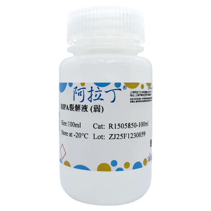
RIPA裂解液 (弱)
有货
BioReagent,for IP,即用型 级 ,适用于对基线干扰要求严格的色谱和分析工作流程。
-20°C储存,避免反复冻融。超低温运输 。请查阅批次 COA 获取详细规格。
SDS、COA、产品数据表及规格说明书均可下载。可通过批号查询获取批次 COA。
在色谱分析、有机合成和交叉偶联反应领域已被 0 篇同行评审文献引用。
阿拉丁RIPA裂解液是一款经典的细胞与组织快速裂解试剂。经本品裂解制备的蛋白样品适用于常规PAGE电泳、Western Blot、免疫沉淀(IP)、免疫共沉淀(Co-IP)及ELISA等多种下游实验。
本产品广泛适用于动物、植物来源的细胞或组织样本,亦可用于真菌与细菌样本。
该中强度RIPA裂解液的主要成分为:50 mM Tris-HCl(pH 7.4)、150 mM NaCl、1% NP-40、0.5%脱氧胆酸钠、0.1% SDS,并添加有氟化钠、EDTA等多种蛋白酶与磷酸酶抑制剂,可有效抑制蛋白质降解,维持蛋白样品完整性。
使用本品裂解所得的蛋白样品,推荐使用BCA法蛋白浓度检测试剂盒进行定量。需要注意的是,由于裂解液中含有较高浓度的去垢剂成分,不宜采用Bradford法对样品进行蛋白浓度测定。
注意事项:
1.为取得最佳的使用效果,尽量避免过多的反复冻融。可以适当分装后使用。
2.所有的样品裂解步骤宜在冰浴或4ºC操作,以尽量减少蛋白降解的可能性。
3.本RIPA裂解液已包含蛋白酶及磷酸酶抑制剂,为达到更佳使用效果,可考虑另外添加PMSF或阿拉丁生产的蛋白酶抑制剂混合物(P301902)。
4.RIPA 裂解液的裂解产物中经常会出现一小团透明胶状物,此为正常现象,该物质为基因组DNA 等形成的复合物。如不检测和基因组DNA紧密结合的蛋白,可以直接离心取上清用于后续实验;若需要检测此类蛋白,则可以通过超声处理打散该透明胶状物,随后离心取上清即可用于后续实验。但如果检测一些常见的转录因子,例如NF-kappaB、p53等,通常不必进行超声处理就可完成检测。
使用说明:
融解RIPA裂解液,混匀。取适当量的裂解液,在使用前数分钟内加入适当量的PMSF (终浓度1mM)或蛋白酶磷酸酶抑制剂混合物。
1.对于贴壁细胞的裂解:
吸除培养液,用PBS洗涤2次,然后吸净残留的液体。按照每50-100万细胞(相当于6孔板的一个孔)加入100-200μl的含抑制剂裂解液,适当吹打,使裂解液和细胞充分接触。通常裂解液接触动物细胞1-2秒后,细胞就会被裂解。植物细胞宜在冰上裂解2-10min。
2.对于悬浮细胞的裂解和准备:
收集细胞。用PBS洗涤2次,然后吸净残留的液体。轻轻vortex或者弹击管底以把细胞尽量分散开。按照每50-100万细胞加入100-200μl的比例加入含裂解液。混匀后置于冰上孵育5~20 min(期间混匀几次),轻弹管底或适当吹打,以充分裂解细胞。充分裂解后应没有明显的细胞沉淀。如果细胞量较多,建议分装成50-100万细胞/管,然后再裂解。大团的细胞较难裂解充分,而少量的细胞由于裂解液容易和细胞充分接触,相对比较容易裂解充分。
3.对于细菌或酵母样品的裂解和准备:
对于1ml菌液或酵母液,离心去上清,用PBS洗涤2次,然后吸净残留的液体。轻轻vortex或者弹击管底以把细菌或酵母尽量分散开。加入100-200μl裂解液,轻轻vortex或者弹击管底以混匀,冰上裂解2-10min。如果希望获得更好的裂解效果,细菌和酵母可以分别使用溶菌酶(lysozyme)和破壁酶(lyticase)消化,然后再使用含抑制剂裂解液进行裂解。
4.对于组织样品的裂解和准备:
把组织剪切成细小的碎片,按照每20mg组织加入约100-200μl裂解液的比例加入裂解液。如果裂解不充分可以适当添加更多的裂解液,如果需要高浓度的蛋白样品,可以适当减少裂解液的用量。然后用玻璃匀浆器或其它匀浆设备进行匀浆。充分匀浆可以确保组织被完全裂解。裂解后4ºC 12000g离心5分钟,取上清,可继续进行蛋白浓度测定。
Aladdin RIPA Lysis Buffer is a classical rapid lysis reagent for cells and tissues. Protein extracts prepared using this product are suitable for various downstream applications, including routine PAGE electrophoresis, Western blotting, immunoprecipitation (IP), co-immunoprecipitation (Co-IP), and ELISA.
This product is widely applicable to cell or tissue samples derived from animal and plant sources, and can also be used for fungal and bacterial samples.
The moderate-strength RIPA Lysis Buffer is composed of 50 mM Tris-HCl (pH 7.4), 150 mM NaCl, 1% NP-40, 0.5% sodium deoxycholate, and 0.1% SDS. It is supplemented with protease and phosphatase inhibitors such as sodium fluoride and EDTA to effectively inhibit protein degradation and maintain sample integrity.
For protein concentration quantification of lysates obtained with this product, the BCA protein assay kit is recommended. It should be noted that due to the high concentration of detergents in the lysis buffer, the Bradford method is not suitable for determining the protein concentration of these samples.
Precautions
1.To ensure optimal performance, minimize repeated freeze-thaw cycles. It is recommended to aliquot the product for use.
2.All sample lysis procedures should be performed on ice or at 4°C to minimize potential protein degradation.
3.This RIPA Lysis Buffer already contains protease and phosphatase inhibitors. For enhanced performance, consider supplementing with additional protease inhibitor cocktail (P301902) manufactured by Aladdin.
4.Lysates prepared with RIPA lysis buffer often contain a small, translucent gelatinous mass, which is a normal phenomenon. This substance consists of complexes formed by genomic DNA and other cellular components. If you are not detecting proteins that tightly bind to genomic DNA, you can simply centrifuge the lysate and use the supernatant for subsequent experiments. If detection of such proteins is required, the translucent mass can be dispersed by sonication, followed by centrifugation to collect the supernatant for downstream applications. However, for the detection of common transcription factors such as NF-kappaB and p53, sonication is generally not necessary.
Instruction for use
Thaw the RIPA lysis buffer and mix thoroughly. Shortly before use, add an appropriate volume of PMSF (to a final concentration of 1 mM) or a protease/phosphatase inhibitor cocktail to a suitable amount of the lysis buffer.
1. For Lysis of Adherent Cells:
Remove the culture medium and wash the cells twice with PBS. Completely remove any residual liquid.Add 100–200 µl of lysis buffer containing inhibitors per 0.5–1 million cells (equivalent to one well of a 6-well plate). Gently pipet or swirl to ensure complete contact between the lysis buffer and cells.Lysis of animal cells typically occurs within 1–2 seconds of contact. For plant cells, lyse on ice for 2–10 minutes.
2. For Lysis and Preparation of Suspension Cells:
Collect the cells by centrifugation. Wash the pellet twice with PBS and completely remove the supernatant.Gently vortex or flick the tube to disperse the cell pellet.Add 100–200 µl of lysis buffer containing inhibitors per 0.5–1 million cells. Mix thoroughly.Incubate the mixture on ice for 5–20 minutes, mixing intermittently by gentle flicking or pipetting.After complete lysis, no significant cell pellet should remain.For larger cell quantities, aliquot into tubes containing 0.5–1 million cells per tube before lysis. Large clumps are difficult to lyse completely, while smaller numbers of cells lyse more efficiently due to better contact with the lysis buffer.
3. For Lysis and Preparation of Bacterial or Yeast Samples:
For 1 ml of bacterial or yeast culture, pellet the cells by centrifugation. Wash the pellet twice with PBS and completely remove the supernatant.Gently vortex or flick the tube to resuspend and disperse the cells.Add 100–200 µl of lysis buffer. Mix by gentle vortexing or flicking.Lyse the cells on ice for 2–10 minutes.For more efficient lysis, bacterial samples can be pretreated with lysozyme and yeast samples with lyticase prior to adding the lysis buffer containing inhibitors.
4. For Lysis and Preparation of Tissue Samples:
Mince the tissue into small fragments.Add approximately 100–200 µl of lysis buffer per 20 mg of tissue.If lysis is insufficient, increase the amount of lysis buffer accordingly. To obtain a more concentrated protein extract, reduce the volume of lysis buffer.Homogenize the mixture using a glass homogenizer or other suitable homogenization equipment. Thorough homogenization is essential for complete tissue lysis.Centrifuge the lysate at 12,000 × g for 5 minutes at 4°C. Collect the supernatant for subsequent protein concentration determination.
| pH | 7.3 – 7.5 |
|---|
通过匹配包装上的批号来查找并下载产品的 COA,每批产品都进行了严格的验证,您可放心使用!
| 批号(Lot Number) | 证书类型 | 货号 |
|---|---|---|
 ZJ25F1230059 |
分析证书 | R1505850 |
| ZJ26F0737664 |
分析证书 | R1505850 |
我们的级别选择指南涵盖了目录中所有的变体的纯度、稳定剂状态和应用适用性。
查看 BioReagent 级别指南 → 查看 Ready-to-use 级别指南 → 查看 for IP 级别指南 →

 危险品化学品经营许可证(带存储)