DCFH-DA 是一种酯化修饰的前体探针,分子两端的乙酸酯基团使其具有良好的脂溶性,能够自由扩散进入活细胞的细胞质中。进入细胞后,胞内非特异性酯酶会迅速水解 DCFH-DA 分子上的两个乙酸酯基团,生成2',7'- 二氯二氢荧光素(DCFH)。DCFH 是水溶性分子,无法自由穿过细胞膜,因此被特异性地滞留在细胞内。DCFH 本身仍然无荧光,但它是一种高度敏感的氧化还原指示剂。当细胞内存在 ROS 时,DCFH 会被氧化生成2',7'- 二氯荧光素(DCF)。DCF 是一种强荧光化合物,其最大激发波长为488nm,最大发射波长为525nm(绿色荧光)。在一定范围内,DCF 的荧光强度与细胞内 ROS 的水平呈线性正相关。
使用方法
1. 装载探针
对于刺激时间较短(通常为2小时以内)的细胞,先装载探针,后用活性氧阳性对照或自己感兴趣的药物刺激细胞。对于细胞刺激时间较长(通常为6小时以上)的细胞,先用活性氧阳性对照或自己感兴趣的药物刺激细胞,后装载探针。
(1) 原位装载探针:本方法仅适用于贴壁培养细胞。按照1:1000用无血清细胞培养基或酌情使用PBS、HBSS等适当溶液稀释DCFH-DA,使终浓度为5 μM(对于不同的样本和处理,DCFH-DA工作液浓度可为0.1-20 μM,需进行预实验确定合适的浓度)。去除细胞培养液,加入适当体积稀释好的DCFH-DA。加入的体积以能充分盖住细胞为宜,通常对于六孔板的一个孔加入稀释好的DCFH-DA不少于1 mL。37ºC细胞培养箱内孵育20分钟。用PBS、HBSS或无血清细胞培养液等适当溶液洗涤细胞三次,以充分去除未进入细胞内的DCFH-DA。通常活性氧阳性对照在刺激细胞15-30分钟后可以显著提高活性氧水平。
(2) 收集细胞后装载探针:按照1:1000用无血清细胞培养基或酌情使用PBS、HBSS等适当溶液稀释DCFH-DA,使终浓度为5 μM(对于不同的样本和处理,DCFH-DA工作液浓度可为0.1-20 μM,需进行预实验确定合适的浓度)。细胞收集后悬浮于稀释好的DCFH-DA中,细胞浓度为一百万至二千万/毫升,37ºC细胞培养箱内孵育20分钟。每隔3-5分钟颠倒混匀一下,使探针和细胞充分接触。用PBS、HBSS或无血清细胞培养液等适当溶液洗涤细胞三次,以充分去除未进入细胞内的DCFH-DA。直接用活性氧阳性对照或自己感兴趣的药物刺激细胞,或把细胞等分成若干份后刺激细胞。通常活性氧阳性对照在刺激细胞20-30分钟后可以显著提高活性氧水平。
2. 设置阳性对照
阳性对照可以按照1:1000的比例使用PBS、HBSS或无血清培养液稀释后使用(工作液每次都要新鲜,不要储存以备将来使用)。例如装载好探针的细胞,直接吸除液体或离心后吸除液体,加入适当体积的按照1:100稀释的阳性对照刺激。通常刺激后30-40分钟内可以观察到非常显著的活性氧水平升高。如果在刺激后40分钟内观察不到活性氧的升高,可以适当提高活性氧阳性对照的浓度。如果活性氧升高得过快,可以适当降低活性氧阳性对照的浓度。
注:阳性对照对不同细胞的效果会有所不同,可根据不同细胞,调节阳性对照稀释(1:400-1:2000)比例和孵育时间。仅在阳性对照孔中加入阳性对照,其余孔不必加入。
3. 检测
对于原位装载探针的样品可以用激光共聚焦显微镜直接观察,或收集细胞后用荧光酶标仪或流式细胞仪检测。对于收集细胞后装载探针的样品可以用荧光酶标仪或流式细胞仪检测,用激光共聚焦显微镜直接观察也可以。
仪器参数为使用488 nm激发波长,525 nm发射波长,实时或逐时间点检测刺激前后荧光的强弱。DCF的荧光光谱和FITC非常相似,可以用FITC的参数设置检测DCF。
注意事项:
(1) 探针装载后,一定要洗净残余的未进入细胞内的探针,否则会导致背景较高。
(2) DCF容易淬灭,孵育完成后的样本最好的2 h之内进行测定,防止荧光减弱。尽量缩短探针装载后到测定所用的时间(刺激时间除外),以减少各种可能的误差。
(3) DCFH-DA和阳性对照避免反复冻融。
(4) 本产品仅限于专业人员的科学研究用,不得用于临床诊断或治疗,不得用于食品或药品,不得存放于普通住宅内。
(5) 为了您的安全和健康,请穿实验服并戴一次性手套操作。
结果展示

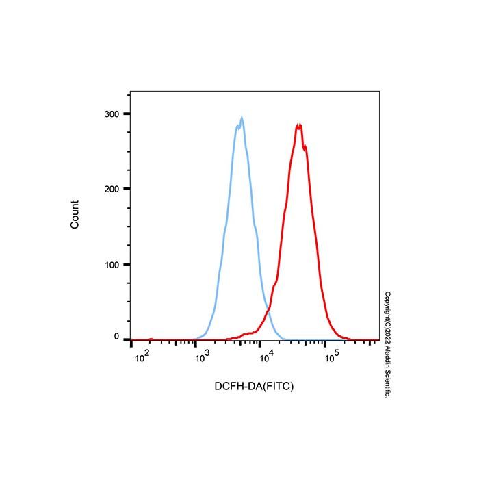
图1:本试剂盒检测K562细胞内活性氧效果如图所示:K562细胞未处理组(蓝色)与 阳性对照处理组(红色)经 DCFH-DA 染色后,流式检测FITC通道荧光强度。结果显示,阳性诱导剂处理可显著提升细胞内超氧阴离子水平,荧光峰明显右移,验证了试剂盒的有效性。

图2:本试剂盒检测HT-29细胞活性氧的效果图。正常的HT-29细胞中探针几乎未被氧化成荧光化合物,细胞中的绿色荧光较弱(图左);使用活性氧阳性对照试剂处理使细胞内活性氧生成大幅增加,DCFH-DA与活性氧反应,细胞中的绿色荧光显著增强(图右)。实际结果会因实验条件、检测仪器等的不同而存在差异,图中效果仅供参考。
DCFH-DA is an ester-modified precursor probe. Ester groups at both ends of its molecule confer favorable lipophilicity, enabling free diffusion across the plasma membrane into the cytoplasm of viable cells. Once inside cells, intracellular non-specific esterases rapidly hydrolyze the two acetate groups of DCFH-DA to produce 2',7'-dichlorodihydrofluorescein (DCFH). As a water-soluble substance, DCFH cannot freely cross cell membranes and is thereby trapped intracellularly. DCFH itself is non-fluorescent yet serves as a highly sensitive redox indicator. In the presence of intracellular ROS, DCFH is oxidized into 2',7'-dichlorofluorescein (DCF), a strongly fluorescent compound with a maximum excitation wavelength at 488 nm and maximum emission wavelength at 525 nm (green fluorescence). Within a certain concentration range, DCF fluorescence intensity displays a positive linear correlation with intracellular ROS content.
Protocol
1. Probe Loading
For short-term cell stimulation (generally within 2 h), load the probe prior to treatment with ROS positive control or test compounds. For long-term stimulation (typically over 6 h), treat cells with positive control or experimental drugs before probe loading.
(1) In-situ Probe Loading (only for adherent cells)
Dilute DCFH-DA at a 1:1000 ratio with serum-free medium, PBS or HBSS to a final working concentration of 5 μM. The usable concentration ranges from 0.1–20 μM for different cell types, which should be optimized via preliminary trials. Discard spent culture medium and add sufficient diluted DCFH-DA solution to fully cover cells; add no less than 1 mL per well for a 6-well plate. Incubate cells at 37 °C in a cell incubator for 20 min. Rinse cells three times with PBS, HBSS or serum-free medium to thoroughly remove extracellular unloaded DCFH-DA. Intracellular ROS levels rise markedly 15–30 min after positive control treatment.
(2) Post-harvest Probe Loading
Dilute DCFH-DA 1:1000 using serum-free medium, PBS or HBSS to reach 5 μM final concentration (optimize 0.1–20 μM via pre-experiment as needed). Resuspend harvested cells in diluted probe solution at a density of 1×10⁶ to 2×10⁷ cells/mL and incubate at 37 °C for 20 min; invert the tube every 3–5 min to ensure full contact between cells and probe. Wash cell pellets three times with PBS, HBSS or serum-free medium to eliminate residual extracellular DCFH-DA. Split cell suspension into aliquots followed by stimulation with positive control or test reagents. Significant ROS elevation can be detected 20–30 min after positive control administration.
2. Positive Control Setup
Dilute positive control reagent 1:1000 in PBS, HBSS or serum-free medium; prepare fresh working solution before each use and discard leftover diluent. Remove supernatant from probed adherent or pelleted suspension cells and add diluted positive control for stimulation. Remarkable ROS upregulation is commonly observed within 30–40 min post-treatment. Increase control concentration if no obvious fluorescence enhancement occurs within 40 min; reduce concentration if ROS surges excessively fast.
Note: Dilution ratio (1:400 to 1:2000) and incubation time shall be adjusted according to distinct cell lines. Add positive control exclusively to designated control wells.
3. Detection
In-situ loaded samples can be directly visualized under laser scanning confocal microscopy or harvested for microplate fluorometer or flow cytometry measurement. Post-harvest loaded samples are compatible with fluorometry, flow cytometry and confocal observation.Set excitation at 488 nm and emission at 525 nm to quantify fluorescence intensity before and after stimulation at single or sequential time points. DCF shares nearly identical fluorescence spectrum with FITC, hence FITC instrument parameters are applicable for DCF detection.
Precautions
1.Thoroughly wash off unincorporated extracellular probe after loading to avoid high background fluorescence.
2.DCF is susceptible to photobleaching; complete sample detection within 2 h after incubation to prevent fluorescence decay and experimental errors. Minimize the interval between probe loading and measurement excluding stimulation duration.
3.Avoid repeated freeze-thaw cycles for DCFH-DA stock and positive control reagents.
4.For research use only; not for clinical diagnosis, therapeutic administration, food or pharmaceutical production; prohibited for household storage.
5.Wear lab coat and disposable gloves during operation for personal safety.
Results
Figure 1: Detection of intracellular ROS in K562 cells using this kit. After DCFH-DA staining, fluorescence intensity was determined via flow cytometry in the FITC channel for untreated K562 cells ( blue ) and positive control-treated cells (red curve). The results show that treatment with the positive inducer markedly increases intracellular superoxide anion levels with an obvious right shift of the fluorescence peak, verifying the validity of the kit.
Figure 2: Fluorescence imaging of HT-29 cellular ROS tested with this kit. Left: untreated normal HT-29 cells display faint green fluorescence with minimal DCFH oxidation. Right: robust green fluorescence is observed in HT-29 after positive control treatment due to massive intracellular ROS accumulation and DCF formation. Actual results vary depending on experimental conditions and detection equipment; images are for reference only.

 ZJ26F0737240


 危险品化学品经营许可证(带存储)