此免疫组化试剂盒采用SABC放大系统,DAB显色。SABC是专为免疫组化和其他免疫检测而设计的,用以显示组织和细胞中抗原分布,链霉亲和素是一种从链霉菌中提取的蛋白质,分子量47000,同亲和素一样,对生物素分子有极高的亲和力,是一般抗原抗体亲和力的一百万倍,亲和素是一个碱性蛋白质(IP=10),经改造后可以转变成中性蛋白质,链霉亲和素等电点接近中性,IP=6.0~6.5,对组织和细胞的非特异吸附很低,基于链霉亲和素的免疫组化方法背景很低,SABC即 StreptAvidin—Biotin Complex,根据研究SABC大约可形成一百个左右的过氧化物酶和五十个左右的链霉亲和素所构成的复合物,大量的酶将保证SABC具有很高的敏感性,SABC兼具高敏感性,低背景和操作简便的优点。
包装清单:
R777868
| Component
| 1 Kit
| Storage
|
R777868A
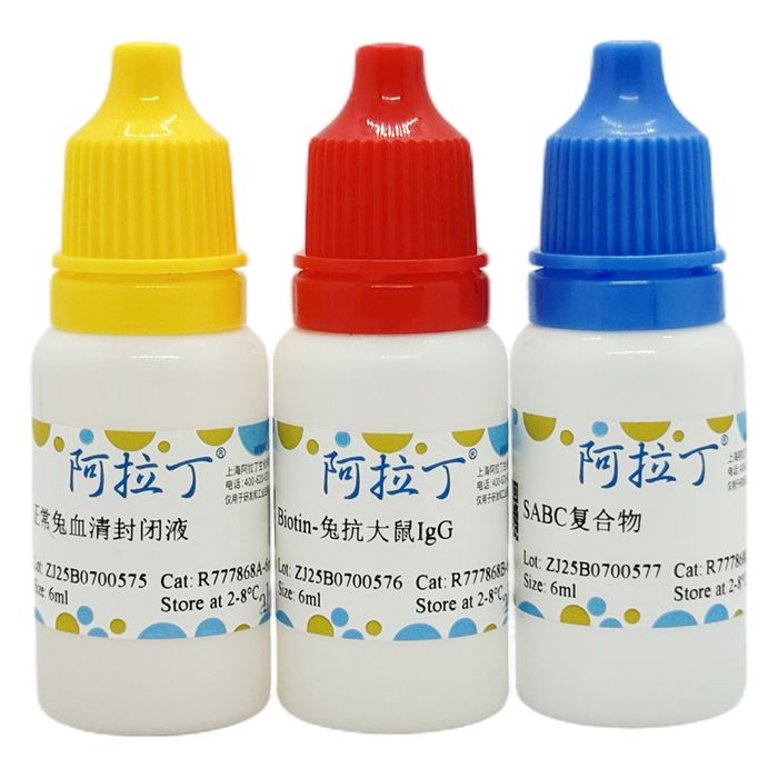
| 正常兔血清封闭液 | 6mL
| 2-8℃
|
R777868B
| Biotin-兔抗大鼠IgG
| 6mL
| 2-8℃
|
R777868C
| SABC复合物
| 6mL
| 2-8℃ |
自备试剂及耗材:
1. 防脱玻片
2. PBS(pH7.2-7.6)
3. EDTA抗原修复液或枸橼酸盐缓冲液
4. 内源性过氧化物酶阻断剂
使用说明:
A.石蜡切片热修复抗原染色程序:
1.制备切片,常规脱蜡至水。
2.滴加内源性过氧化物酶阻断剂,室温5-10分钟以灭活内源性酶。蒸馏水洗3次。
3.热修复抗原:将切片浸入EDTA抗原修复液或枸橼酸盐缓冲液,电炉或微波炉加热至沸腾后断电,间隔5-10分钟后,反复1-2次。冷却后PBS洗涤1-2次。
4. 滴加5%BSA封闭液,室温20分钟。甩去多余液体, 不洗。
5. 滴加适当稀释的一抗,37 ℃ 1小时左右或20℃时2小时左右。也可4℃过夜。PBS洗2分钟×3次。
(一抗的稀释度、孵育时间和温度与染色强度、背景有直接关系。一般来说,阳性染色强度不够时,可提高一抗浓度和延长孵育时间;背景过高时,可降低一抗浓度和缩短孵育时间。)
6. 滴加biotin山羊抗大鼠IgG,20-37℃ 20分钟。PBS洗2分钟×3次。
7. 滴加试剂SABC,20-37℃ 20分钟。PBS洗5分钟×4次。
8. DAB显色:使用DAB显色剂。取1ml蒸馏水,加试剂盒中DAB显色剂A,B,C试剂各1滴,混匀后加至切片。
室温显色, 镜下控制反应时间,一般在1-15分钟之间,蒸馏水洗涤。
9.苏木素轻度复染。脱水,透明,封片。显微镜观察。
B. 石蜡切片酶消化程序:
以下面的步骤代替A程序中的第3步:滴加复合消化液5-10分钟。蒸馏水洗3次。也可以使用0.1%的胰蛋白酶作消化液。
C. 石蜡切片不消化/不修复程序:
对于不需要微波修复或消化的抗原,省略A程序中的第4步即可。
D.血涂片,细胞和冰冻切片染色程序:
1. 载玻片经防脱片剂处理(Poly-L-Lysine)。抗凝血经分层离心后涂片;培养细胞也可涂片或贴片生长;冰冻切片室温风扇吹干。
2. 固定方案首选4%多聚甲醛或10%福尔马林固定60-90分钟。
3. 30%H₂O₂ 1份+纯甲醇50份混合,室温浸泡30分钟,以灭活内源性过氧化物酶。蒸馏水洗1-2次。
其余步骤和石蜡切片5-10步相同。
如果冰冻切片直接染色结果不理想时,可以参照石蜡切片对切片进行热修复,方法和石蜡切片3-9步相同。
注意事项:
1. 如果染色背景过高,在SABC反应之后,DAB显色之前,用加有0.01—0.02% Tween-20的PBS(pH7.2-7.6)
洗切片4次,单纯PBS洗2次,然后DAB显色。
2. 热修复抗原首先推荐使用EDTA抗原修复。
This immunohistochemistry kit adopts the SABC amplification system with DAB chromogenic detection. SABC is designed for immunohistochemistry and other immunoassays to visualize antigen distribution in tissues and cells.
Streptavidin is a protein extracted from Streptomyces with a molecular weight of 47000. Similar to avidin, it possesses extremely high affinity for biotin, approximately one-million-fold higher than typical antigen-antibody affinity. Avidin is a basic protein (IP = 10) and can be modified into a neutral form. Streptavidin has a near-neutral isoelectric point (IP = 6.0-6.5) and exhibits very low non-specific adsorption to tissues and cells, which enables low-background staining in streptavidin-based immunohistochemical methods.
SABC stands for StreptAvidin-Biotin Complex. Studies show each SABC complex contains roughly 100 peroxidase molecules and around 50 streptavidin molecules. The abundant enzyme moiety delivers high assay sensitivity. Therefore, the SABC method features high sensitivity, low background and simple operation.
Kit Contents
R777868 | Components | 1 Kit | Storage |
R777868A | Normal Rabbit Serum Blocking Solution | 6mL | 2-8℃. |
R777868B | Biotin-Rabbit Anti Rat IgG | 6mL | 2-8℃. |
R777868C | SABC Complex | 6mL | 2-8℃. |
Required Reagents & Consumables (Not Supplied)
1. Adhesion-coated glass slides
2. PBS (pH 7.2-7.6)
3. EDTA antigen retrieval buffer or citrate buffer
4. Endogenous peroxidase blocking reagent
Operating Procedures
A. Heat-Induced Antigen Retrieval for Paraffin Sections
1. Prepare sections and perform routine de-paraffinization to hydration.
2. Apply endogenous peroxidase blocking reagent; incubate at room temperature for 5-10 min to block endogenous peroxidase activity. Rinse with distilled water 3 times.
3. Heat-induced antigen retrieval: Immerse sections in EDTA antigen retrieval buffer or citrate buffer. Heat in microwave or electric heater until boiling, then power off. Keep interval of 5-10 min and repeat heating-cooling cycle 1-2 times. Cool down, then wash with PBS 1-2 times.
4. Add Normal Rabbit Serum Blocking Solution . Incubate at room temperature for 20 min. Remove excess liquid; do not wash.
5. Add appropriately diluted primary antibody. Incubate at 37 ℃ for ~1h, or at 20 ℃ for ~2h; alternatively incubate at 4 ℃ overnight. Wash with PBS for 2 min × 3 times.
Note: Primary-antibody dilution, incubation temperature and duration directly affect staining intensity and background. If positive signal is weak, increase primary-antibody concentration or prolong incubation time. If high background occurs, reduce primary-antibody concentration or shorten incubation time.
6. Add Biotin-Rabbit Anti Rat IgG. Incubate at 20-37 ℃ for 20 min. Wash with PBS for 2 min × 3 times.
7. Add SABC complex reagent. Incubate at 20-37 ℃ for 20 min. Wash with PBS for 5 min × 4 times.
8. DAB chromogenic reaction: Prepare DAB working solution: add 1 drop each of DAB-A, DAB-B and DAB-C reagents into 1 mL distilled water and mix well. Apply onto sections. Develop at room temperature and monitor reaction under microscope (typically 1-15 min). Terminate reaction by rinsing with distilled water.
9. Counterstain lightly with hematoxylin. Dehydrate, clear, mount slides, and observe under microscope.
B. Enzymatic Digestion Protocol for Paraffin Sections
Replace Step 3 in Protocol A with the following step:
Add compound digestive solution and incubate for 5-10 min. Rinse with distilled water 3 times. Alternatively, 0.1 % trypsin solution can be used for digestion.
C. Protocol without Digestion / Antigen Retrieval for Paraffin Sections
For antigens that do not require microwave retrieval or enzymatic digestion, skip Step 3 in Protocol A.
D. Staining for Blood Smears, Cell Smears and Frozen Sections
1. Use poly-L-lysine-coated adhesive slides. Prepare blood smears from anticoagulated blood by density-gradient centrifugation; use cultured cell smears or adherent cell monolayers. Air-dry frozen sections with fan at room temperature.
2. Fix specimens, preferably with 4 % paraformaldehyde or 10 % formalin for 60-90 min.
3. Prepare blocking solution: mix 1 volume of 30 % H₂O₂ with 50 volumes of pure methanol. Immerse slides at room temperature for 30 min to inactivate endogenous peroxidase. Rinse with distilled water 1-2 times.
4. Follow Steps 5-9 described in Protocol A for subsequent staining.
5. If unsatisfactory results are obtained from direct staining of frozen sections, perform heat-induced antigen retrieval as described in Steps 3-9 of Protocol A.
Important Notes
1. In case of high staining background: after SABC incubation and before DAB development, wash slides 4 times with PBS (pH 7.2-7.6) containing 0.01-0.02 % Tween-20, then wash twice with plain PBS before DAB chromogenic reaction.
2. EDTA antigen retrieval buffer is the preferred choice for heat-induced antigen retrieval.

 ZJ25F0723942


 危险品化学品经营许可证(带存储)