Succinate dehydrogenase (SDH) is located on the inner side of the mitochondrial ridge membrane and binds to the inner membrane of the mitochondria. SDH is the most membrane-binding enzyme among the three carboxylic acid cycles and is the hallmark enzyme of mitochondria. The active reaction site of SDH enzyme was blue-purple granules, located in cardiomyocytes and cell membranes, and the interstitium of cardiomyocytes was not colored. When the enzyme activity was high, the particles were dense and coarse, the enzyme activity of young animals was significantly higher than that of young and old animals, and the activity of SDH was significantly reduced when the myocardial cells were injured by ischemia or hypoxia. Operation process: 1 Sample handling 1.1 For paraffin sections: Dewaxing in xylene for 2×5min; Anhydrous ethanol 5 min, 90% ethanol 2 min, 70% ethanol 2 min; Rinse with PBS for 2 min. 1.2 For frozen sections: Rinse with PBS for 2 min. 2 succinate dehydrogenase stain solution 2.1 Dye succinate dehydrogenase solution in a wet box at 37°C for 2h; 2.2 Rinse slices with running water, 2×3~5min; 2.3 Use absorbent paper to absorb excess water on the section; 2.4 Dehydration with 100% ethanol for 3×1~3min; 2.5xylene transparent, 2 x 5min, sealed with resin. Dyeing result: The active reaction site of myocardial SDH enzyme was blue-purple granule, distributed in myocardial cells and cell membrane. Note: 1 A fluorescence microscope or laser confocal microscope that can observe blue fluorescence is required. 2 Prepare PBS, fixing solution and anti-fluorescence quenching sealing solution. 3 When using this kit for the first time, it is recommended to take 1 to 2 samples for pre-test. Warm reminder: For your own safety, please take good protection before using the reagent, such as wearing a lab coat, gloves, etc.
规格
产品名称
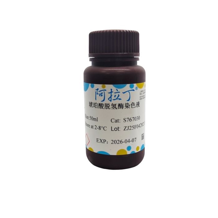
琥珀酸脱氢酶染色液
别名
琥珀酸脱氢酶染色液
英文别名
Succinate dehydrogenase solution; SDH
规格或纯度
BioReagent, 即用型, 适用于免疫组织化学
分子类型
酶
产品线
生物产品
储存与运输
物理形态
液体
储存条件
2-8°C储存,避光
运输条件
低温运输
稳定性与储存
Store at 2-8℃ long term (12 months). Upon receipt, it is recommended to aliquot. Store in the dark.

 ZJ26F0434647


 危险品化学品经营许可证(带存储)