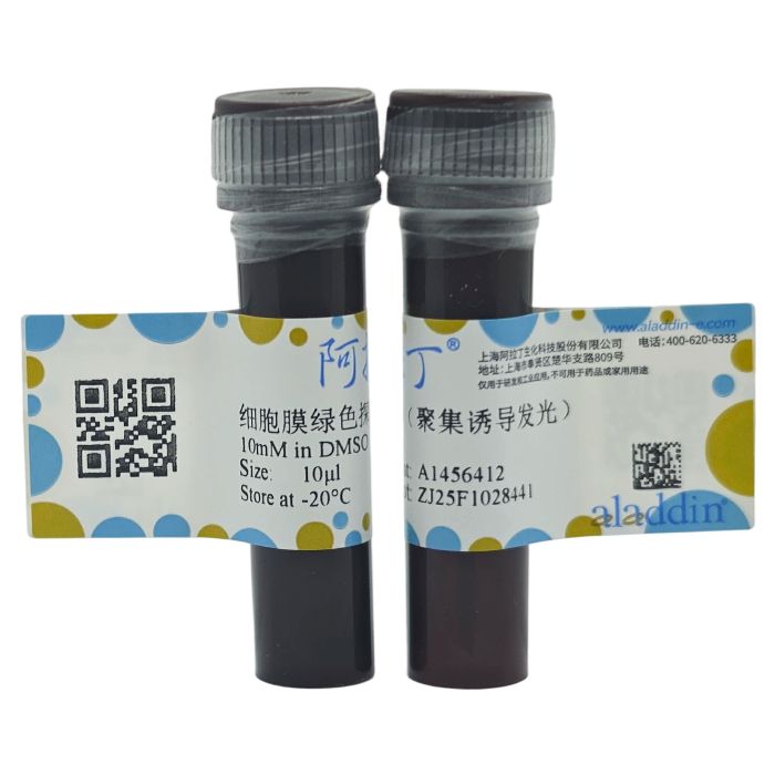
细胞膜绿色探针(聚集诱导发光)是一种基于AIE原理开发的四苯基乙烯类细胞膜探针,具有典型的AIE特性。产品只需简单 地和细胞孵育,即可标记细胞膜。
产品特性
细胞膜绿色探针(聚集诱导发光)具有良好的聚集诱导发光特性,可以特异性标记多种细胞的细胞膜,而在与细胞膜结合前 后由于聚集状态的变化,其荧光强度会产生极为明显的变化,而未与细胞膜结合的荧光探针基本不会发射出荧光 信号。

图 1. HeLa细胞上的激光共聚焦成像效果图
与常见染料不同,
细胞膜绿色探针(聚集诱导发光)可通过405 nm激发波长激发,并且斯托克斯位移大,可以明显区别与其 他染料,减少成像中串色的可能性。同时
细胞膜绿色探针(聚集诱导发光)的生物相容性好,成像浓度高,可在多次扫描的状态 仍然保证稳定的荧光信号输出,非常适合多次成像。
产品性质
| 化学式(Formula) | C₄₄H₃₉IN₂O₂ | | 分子量(Molecular Weight) | 754.71 g/mol | | 纯度(Purity) | >98%(HPLC) | | 工作浓度(Working Concentration) | 5 - 10 μM | | 半峰宽(Full width at half maximum) | 560 nm - 700 nm | | 最大吸收 / 发射波长(nm) | λabs = 370 nm / Em = 675 nm |
|
产品优势
1、细胞毒性相对较小,可以进行活细胞成像;
2、光稳定性好,抗光漂白能力强;
3、背景噪点低,可免洗,快速成像。
实验方法
1、染料储备液准备:将染料储备液适当分装后避光保存于-20℃或更低温度,储备液浓度为10mM;
2、染料工作液准备:取1µL
细胞膜绿色探针(聚集诱导发光)储备液加入到1-2 mL 细胞培养液或适当的缓冲液中(例如PBS), 得到终浓度5-10 μM 的
细胞膜绿色探针(聚集诱导发光)工作液。
3、细胞染色。适量的工作染液孵育贴壁细胞 10 - 15 min后(最好置于细胞培养箱中),用共聚焦荧光显 微镜或荧光显微镜观察,激发波长设为 405 nm;收集 540 - 590 nm的信号。
注意事项
1、使用前请先短暂离心,推荐在染色后一小时内观察;
2、第一次使用时请先配置好母液后分装,于-20℃保存,避免反复冻融;
3、为了您的安全和健康,请穿实验服并戴一次性手套操作;
4、本品仅适用于科研用途。
Membrane Green Probe ( AIE) is a tetraphenylethylene-based cell membrane probe developed based on the AIE principle. It has typical AIE characteristics. The product only needs to be simply incubated with cells to label the cell membrane.
Product Characteristics
Membrane Green Probe ( AIE) has excellent aggregation-induced emission properties and can specifically label the cell membranes of various cells. Before and after binding to the cell membrane, due to changes in the aggregation state, its fluorescence intensity will undergo extremely obvious changes, while the fluorescent probes that do not bind to the cell membrane basically do not emit fluorescent signals.
Figure 1. Laser confocal imaging effect diagram on HeLa cells
Different from common dyes,
Membrane Green Probe ( AIE)
can be excited by a 405 nm excitation wavelength, and has a large Stokes shift, which can clearly distinguish it from other dyes, reducing the possibility of crosstalk in imaging. At the same time,
Membrane Green Probe ( AIE) has good biocompatibility and high imaging concentration, and can still ensure stable fluorescence signal output in the state of multiple scans, making it very suitable for multiple imaging.
Product Properties
| Molecular Weight | 754.71 g/mol | | Purity | > 98% (HPLC) | | Working Concentration | 5 - 10 μM | | Full width at half maximum | 560 nm - 700 nm | | Maximum absorption/emission wavelength (nm) | λabs = 370 nm / Em = 675 nm |
|
Product Advantages
1.Relatively low cytotoxicity, suitable for live cell imaging;
2.Good photostability and strong anti-photobleaching ability;
3.Low background noise, no need for washing, enabling rapid imaging.
Experimental Methods
1.Preparation of stock dye solution: Appropriately aliquot the stock dye solution and store it in the dark at -20°C or lower. The concentration of the stock solution is 10 mM;
2.Preparation of working dye solution: Take 1 μL of
Membrane Green Probe ( AIE)
stock solution and add it to 1-2 mL of cell culture medium or an appropriate buffer solution (such as PBS) to obtain an
Membrane Green Probe ( AIE)
working solution with a final concentration of 5-10 μM;
3.Cell staining: Incubate adherent cells with an appropriate amount of the working solution for 10-15 minutes (preferably in a cell culture incubator). Observe with a confocal fluorescence microscope or a fluorescence microscope. Set the excitation wavelength to 405 nm and collect signals at 540-590 nm.
Precautions
1.Centrifuge briefly before use. It is recommended to observe within one hour after staining;
2.When using for the first time, prepare the mother solution first, aliquot it, and store at -20°C to avoid repeated freezing and thawing;
3.For your safety and health, please wear a lab coat and disposable gloves;
4.This product is only for scientific research use.

 ZJ25F1028441


 危险品化学品经营许可证(带存储)