
计算溶液所需的质量、体积或浓度。
有货
BioReagent,生物染色剂,用于显微镜 级 ,适用于对基线干扰要求严格的色谱和分析工作流程。
2-8°C储存,避光。低温运输 。请查阅批次 COA 获取详细规格。
SDS、COA、产品数据表及规格说明书均可下载。可通过批号查询获取批次 COA。
在色谱分析、有机合成和交叉偶联反应领域已被 1 篇同行评审文献引用。
脂质 (Lipid) 是中性脂肪、类脂及其衍生物的总称,不溶于水而溶于有机溶剂。人体的脂质主要有两种:一是储存脂质,如中性脂肪 (即甘油三酯),主要分布于皮下、肾、 胰腺等部位;二是结构脂质,如类脂 (磷脂、糖脂、胆固醇等),主要分布于细胞内。脂滴是细胞内中性脂肪的主要贮存场所,在正常情况下,除脂肪细胞之外,其他细胞在光学显微镜下几乎看不到脂滴,如果细胞质内出现大量脂滴即指示为脂肪变性,常见于肝细胞、心肌细胞、肾曲管上皮细胞等。
油红O是一种脂溶性偶氮染料,是很强的脂溶剂和染脂剂,能特异性地使组织和细胞内的中性甘油三脂、类脂、脂蛋白等染色,效果要优于传统的苏丹Ⅱ、苏丹Ⅲ、苏丹Ⅳ、 苏丹黑B等。当用油红O染色时,油红O在组织细胞内脂质的溶解度比在原溶剂中的溶解度更大,所以油红O就从有机溶剂转移入脂质而使之染色。
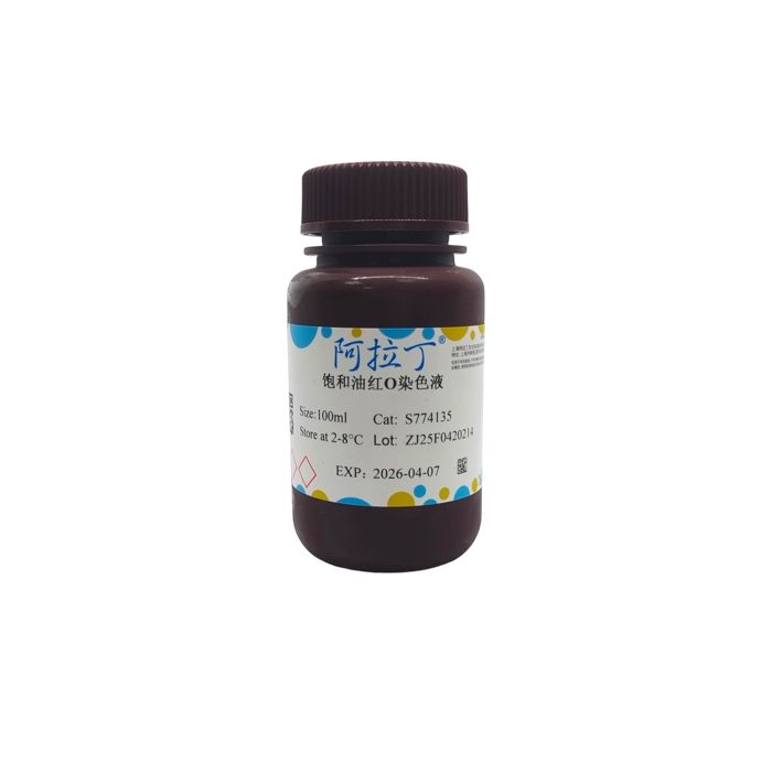
饱和油红O染色液 (Saturated Oil Red O Staining Solution) 可以通过显示细胞内的脂滴来反应组织器官的脂肪变性和类脂的异常沉着;也可以鉴别脂肪组织中所发生的肿瘤及其性质。本产品既可以用于组织染色又可以用于分析细胞样品中脂质状况。根据脂质的浓度不同,脂质阳性染色结果可呈橘黄至红色。本产品选用优质的进口原料,染色清晰而稳定,操作便捷。20 mL饱和油红O染色液可以配置成大约33 mL的工作液,100 mL饱和油红O染色液可以配置成大约166 mL的工作液。
使用说明:
(一)工作液的配制
按照饱和油红O染色液:蒸馏水= 3:2的比例配成油红O工作液,混匀,室温放置 5-10min,过滤后使用。【油红O染色工作液不稳定,不宜提前配制】
(二)染色程序
1、冰冻切片:
1.1 制作好的冰冻切片不固定或用10%中性福尔马林固定10min,蒸馏水充分洗涤。
1.2 60%异丙醇浸洗20-30s。
1.3 油红O染色工作液染色10-15min。【可根据染色结果和要求调整时间】
1.4 60%异丙醇分化至间质清晰,蒸馏水中稍微清洗。
1.5 Mayer苏木素染色液复染核1-5min。【可根据染色结果和要求调整时间】
1.6 (可选) 1%盐酸溶液稍微分化一下。
1.7 (可选) 自来水漂洗10min或稀碳酸锂溶液中促蓝。
1.8 蒸馏水中稍微清洗。
1.9 用滤纸吸干周围水分,甘油明胶封片并在显微镜下观察。
2、培养细胞:
2.1 移除细胞培养基,PBS洗两次,加10%中性福尔马林固定液固定20-30min。
2.2 弃去固定液,用蒸馏水洗2次。
2.3 60%异丙醇浸洗5min。
2.4 弃去 60%异丙醇后加入油红O染色工作液,覆盖住底板即可,浸染10-20min。【可根据染色结果和要求调整时间】
2.5 弃去染色液,蒸馏水洗2-5次,直到无多余染液。
2.6 Mayer苏木素染色液复染核1-2min。【可根据染色结果和要求调整时间】弃去染液后水洗2-5次。
2.7 1%盐酸溶液分化1min,弃去。
2.8 加入蒸馏水覆盖细胞并在显微镜下观察。
3、细胞涂片:
3.1 制备新鲜骨髓、血液涂片,置于10%中性福尔马林固定10-15min。
3.2 取出涂片,放于流通的空气中10-15min。
3.3 放入油红O染色工作液浸染15min。【可根据染色结果和要求调整时间】
3.4 60%异丙醇分化20-30s,流水冲洗,蒸馏水稍微清洗。
3.5 Mayer苏木素染色液复染核1-2min。【可根据染色结果和要求调整时间】
3.6 1%盐酸溶液分化1min,蒸馏水稍微清洗。
3.7 用滤纸吸干周围水分,甘油明胶封片并在显微镜下观察。
(三)染色结果
| 中性脂肪 | 橘黄色至红色 |
| 磷脂 | 粉红色 |
| 细胞核 | 蓝色 |
注意事项:
1、油红O染色工作液不够稳定,易产生沉淀,不宜提前配制。工作液可以重复利用, 但如果当天没有用完需舍弃。
2、有机溶剂会溶解样品中的脂质,因此固定样品时不采用含有乙醇的固定液 (如需要固定可采用10%中性福尔马林或10%福尔马林),也不采用石蜡切片,需用冰冻切片 或碳蜡切片。
3、如果做脂肪组织冰冻切片,冰冻温度比普通要低,切片厚度不可太薄,过薄的切片常会使脂质丢失,一般厚度为6-10µm。
4、完全成熟饱满的脂肪细胞容易破裂,脂滴泄漏,所以压片、切片时应注意。
5、甘油明胶封片的样本,保存时间不长。如需长期保存,可以在盖玻片与载玻片交界的边缘用中性树胶封闭。
6、油红O染色时应避免试剂挥发过多,否则易形成背景沉淀。染色时染色液应充分覆盖组织细胞。
7、60%异丙醇分化,可在镜下控制至脂肪组织呈鲜红色,间质无色时为度。建议使用苏木素复染,增加染色对比度。
8、第一次使用本试剂时建议先取1-2个样品做预实验。
9、本试剂为油红O的饱和溶液,因此有少量沉淀为正常现象。使用时,应在配制成工作液后自行过滤。
10、为了您的安全和健康,请穿实验服并戴一次性手套操作。
Lipids are a general term for neutral fats, lipoids and their derivatives, which are insoluble in water but soluble in organic solvents. There are two major categories of lipids in the human body. The first is storage lipids, represented by neutral fats (triglycerides), which are mainly distributed in subcutaneous tissue, kidneys, pancreas and other tissues. The second category is structural lipids, namely lipoids including phospholipids, glycolipids, cholesterol and more, predominantly located inside cells.
Lipid droplets serve as the primary intracellular storage compartment for neutral fats. Under physiological conditions, lipid droplets are barely visible under light microscopy in all cell types except adipocytes. Massive accumulation of lipid droplets in the cytoplasm indicates fatty degeneration, which is commonly observed in hepatocytes, cardiomyocytes, renal tubular epithelial cells and other cell types.
Oil Red O is a lipophilic azo dye with powerful liposolvent and lipid-staining properties. It specifically stains intracellular neutral triglycerides, lipoids, lipoproteins and other lipid components in tissues and cells, delivering superior staining performance compared with classic dyes such as Sudan II, Sudan III, Sudan IV and Sudan Black B. During Oil Red O staining, the dye exhibits higher solubility in cellular lipids than in its original organic solvent, driving the migration of Oil Red O from the solvent into lipid compartments to produce distinct staining.
Saturated Oil Red O Staining Solution detects fatty degeneration and abnormal deposition of lipoids in tissues and organs by visualizing intracellular lipid droplets. It can also identify tumors arising from adipose tissue and determine their pathological properties. This product is compatible with both tissue section staining and lipid profiling analysis of cellular samples. Lipid-positive staining ranges from orange-yellow to deep red depending on lipid abundance. Manufactured with high-quality imported raw materials, this reagent delivers clear, stable staining results with simple operating procedures. Approximately 33 mL of working solution can be prepared from 20 mL saturated Oil Red O stock solution, and 100 mL saturated stock solution yields roughly 166 mL working solution.
Instructions for Use
I. Preparation of Working Solution
Mix saturated Oil Red O staining solution with distilled water at a volume ratio of 3:2 to prepare Oil Red O working solution. Homogenize the mixture, let it stand at room temperature for 5–10 minutes, and filter before use.
Note: Oil Red O working solution is unstable and shall not be prepared in advance.
II. Staining Protocols
1. Frozen Sections
1.1 Fix prepared frozen sections for 10 minutes with 10% neutral formalin, or skip fixation entirely; rinse thoroughly with distilled water.
1.2 Immerse slides in 60% isopropanol for 20–30 seconds.
1.3 Stain with Oil Red O working solution for 10–15 minutes. Adjust staining time according to expected staining intensity and experimental requirements.
1.4 Differentiate with 60% isopropanol until stroma is clearly visible, then briefly rinse with distilled water.
1.5 Counterstain nuclei with Mayer’s hematoxylin staining solution for 1–5 minutes. Adjust staining time as needed.
1.6 (Optional) Briefly differentiate slides with 1% hydrochloric acid solution.
1.7 (Optional) Rinse with running tap water for 10 minutes or bluing in dilute lithium carbonate solution.
1.8 Briefly rinse with distilled water.
1.9 Blot excess liquid around sections with filter paper, mount slides with glycerin gelatin, and observe under a microscope.
2. Cultured Cells
2.1 Aspirate cell culture medium, wash cells twice with PBS, and fix cells with 10% neutral formalin fixative for 20–30 minutes.
2.2 Discard fixative and rinse cells twice with distilled water.
2.3 Immerse cells in 60% isopropanol for 5 minutes.
2.4 Remove 60% isopropanol, add sufficient Oil Red O working solution to fully cover the cell substrate, and incubate for 10–20 minutes. Adjust staining time according to expected staining intensity and experimental requirements.
2.5 Discard staining solution, rinse cells with distilled water 2–5 times to remove residual dye.
2.6 Counterstain nuclei with Mayer’s hematoxylin staining solution for 1–2 minutes. Adjust staining time as needed; rinse cells 2–5 times with distilled water after discarding dye solution.
2.7 Differentiate with 1% hydrochloric acid solution for 1 minute, then discard the solution.
2.8 Add distilled water to cover cells and observe under a microscope.
3. Cell Smears
3.1 Prepare fresh bone marrow or blood smears, and fix smears in 10% neutral formalin for 10–15 minutes.
3.2 Take out smears and air-dry in circulating air for 10–15 minutes.
3.3 Immerse smears in Oil Red O working solution for 15 minutes. Adjust staining time according to expected staining intensity and experimental requirements.
3.4 Differentiate with 60% isopropanol for 20–30 seconds, flush with running water, then briefly rinse with distilled water.
3.5 Counterstain nuclei with Mayer’s hematoxylin staining solution for 1–2 minutes. Adjust staining time as needed.
3.6 Differentiate with 1% hydrochloric acid solution for 1 minute, then briefly rinse with distilled water.
3.7 Blot excess liquid around smears with filter paper, mount with glycerin gelatin, and observe under a microscope.
III. Staining Results
|
Precautions
1.Oil Red O working solution is unstable and prone to precipitate; it must not be prepared in advance. The working solution can be reused, but any leftover solution must be discarded at the end of the day.
2.Organic solvents dissolve intracellular lipids. Therefore, fixatives containing ethanol are prohibited for sample fixation. Use 10% neutral formalin or 10% formalin if fixation is required. Paraffin sections are not applicable; frozen sections or carbowax sections should be adopted instead.
3.For frozen sections of adipose tissue, a lower freezing temperature is required compared with regular specimens. Sections cannot be cut too thin, as excessive thinness will cause lipid loss. The recommended section thickness ranges from 6 to 10 µm.
4.Fully mature and plump adipocytes are fragile and prone to rupture, resulting in lipid droplet leakage. Extra care should be taken during slide pressing and sectioning.
5.Specimens mounted with glycerin gelatin have a short shelf life. For long-term preservation, seal the edges between the coverslip and slide with neutral balsam.
6.Excessive evaporation of staining reagent during incubation will lead to background precipitates. Ensure tissues and cells are fully submerged in staining solution throughout the staining process.
7.Differentiate with 60% isopropanol under microscopic observation until adipose tissue turns bright red and the stroma appears colorless. Counterstaining with hematoxylin is recommended to improve staining contrast.
8.A pilot test with 1–2 specimens is recommended for first-time use of this reagent.
9.This product is a saturated Oil Red O stock solution, so a small amount of precipitate is normal. Filtration is mandatory after preparing the working solution before use.
10.For personal safety and health protection, wear a lab coat and disposable gloves during all operations.
通过匹配包装上的批号来查找并下载产品的 COA,每批产品都进行了严格的验证,您可放心使用!
| 批号(Lot Number) | 证书类型 | 货号 |
|---|---|---|
 ZJ25F1230706 |
分析证书 | S774135 |
| ZJ26F0838327 |
分析证书 | S774135 |
| ZJ26F0838330 |
分析证书 | S774135 |
| ZJ26F0838329 |
分析证书 | S774135 |
| ZJ26F0838328 |
分析证书 | S774135 |
| ZJ25F0725001 |
分析证书 | S774135 |
| ZJ25F0928121 |
分析证书 | S774135 |
| ZJ25F0928120 |
分析证书 | S774135 |
| ZJ25F0725000 |
分析证书 | S774135 |
| ZJ25F0723938 |
分析证书 | S774135 |
| ZJ25F0723937 |
分析证书 | S774135 |
| ZJ25F0420214 |
分析证书 | S774135 |
我们的级别选择指南涵盖了目录中所有的变体的纯度、稳定剂状态和应用适用性。
查看 BioReagent 级别指南 → 查看 Biological Stain 级别指南 → 查看 for Microscopy 级别指南 →

 危险品化学品经营许可证(带存储)