
计算溶液所需的质量、体积或浓度。
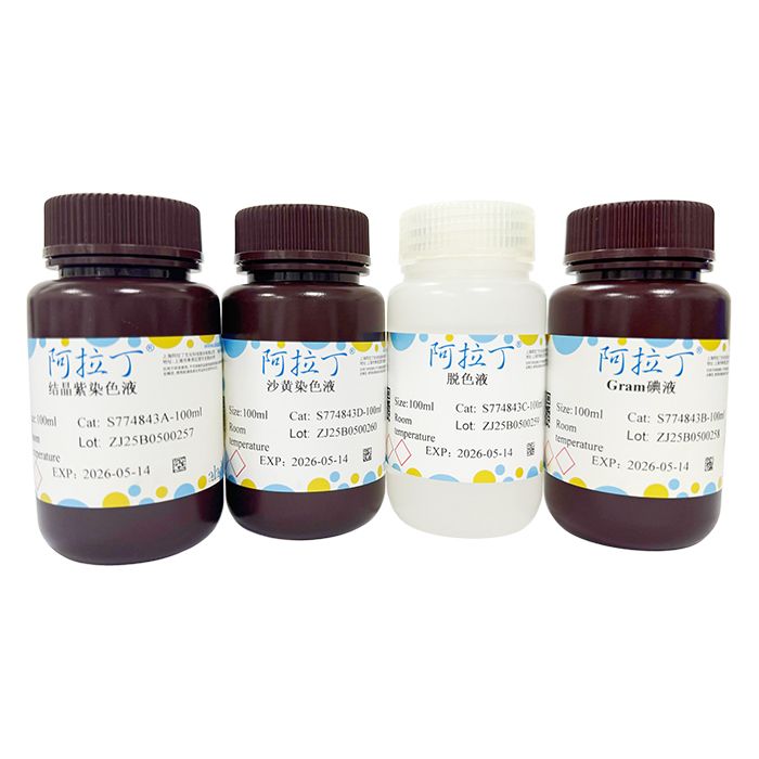
标准革兰氏染色试剂盒
有货
BioReagent,生物染色剂,用于显微镜 级 ,适用于对基线干扰要求严格的色谱和分析工作流程。
避光,室温。常规运输 。请查阅批次 COA 获取详细规格。
SDS、COA、产品数据表及规格说明书均可下载。可通过批号查询获取批次 COA。
在色谱分析、有机合成和交叉偶联反应领域已被 0 篇同行评审文献引用。
革兰氏染色法是细菌学中广泛使用的一种鉴别染色法,亦是一种复染法。未经染色的细菌,由于其不周围环境折光率差别甚小,故在显微镜下极难观察。染色后细菌与环境形成鲜明对比,可以清楚地观察到细菌的形态、排列及某些结构特征,用以分类鉴定。通过此法染色可将细菌鉴别为革兰阳性菌 (G+) 和革兰阴性菌 (G-) 两大类。细菌的不同显色反应是由于细胞壁对乙醇的通透性和抗脱色能力的差异,主要是肽聚糖层厚度和结构决定的。经结晶紫染色的细胞用碘液处理后形成不溶性复合物,乙醇能使它脱色。在革兰阴性细胞染色中,乙醇或丙酮破坏了胞壁外膜、损伤肽聚糖层和细胞质膜,结晶紫和碘复合物从细胞中渗漏出来,当再用其他染色液复染时,显现红色。红色染料虽然也能进入已染成紫色的G+细胞,但被紫色盖没,所以红色显示不出来。在革兰阳性细胞染色中,乙醇还能使厚的肽聚糖层脱水,导致孔隙变小,由于结晶紫和碘复合物分子太大,不能通过细胞壁,不易脱色,所以保持着紫色。
标准革兰氏染色液采用最经典的革兰染色配方,临床标本直接涂片,背景干净,胞核胞质对比强烈,胞内吞噬体清晰易辨认,细菌染色特征典型。
| S774843 | Component | 4×10 mL | 4×100 mL | 4×250 mL | 4×500 mL | Storage |
| S774843A | 结晶紫染色液 | 10 mL | 100 mL | 250 mL | 500 mL | RT. Store in the dark |
| S774843B | Gram碘液 | 10 mL | 100 mL | 250 mL | 500 mL | RT. Store in the dark |
| S774843C | 脱色液 | 10 mL | 100 mL | 250 mL | 500 mL | RT. |
| S774843D | 沙黄染色液 | 10 mL | 100 mL | 250 mL | 500 mL | RT. Store in the dark |
使用说明:
1、涂片:取待检细菌,于载玻片中央涂成薄层或者在载玻片上滴加少许无菌水,取菌与水混合均匀,涂成一薄层。
2、干燥:涂片后在室温下自然干燥,也可在酒精灯上略加温,使之迅速干燥。
3、固定:手持载玻片一端,标本面朝上,在酒精灯的火焰外侧快速来回移动3-5次,每次1s,温度不宜过高,防止菌体蛋白变性,放置待凉后染色。也可以用甲醇或乙醇固定。
4、初染:滴加结晶紫染色液染色1-2min,清水冲洗去染色液。
5、媒染:滴加 Gram 碘液,并覆盖载玻片,室温放置1-2min,水洗。
6、脱色:滴加脱色液,摇动10-30s,直至流下的脱色液不出现紫色时为止,立即用水冲去脱色液,终止反应。
7、复染:滴加沙黄染色液染色30-60s,水洗。
8、干燥、镜检:置油镜观察。
注意事项:
1、涂片之前,应事先在背面做好圆圈标记,以便判断后续试验的位置。
2、取细菌时,应注意自我防护,拔试管塞时,应将试管口通过火焰略加烧灼,最后将接种环在火焰上烧灼灭菌。
3、加热固定涂片时,应注意玻片勿太靠近火焰,一般要求玻片温度不超过60°C,以玻片背面触及手背皮肤不觉过烫为宜。
4、革兰氏染色的关键在于严格掌握脱色程度,脱色时间应根据经验判断。脱色过度,阳性菌可被误染为阴性菌;脱色不够,阴性菌可被误染为阳性菌。
5、待检细菌培养时间会影响染色,阳性菌培养时间过长或已死亡或细菌溶解,常呈阴性。
6、为了您的安全和健康,请穿实验服并戴一次性手套操作。
染色结果:
| 革兰氏阳性菌 | 蓝色至紫色 |
| 革兰氏阴性菌 | 红色 |
Gram staining is a widely used differential staining technique in bacteriology, and it is also a type of counterstaining method. Unstained bacteria are extremely difficult to observe under a microscope because their refractive index differs minimally from the surrounding environment. After staining, a sharp contrast forms between bacteria and the background, allowing clear visualization of bacterial morphology, arrangement, and certain structural features for taxonomic identification. This staining method categorizes bacteria into two major groups: Gram-positive (G⁺) and Gram-negative (G⁻). The divergent color outcomes stem from differences in cell wall permeability to ethanol and resistance to decolorization, primarily determined by the thickness and structure of the peptidoglycan layer. Cells stained with crystal violet form an insoluble complex upon treatment with iodine solution, which is susceptible to decolorization by ethanol. For Gram-negative cells: ethanol or acetone disrupts the outer cell wall membrane, damages the peptidoglycan layer and cytoplasmic membrane, enabling the crystal violet–iodine complex to leak out of the cells. Subsequent counterstaining with another dye yields a red color. Although the red stain can penetrate pre-stained purple Gram-positive cells, the deep purple masks the red hue completely. For Gram-positive cells: ethanol dehydrates the thick peptidoglycan layer and shrinks its pores. The large molecular size of the crystal violet–iodine complex prevents it from passing through the cell wall, rendering the cells resistant to decolorization and retaining a purple color.
The standard Gram staining solution adopts the classic traditional Gram stain formula. It delivers clean background for direct smears of clinical specimens, strong contrast between cell nuclei and cytoplasm, easily distinguishable intracellular phagosomes, and typical characteristic staining patterns of bacteria.
| S774843 | Component | 4×10 mL | 4×100 mL | 4×250 mL | 4×500 mL | Storage |
| S774843A | Crystal Violet Stain Solution | 10 mL | 100 mL | 250 mL | 500 mL | RT. Store in the dark |
| S774843B | Gram’s Iodine Solution | 10 mL | 100 mL | 250 mL | 500 mL | RT. Store in the dark |
| S774843C | Decolorizing Solution | 10 mL | 100 mL | 250 mL | 500 mL | RT. |
| S774843D | Safranin Stain Solution | 10 mL | 100 mL | 250 mL | 500 mL | RT. Store in the dark |
Instructions for Use:
1. Smear preparation: Take the test bacteria and spread a thin layer in the center of a glass slide. Alternatively, drop a small amount of sterile water onto the slide, mix the bacteria thoroughly with the water, and spread into a thin film.
2. Drying: Air-dry the smear at room temperature, or heat gently over an alcohol lamp for rapid drying.
3. Fixation: Hold one end of the slide with the specimen side upward. Pass the slide quickly back and forth 3–5 times beside the flame of an alcohol lamp, 1 second each pass. Avoid excessive heat to prevent denaturation of bacterial proteins. Let the slide cool completely before staining. Methanol or ethanol can also be used for fixation.
4. Primary staining: Add Crystal Violet Stain Solution and stain for 1–2 minutes, then rinse off the stain with clean water.
5. Mordanting: Cover the slide completely with Gram’s Iodine Solution, incubate at room temperature for 1–2 minutes, then rinse with water.
6. Decolorization: Apply Decolorizing Solution and rock the slide for 10–30 seconds until the runoff decolorant no longer turns purple. Immediately flush with water to stop the reaction.
7. Counterstaining: Add Safranin Stain Solution, stain for 30–60 seconds, then rinse with water.
8. Drying and microscopic examination: Dry the slide and observe under an oil immersion objective.
Precautions:
1. Draw a circular mark on the back of the slide in advance before smearing to locate the specimen area during subsequent procedures.
2. Wear proper personal protective equipment when handling bacteria. Flame the mouth of the test tube briefly after removing the stopper, and finally sterilize the inoculating loop completely by flaming.
3. Do not hold the slide too close to the flame during heat fixation. The slide temperature should not exceed 60 °C; the back of the slide should feel warm but not hot when touched against the back of your hand.
4. Decolorization control is the critical step for Gram staining, and decoloring duration relies on operational experience. Over-decolorization may falsely render Gram-positive bacteria Gram-negative; insufficient decolorization can misclassify Gram-negative bacteria as Gram-positive.
5. Culture age affects staining results. Gram-positive strains may appear Gram-negative if cultured for too long, or when cells are dead or lysed.
6. Wear a lab coat and disposable gloves throughout operation for personal safety and health protection.
Staining Results:
| Gram-positive bacteria | Blue to purple |
| Gram-negative bacteria | Red |
通过匹配包装上的批号来查找并下载产品的 COA,每批产品都进行了严格的验证,您可放心使用!
| 批号(Lot Number) | 证书类型 | 货号 |
|---|---|---|
 ZJ26F0737260 |
分析证书 | S774843 |
| ZJ26F0737261 |
分析证书 | S774843 |
| ZJ26F0333308 |
分析证书 | S774843 |
| ZJ26F0333309 |
分析证书 | S774843 |
| ZJ25F0927325 |
分析证书 | S774843 |
| ZJ25F0825985 |
分析证书 | S774843 |
| ZJ25F0521503 |
分析证书 | S774843 |
我们的级别选择指南涵盖了目录中所有的变体的纯度、稳定剂状态和应用适用性。
查看 BioReagent 级别指南 → 查看 Biological Stain 级别指南 → 查看 for Microscopy 级别指南 →

 危险品化学品经营许可证(带存储)