产品介绍
丙氨酸转氨酶 (Alanine aminotransferase, ALT),又称丙氨酸氨基转移酶、谷丙转氨酶 (Glutamate pyruvate transaminase, GPT),是一种磷酸吡哆醛 (维生素B6) 依赖性酶,可催化丙氨酸与α-酮戊二酸之间可逆转氨基反应,生成丙酮酸和谷氨酸,在氨基酸代谢、葡萄糖丙氨酸循环和糖异生过程中发挥着重要作用。ALT广泛分布于肝脏、骨骼肌等多种组织和体液中,主要存在于肝脏。在健康者体内,血清ALT的水平很低,当肝细胞或某些组织 (如心肌、肌肉) 受损时,如病毒性或中毒性肝炎、传染性单核 细胞增多症及梗阻性黄疸等,ALT会进入血液引起血清中ALT浓度的显著升高,即使1%的肝细胞损伤也可以引起血液中ALT浓度升高1倍以上。因此,ALT是评估肝细胞损伤 (Liver injury) 或肝脏功能是否正常的高灵敏度特异性指标,其活性检测对于临床相关疾病的诊断、预后监测和治疗具有重要意义。
在丙氨酸转氨酶 (Alanine aminotransferase, ALT) 的催化作用下,丙氨酸 (Alanine) 与α-酮戊二酸 (α-Ketoglutarate, α-KG) 反应生成谷氨酸 (Glutamate) 和丙酮酸 (Pyruvic acid),在黄素腺嘌呤二核苷酸 (Flavin adenine dinucleotide, FAD)、硫胺素焦磷酸 (Thiamine pyrophosphate, TPP) 和镁离子的存在下,生成的丙酮酸在丙酮酸氧化酶 (Pyruvate oxidase, PO) 的作用下和氧气发生氧化反应生成乙酰磷酸 (Acetyl phosphate)、CO₂ 和 H₂O₂ ,再通过检测 H₂O₂ 与 Amplex Red的反应产物试卤灵 (Resorufin)的荧光强度或吸光度来最终计算样品中丙氨酸转氨酶的酶活力。试卤灵的荧光强度或吸光度与样品中丙氨酸转氨酶的酶活力成正比。
本试剂盒可采用分光光度法和荧光法两种方法进行检测,具有检测灵敏度高、线性范围宽、样品用量少等特点。在样品体积为 20 µL 时,采用分光光度法检测在50-500 μM 浓度范围内有良好的线性关系;采用荧光法检测在5-200 μM 浓度范围内有良好的线性关系。
A1510316 | 组分 | 物理外观 | 100 T | 500 T | 储存条件 | 单次测试使用量 | A1510316A | 酶混合液 | 液体 | 1.0 mL | 5.0 mL | -20℃.避光保存. | 10.0 μL | | A1510316B | 辅因子混合液 | 液体 | 1.0 mL | 5.0 mL | -20℃.避光保存. | 10.0 μL | | A1510316C | Amplex Red | 液体 | 0.2 mL | 1.0 mL | -20℃.避光保存. | 2.0 μL | | A1510316D | 底物 | 液体 | 0.8 mL | 4.0 mL | -20℃.避光保存. | 8.0 μL | | A1510316E | 丙酮酸标准品 (10 mM) | 液体 | 0.1 mL | 0.5 mL | -20℃.避光保存. | 按需 | | A1510316F | 检测缓冲液 | 液体 | 20.0 mL | 100.0 mL | -20℃. | 50 μL | |
|
使用方法
1. 试剂盒的准备
1) 融解Amplex Red和检测缓冲液 ,平衡至室温后混匀备用。其它试剂存放于冰浴备用,使用完毕后宜立即按照试剂盒要求的条件保存。
2.标准曲线的测定
1) 丙酮酸标准曲线的设置,吸光度或荧光检测可选取其中的一种,对于样品量较少或浓度较低的情况,优先推荐采用荧光检测。
a) 吸光度法检测
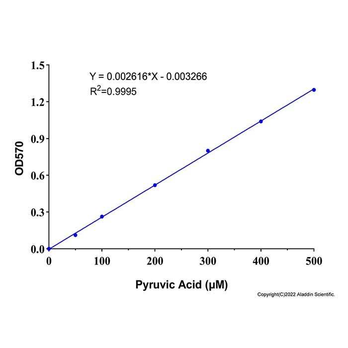
取5 μL丙酮酸标准溶液 (10 mM) ,加入95 μL检测缓冲液,混匀,配制成浓度为500 μM的丙酮酸标准溶液。分别取500 μM的丙酮酸标准溶液0、2、4、8、12、16、20 μL加入96孔板的标准品孔中,并用检测缓冲液补足至20 μL,此时,标准曲线的浓度分别为0、50、100、200、300、400、500 μM。
注:吸光度检测时建议使用透明96孔板
b) 荧光法检测。取2 μL丙酮酸标准溶液 (10 mM) ,加入98 μL检测缓冲液 混匀,配制成浓度为200 μM的丙酮酸标准溶液。分别取200 μM的丙酮酸标准溶液0、0.5、1、2、5、10、20 μL加入96孔板的标准品孔中,并相应地用检测缓冲液补足至20 μL ,此时,标准曲线的浓度分 别为0、5、10、20、50、100、200 μM。
注:荧光检测时建议使用96孔黑板
3. 样本制备
推荐使用新鲜样本,若不立即实验,-80℃可保存 1 个月,使用时冰上解冻。
1) 动物组织用冷 PBS 清洗组织,尽可能去除血液。吸干水分,称取 0.1 g,加入1 mL 预冷的裂解液,冰浴匀浆。10000 g,4℃离心 5 min,取上清置冰上待测。
2) 植物组织称取约 0.1 g 样本,加入 1 mL 预冷的裂解液捣碎,冰浴超声破碎 5 min(功率 20% 或 200 W,超声 3 s,间隔 7 s,重复 30 次),10000 g,4℃离心 5 min,取上清置冰上待测。
3) 细胞(菌)收集 1×10⁶ 个细胞或细菌,用冷 PBS 清洗后,加入 0.1-0.2 mL预冷的裂解液冰浴匀浆或超声破碎 5 min。12000 g,4℃离心 5 min,取上清置冰上待测。
4) 血浆、血清、尿液等生物体液去除蛋白后取上清:可使用 10 kDa 超滤管过滤取滤液;或将样本:30% ZnSO₄溶液 = 20:1 混合涡旋,10000 g 室温离心 5 min,取上清。
注1:裂解液需自行制备,不影响酶活检测即可,可使用PBS。
4. Amplex Red反应工作液的配制:
按照每个反应80 µL的体积配制适量的Amplex Red反应工作液。 均匀混合50 μL检测缓冲液 、2 μL Amplex Red 、10 µL 酶溶液 、10 µL 辅因子混合液 、8 µL 底物 ,即可配制成80 µL Amplex Red反应工作液。根据待检测样品 (包括标准品) 的数量,配制适量的Amplex Red反应工作液。配制好的 Amplex Red反应工作液置于4ºC或冰浴避光保存,可以在当天使用,建议尽量现配现用。
5. 样本中酶活性测定
1) 按以下体系加样:
试剂 (µL) | 对照孔 | 测定孔 |
Amplex Red | 2 | 2 |
酶混合液 | 10 | 10 |
辅因子混合液 | 10 | 10 |
底物 | / | 8 |
样品 | 20 | 20 |
检测缓冲液 | 58 | 50 |
注1:由于酶溶液的用量较少且易沉降,必须注意在使用前先轻轻离心一下,然后适当混匀后再使用。
注2:丙酮酸及H₂O₂ 的存在会对丙氨酸转氨酶的检测产生干扰。如果样品含有丙酮酸或者H₂O₂ , 须同时设置背景对照孔,加入不含底物的Amplex Red反应工作液,即配制Amplex Red反应工作液时8 µL底物用检测工作液替代。
注3:为确保样品数值在标准曲线范围内,建议设置多个稀释倍数,以确定样品中大致酶活,如果数值不在标准曲线范围内,请使用检测工作液稀释。样品总稀释倍数记为n (例如本步骤中对样品进行10倍稀释,加入的 ‘‘稀释后的样品’’为20 µL,则n=10)。
2) 反应体系充分混匀后37℃避光孵育20-30 min。 吸光度法检测,读取570 nm ,记为A;如果使用荧光检测,设置激发波长为560 nm,发射波长为590 nm进行荧光强度检测,记为RFU 。
注1:为取得最佳的检测效果,反应时间可以根据待测样品中的丙氨酸转氨酶活性进行调整,但是必须确保读数在标准曲线范围内。对于丙氨酸转氨酶活性较高的样品,建议测定总时间为20分钟或30分钟,对应的测定间隔时间设为2分钟或5分钟;对于丙氨酸转氨酶活性较低的样品,可以延长测定总时间为45至60分钟,对应的测定间隔时间设为5或10分钟。也可以连续测定30分钟,每隔1或2分钟测定1次,最后取检测数据中前期一段时间内呈现线性的数据用于分析和计算。
注2:随着反应时间的延长,高浓度标准品孔的信号值可能会达到平台期,从而导致其不在标准曲线的线性范围内,此时可以舍去异常点的数据,取在线性范围内的数据来拟合标准曲线。
6. 结果计算
建立标准曲线,将A或RFU带入标准曲线,即可算出在反应时间内样品中丙氨酸转氨酶催化产生的丙酮酸的浓度(B),如果样品的背景对照信号比较高,样品的信号值应减去样品背景对照的信号值。丙酮酸标准曲线,吸光度检测50-500 μM浓度范围内有良好的线性关系,荧光检测5-200 μM浓度范围内有良好的线性关系。丙氨酸转氨酶活力的计算公式如下:
ALT (U/L)=B×n/T
注:B为根据标准曲线确定的丙酮酸浓度浓度 (μM);
n为样品总稀释倍数;
T为的反应时间(min)。
以标准品浓度为横坐标 , A 为纵坐标绘制标准曲线,计算样品丙氨酸转氨酶活性。
丙氨酸转氨酶活性单位的定义为:1个酶活力单位(unit, U)为在37ºC条件下,每分钟催化产生1.0 μmol丙酮酸所需的酶量。
注意事项
1) Amplex Red 试剂光敏、易氧化,全程避光,开瓶配制后尽快使用。
2) 丙氨酸转氨酶浓度过高会导致信号下降,样品需提前稀释。
3) 反应体系避免含 DTT、2 - 巯基乙醇等还原剂,否则干扰检测。
4) 每次实验均需重新配制标准品并做标准曲线。
5) 为了您的安全和健康,请穿实验服并戴一次性手套操作。
6) 本产品仅限于专业人员的科学研究用,不得用于临床诊断或治疗,不得用于食品或药品。
Product Introduction
Alanine aminotransferase (ALT), also known as glutamate pyruvate transaminase (GPT), is a pyridoxal phosphate (vitamin B6)-dependent enzyme. It catalyzes the reversible transamination reaction between alanine and α-ketoglutarate to produce pyruvic acid and glutamate, and plays a vital role in amino acid metabolism, the glucose-alanine cycle and gluconeogenesis. ALT is widely distributed in various tissues and body fluids including the liver and skeletal muscle, with the highest abundance in the liver.
Serum ALT levels remain very low in healthy individuals. When liver cells or other tissues (such as cardiac muscle and skeletal muscle) are damaged - for instance, in viral or toxic hepatitis, infectious mononucleosis, obstructive jaundice and other conditions - ALT leaks into the bloodstream and causes a dramatic elevation of serum ALT concentration. Even damage to merely 1% of hepatocytes can result in a more than twofold increase in blood ALT levels. Therefore, ALT serves as a highly sensitive and specific biomarker for evaluating liver injury and normal liver function. The measurement of ALT activity is of great significance for clinical diagnosis, prognosis monitoring and treatment of related diseases.
Catalyzed by alanine aminotransferase (ALT), alanine reacts with α-ketoglutarate (α-KG) to generate glutamate and pyruvic acid. In the presence of flavin adenine dinucleotide (FAD), thiamine pyrophosphate (TPP) and magnesium ions, the resulting pyruvic acid undergoes oxidation with oxygen under the action of pyruvate oxidase (PO), yielding acetyl phosphate, carbon dioxide (CO₂) and hydrogen peroxide (H₂O₂). The enzyme activity of ALT in samples is finally quantified by measuring the absorbance or fluorescence intensity of resorufin, the reaction product of H₂O₂ and Amplex Red. The absorbance and fluorescence intensity of resorufin are directly proportional to the ALT activity in the sample.
The kit supports both absorbance and fluorescence detection, featuring high sensitivity, wide linear range, and low sample consumption. With a sample volume of 20 µL:
- Absorbance assay: good linearity from 50-500 µM
- Fluorescence assay: good linearity from 5-200 µM
A1510315 | Component | Appearance | 100 T | 500 T | Storage conditions | Quantity Per Test | A1510315A | Enzyme Solution | Liquid | 1.0 mL | 5.0 mL | -20℃.Store in the dark. | 10.0 μL | A1510315B | Cofactor | Liquid | 1.0 mL | 5.0 mL | -20℃.Store in the dark. | 10.0 μL | A1510315C | Amplex Red | Liquid | 0.2 mL | 1.0 mL | -20℃.Store in the dark. | 2.0 μL | A1510315D | Substrate | Liquid | 0.8 mL | 4.0 mL | -20℃.Store in the dark. | 8.0 μL | A1510315E | Pyruvic Acid Standard (10 mM) | Liquid | 0.1 mL | 0.5 mL | -20℃.Store in the dark. | As needed | A1510315F | Assay Buffer | Liquid | 20.0 mL | 100.0 mL | -20℃. | 50 μL |
|
Usage Method
1. Kit Preparation
1) Thaw Amplex Red and Assay Buffer, equilibrate them to room temperature and mix well for later use. Keep other reagents on ice until use. Immediately store all reagents under the conditions specified in the kit manual after the experiment is completed.
2. Standard Preparation
1) Preparation of pyruvic Acid Standard curve. Either absorbance detection or fluorescence detection can be selected. Fluorescence detection is recommended preferentially for samples with low volume or low concentration.
a) Absorbance Detection
Transfer 5 µL of 10 mM Pyruvic Acid Standard and mix with 95 µL Assay Buffer to prepare a 500 µM pyruvic acid standard solution. Pipette 0、2、4、8、12、16、and 20 µL of the 500 µM oxaloacetic acid standard solution into the standard wells of a 96-well plate respectively, then top up each well to 20 µL with ALT Assay Buffer. The corresponding concentrations of the standard curve are 0、50、100、200、300、400、and 500 µM.
Note: A transparent 96-well plate is recommended for absorbance measurement.
b) Fluorescence Detection
Transfer 2 µL of 10 mM Pyruvic Acid Standard and mix with 98 µL Assay Buffer to prepare a 200 µM pyruvic acid standard solution. Pipette 0, 0.5, 1, 2, 5, 10 and 20 µL of the 200 µM oxaloacetic acid standard solution into the standard wells of a 96-well plate respectively, then top up each well to 20 µL with Assay Buffer. The corresponding concentrations of the standard curve are 0, 5, 10, 20, 50, 100 and 200 µM.
Note: A black 96-well plate is recommended for fluorescence measurement.
3. Sample Preparation
Fresh samples are recommended. If not assayed immediately, store samples at –80℃ for up to 1 month; thaw on ice before use.
1) Animal Tissue
Rinse tissue with cold PBS to remove blood; blot dry. Weigh 0.1 g, add 1 mL of lysis buffer, and homogenize on ice. Centrifuge at 10,000 g, 4℃ for 5 min. Collect supernatant on ice for assay.
2) Plant Tissue
Weigh ~0.1 g sample, add 1 mL of cold lysis buffer, and homogenize. Sonicate on ice for 5 min (20% or 200 W; 3 sec on, 7 sec off; 30 cycles). Centrifuge at 10,000 g, 4℃ for 5 min. Collect supernatant on ice for assay.
3) Cells / Bacteria
Collect 1×10⁶ cells or bacteria; wash with cold PBS. Add 0.1-0.2 mL of cold lysis buffer, then homogenize or sonicate on ice for 5 min. Centrifuge at 12,000 g, 4℃ for 5 min. Collect supernatant on ice for assay.
4) Plasma, Serum, Urine, and Other Biological Fluids
Deproteinize before use:Filter through a 10 kDa ultrafiltration tube and collect filtrate; or
Mix sample : 30% ZnSO₄ solution = 20:1, vortex, centrifuge at 10,000 g, room temperature for 5 min, collect supernatant.
Note 1: Lysis buffer needs to be prepared by the user. Any formulation that does not interfere with enzyme activity measurement is acceptable; PBS can be used as an alternative.
4. Detection Working Solution Preparation
Prepare an appropriate volume of Amplex Red working solution based on a reaction volume of 80 µL per test. Mix 50 µL Assay Buffer, 2 µL Amplex Red, 10 µL Enzyme Solution , 10 µL Cofactor and 8 µL Substrate thoroughly to prepare 80 µL Amplex Red working solution. Prepare sufficient working solution according to the total number of samples to be tested (including standards).
The prepared Amplex Red working solution should be stored at 4 °C or on ice under light protection and used within the same day. Fresh preparation prior to use is highly recommended.
5. Measurement of Sample Enzyme Activity
1) Set up reactions as follows:
Reagent (μL) | Control Well | Sample Well |
Amplex Red | 2 | 2 |
Enzyme Solution | 10 | 10 |
Substrate | / | 8 |
Cofactor | 10 | 10 |
Sample | 20 | 20 |
Detection Working Solution | 58 | 50 |
Note 1: The enzyme solution is used in a small volume and prone to sedimentation. Briefly centrifuge the tube gently before use and mix thoroughly prior to pipetting.
Note 2: Pyruvic acid and H₂O₂ interfere with ALT measurement. If samples contain any of the above substances, background control wells must be set up in parallel, loaded with Amplex Red working solution without Substrate. Specifically, replace the 8 μL Substrate with ALT Assay Buffer when preparing the Amplex Red working solution.
Note 3: To guarantee sample readings fall within the linear range of the standard curve, prepare serial dilutions to estimate the approximate ALT activity in samples. If readings exceed the linear range, dilute samples with ALT Assay Buffer. The total dilution factor of the sample is denoted as n (e.g., if the sample is diluted 10-fold and 20 μL of diluted sample is added, n = 10).
2) Thoroughly mix the reaction system and incubate at 37 °C protected from light for 20-30 min. Absorbance detection is performed by measuring the absorbance at 570 nm, denoted as A. For fluorescence detection, the excitation wavelength is set to 560 nm and the emission wavelength to 590 nm to record the relative fluorescence unit (RFU).
Note 1: Incubation time can be adjusted according to ALT activity in test samples to achieve optimal detection performance, provided all readings stay within the standard curve linear range. For samples with high ALT activity: total incubation time of 20 or 30 min is recommended, with measurement intervals of 2 or 5 min respectively. For samples with low ALT activity: extend total incubation time to 45 - 60 min, with measurement intervals of 5 or 10 min respectively. Alternatively, continuous detection can be performed over 30 min at 1 or 2 min intervals. Only data showing linearity in the early reaction phase shall be adopted for analysis and calculation.
Note 2: Prolonged incubation may cause signal plateau in high-concentration standard wells, making data points out of the linear range. Abnormal plateau points can be excluded, and only linear-range data shall be used for standard curve fitting.
6. Data Analysis
1) Construct a standard curve, then substitute the absorbance (A) or relative fluorescence unit (RFU) value into the curve to calculate the concentration of oxaloacetic acid (B) generated by Alanine Aminotransferase catalysis in the sample within the reaction duration. If the background control signal of the sample is high, subtract the background signal value from the raw sample signal. For the pyruvic acid standard curve, good linearity is obtained from 50-500 μM via absorbance measurement, and from 5-200 μM via fluorescence measurement.
The formula for calculating Alanine Aminotransferase activity is as follows:
- ALT activity (U/L) = =B×n/T
- B = Pyruvic acid concentration (μM) calculated from the standard curve
- n = total dilution factor of the sample
- T = reaction incubation time (min)
- Plot the standard curve with standard concentration as the X-axis and absorbance (A) as the Y-axis to calculate ALT activity in samples.
Definition of Alanine Aminotransferase Activity Unit
- Definition of alanine aminotransferase (ALT) activity unit: One unit (U) refers to the amount of ALT enzyme that catalyzes the production of 1.0 μmol pyruvic acid per minute at 37 °C.
Precautions
1. Amplex Red is light‑sensitive and easily oxidized; protect from light throughout and use promptly after opening/preparation.
2. Excessively high Alanine Aminotransferase concentration reduces signal; dilute samples before assay if needed.
3. Avoid reductants such as DTT or 2‑mercaptoethanol in the reaction, which interfere with detection.
4. Fresh standards and a new standard curve must be used in every experiment.
5. Wear lab coat and disposable nitrile gloves during operation for personal safety and health protection.
6. This product is for research use only by qualified laboratory personnel. It shall not be used for clinical diagnosis, clinical therapy, food or pharmaceutical applications.

 ZJ26F0838353


 危险品化学品经营许可证(带存储)