产品介绍
丙氨酸氨基转氨酶,旧称谷丙转氨酶,缩写为 ALT或GPT广泛存在于动植物、微生物和培养细胞中,催化氨基酸和酮酸转氨基反应,在氨基酸代谢中具有重要作用。哺乳动物肝细胞内的 ALT 浓度远高于血清,当肝细胞坏死,ALT 释放到血液中,血清 ALT活性显著增高。因此,ALT 被世界卫生组织推荐为肝功能损害最敏感的检测指标。
本产品的检测原理是ALT催化丙氨酸与α-酮戊二酸发生转氨基反应,生成丙酮酸和谷氨酸;加入 2,4-二硝基苯肼溶液,不仅终止上述反应,而且与丙酮酸反应形成丙酮酸二硝基苯腙,在碱性溶液中显棕红色。通过在520 nm 读取吸光值并计算出该酶活力大小。
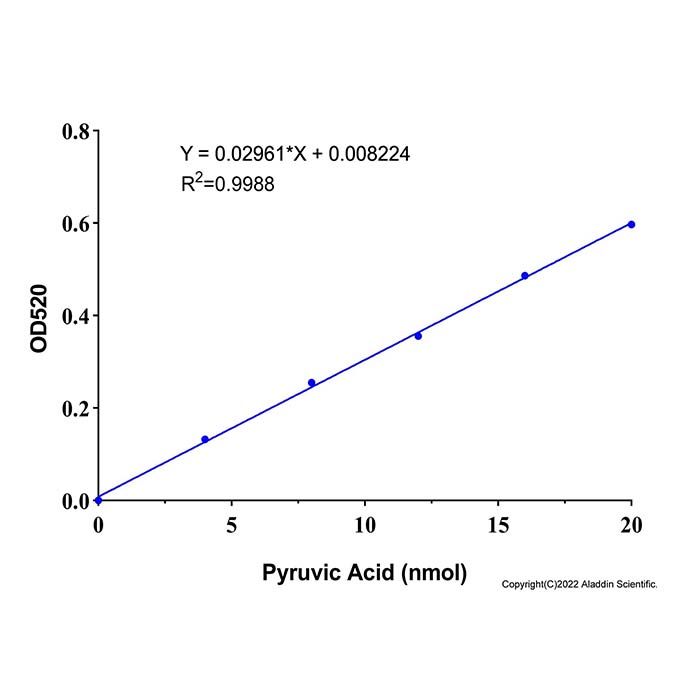
本试剂盒可采用分光光度法进行检测,具有检测灵敏度高、线性范围宽、样品用量少等特点。在样品体积为10 µL 时,采用分光光度法检测在4-20 nmol 浓度范围内有良好的线性关系。
组分货号 | 组件 | 物理外观 | 100 T | 储存 | 单次测试使用量 |
A1515821A | 终止液 | 液体 | 20.0 mL | 2-8℃. | 200.0 μL |
A1515821B | 显色液 | 液体 | 2.0 mL | 2-8℃. 避光 | 20.0 μL |
A1515821C | 丙酮酸标准品 (10 mM) | 液体 | 0.2 mL | 2-8℃. 避光 | 按需 |
A1515821D | 底物 | 液体 | 2.0 mL | 2-8℃. 避光 | 20.0 μL |
A1515821E | 检测液 | 液体 | 50.0 mL | 2-8℃. | 按需 |
使用方法
建议正式实验前,选取 2 个样本做预测定,了解实验样品情况,熟悉流程, 避免样本和试剂浪费。
1.标准曲线的测定
1) 取丙酮酸标准品 (10 mM) ,稀释成以下浓度:0,0.4,0.8,1.2,1.6,2.0 μmol/mL丙酮酸标准液 。也可根据实际来调整浓度。
2) 取各个梯度的丙酮酸标准液 ,进行标准曲线的测定: 10 μL丙酮酸标准液 +20 μL 底物 +20 μL 显色液 ,混匀,于 37℃孵育 10 min ;再加 200 μL 终止液 ,混匀,25℃孵育10 min,于 520 nm 处测定吸光值 A,依据结果即可制作标准曲线。
注: 以对应的丙酮酸量 (nmol) 作为X轴,以△A为纵坐标Y,即不同浓度标品读数后均扣除0 μmol/mL点对应信号值后为纵坐标。
2. 样本制备
推荐使用新鲜样本,若不立即实验,-80℃可保存 1 个月,使用时冰上解冻。
1) 动物组织用冷 PBS 清洗组织,尽可能去除血液。吸干水分,称取 0.1 g,加入1 mL (V) 预冷的检测液,冰浴匀浆。10000 g,4℃离心 5 min,取上清置冰上待测。
2) 植物组织称取约 0.1 g 样本,加入 1 mL (V) 预冷的检测液捣碎,冰浴超声破碎 5 min(功率 20% 或 200 W,超声 3 s,间隔 7 s,重复 30 次),10000 g,4℃离心 5 min,取上清置冰上待测。
3) 细胞 (菌) 收集 5×10⁶ 个细胞或细菌,用冷 PBS 清洗后,加入 1.0 m (V) L预冷的检测液冰浴匀浆或超声破碎 5 min。12000 g,4℃离心 5 min,取上清置冰上待测。
4) 血浆、血清、尿液等生物体液:直接检测。若浑浊,离心后取上清检测。
3. 样品测定
1) 酶标仪预热 30 min 以上,调节波长至 520 nm。
2) 可先做 2 个样本预测定,熟悉操作过程,并依据测出的吸光值 A 是否超出标准曲线的线性范围来做调整 。
3) 所有试剂可于 37℃孵育 5-10 min,在 96 孔板中按以下体系加样。
试剂 (µL) | 对照孔 | 测定孔 |
样本 (V₁) | - | 10 |
底物 | 20 | 20 |
混匀,于 37℃孵育 30 min | | |
显色液 | 20 | 20 |
样本 ( V₁) | 10 | - |
混匀,于 37℃孵育 10 min | | |
终止液 | 200 | 200 |
混匀,25℃孵育 10 min,于 520 nm 处测定吸光值 A,△A= A测定 - A对照 (每个样本做一个自身对照)。 | | |
注:若测定超过 0.8 ,可降低样本V₁量 (如 5 μL,另外 5 μL 用蒸馏水补齐,总体积 10 μL 保持不变)。则改变后的加样体积 V₁需代入计算公式重新计算,或者稀释后再进行倍数计算。
4. 结果计算
1) 建立标准曲线,以丙酮酸标准液摩尔质量 (nmol) 为X轴,以△A为纵坐标Y,根据吸光度建立的标准曲线计算对应样品的X数值:
a) 按样本蛋白浓度计算酶活:
酶活定义:每毫克组织蛋白每分钟催化产生 1 nmol 丙酮酸定义为一个酶活力单位。
ALT/GPT (nmol/min/mg prot)=X÷(V₁× Cₚᵣ)÷T
b) 按样本鲜重计算酶活 :
酶活定义:每克组织每分钟催化产生 1 nmol 丙酮酸定义为一个酶活力单位。
ALT/GPT (nmol/min/g 鲜重)=X÷(W×V₁÷V)÷T
c) 按细菌或细胞数量计算酶活 :
酶活定义:每1万个细菌或细胞每分钟催化产生 1 nmol 丙酮酸定义为一个酶活力单位。
ALT/GPT (nmol/min/ 10⁴cell)=X÷(500×V₁÷V)÷T
d) 血清 (浆)活力计算酶活 :
酶活定义:每毫升血清 (浆) 每分钟催化产生 1 nmol 丙酮酸定义为一个酶活力单位。
ALT/GPT (nmol/min/mL)=X÷V₁÷T
注:
V-加入提取液体积,1 mL ;
V₁-加入样本体积,0.01 mL ;
T-反应时间,30 min ;
W-样品质量,g ;
500-细胞或细菌总数,万 ;
Cₚᵣ-样本蛋白质浓度,mg/mL;
注意事项
1) 显色液光敏、易氧化,全程避光,开瓶配制后尽快使用。
2) 丙氨酸氨基转氨酶浓度过高会导致信号下降,样品需提前稀释。
3) 检测样本前需建立自己配制标准品并做标准曲线,检测1-2次即可。
4) 为了您的安全和健康,请穿实验服并戴一次性手套操作。
5) 本产品仅限于专业人员的科学研究用,不得用于临床诊断或治疗,不得用于食品或药品。
Product Introduction
Alanine aminotransferase, formerly known as glutamic‑pyruvic transaminase, is abbreviated as ALT or GPT. It is widely present in animals, plants, microorganisms and cultured cells. It catalyzes the transamination reaction between amino acids and ketoacids and plays a vital role in amino‑acid metabolism. The concentration of ALT in mammalian hepatocytes is much higher than that in serum. Upon hepatocyte necrosis, ALT is released into the bloodstream, resulting in a marked increase in serum ALT activity. Accordingly, ALT is recommended by the World Health Organization as the most sensitive biomarker for liver function injury. The detection principle of this product is described as follows: ALT catalyzes the transamination reaction between alanine and α‑ketoglutarate to produce pyruvate and glutamate. Addition of 2,4‑dinitrophenylhydrazine (DNPH) solution not only terminates the above reaction, but also reacts with pyruvate to form pyruvate dinitrophenylhydrazone, which yields a reddish‑brown color in alkaline solution. The enzyme activity is determined by measuring absorbance at 520 nm.
This kit is designed for spectrophotometric detection and features high assay sensitivity, broad linear range and low sample consumption. With a sample volume of 10 µL, the spectrophotometric method exhibits good linearity within the concentration range of 4‑20 nmol.
Component No. | Component Name | Appearance | 100 T | Storage conditions | Quantity Per Test |
A1515821A | Reaction‑Stopping Reagent | Liquid | 20.0 mL | 2-8℃ . | 200.0 μL |
A1515821B | Chromogenic Solution | Liquid | 2.0 mL | 2-8℃ .Store in the dark | 20.0 μL |
A1515821C | Pyruvic Acid Standard (10 mM) | Liquid | 0.2 mL | 2-8℃ .Store in the dark | As needed |
A1515821D | Substrate | Liquid | 2.0 mL | 2-8℃ .Store in the dark | 20.0 μL |
A1515821E | Assay Buffer | Liquid | 50.0 mL | 2-8℃ | As needed |
Usage Method
It is recommended to perform a preliminary assay with 2 samples prior to formal experiments to characterize sample properties and familiarize yourself with the procedure, so as to avoid waste of samples and reagents.
1. Determination of standard curve
1) Dilute pyruvate acid standard (10 mM) to obtain working standards at concentrations of 0, 0.4, 0.8, 1.2, 1.6, 2.0 μmol/mL pyruvate standard solutions . Concentrations may be adjusted according to practical conditions.
2) Take pyruvate standard solutions of each gradient for standard‑curve determination: mix 10 μL pyruvate standard solution, 20 μL substrate and 20 μL chromogenic solution, incubate the mixture at 37 °C for 10 min. Add 200 μL Reaction‑Stopping Reagent , mix thoroughly and incubate at 25 °C for 10 min. Measure the absorbance (A) at 520 nm. The standard curve can be plotted based on the obtained results.
Note: Use the corresponding amount of pyruvate (nmol) as the X‑axis, and ΔA as the Y‑axis. ΔA is calculated by subtracting the signal value of the 0 μmol/mL standard from the absorbance reading of each standard concentration.
2. Sample Preparation
Fresh samples are recommended. If not assayed immediately, store samples at -80℃ for up to 1 month; thaw on ice before use.
1) Animal Tissue
Rinse tissue with cold PBS to remove blood; blot dry. Weigh 0.1 g, add 1 mL of cold Assay Buffer. and homogenize on ice. Centrifuge at 10,000 g, 4℃ for 5 min. Collect supernatant on ice for assay.
2) Plant Tissue
Weigh - 0.1 g sample, add 1 mL of cold Assay Buffer (V) , and homogenize. Sonicate on ice for 5 min (20% or 200 W; 3 sec on, 7 sec off; 30 cycles). Centrifuge at 10,000 g, 4℃ for 5 min. Collect supernatant on ice for assay.
3) Cells / Bacteria
Collect 5×10⁶ cells or bacteria; wash with cold PBS. Add 1.0 mL of cold Assay Buffer , then homogenize or sonicate on ice for 5 min. Centrifuge at 12,000 g, 4℃ for 5 min. Collect supernatant on ice for assay.
4) Plasma, Serum, Urine, and Other Biological Fluids
Biological fluids such as plasma, serum and urine can be assayed directly. If turbidity is observed, collect the supernatant after centrifugation for detection.
3. Measurement of Sample Enzyme Activity
1) Warm‑up the microplate reader for no less than 30 min and set the wavelength to 520 nm.
2) A preliminary assay of 2 samples may be performed first to familiarize with the operating procedure. Adjust experimental conditions according to whether the measured absorbance (A) exceeds the linear range of the standard curve.
3) All reagents may be pre‑incubated at 37 °C for 5‑10 min. Pipette reagents into the 96‑well microplate following the system below:
Reagent (µL) | Control Well | Sample Well |
Sample (V₁) | - | 10 |
Substrate | 20 | 20 |
The mixture was mixed thoroughly and incubated at 37 °C for 30 minutes. |
|
|
Chromogenic Solution | 20 | 20 |
Sample (V₁) | 10 | - |
The mixture was mixed thoroughly and incubated at 37 °C for 10 minutes. |
|
|
Reaction‑Stopping Reagent | 200 | 200 |
Mix thoroughly and incubate the reaction mixture at 25 °C for 10 min. Measure the absorbance A at 520 nm. Calculate
ΔA = Aₛₐₘₚₗₑ - AControl(one self‑control per sample). |
Note : If the measured absorbance A exceeds 0.8, reduce the sample volume V₁ (e.g. use 5 μL sample and make up the remaining 5 μL with distilled water to keep the total volume constant at 10 μL). The adjusted sample‑loading volume V₁ shall be substituted into the calculation formula for recalculation, or the dilution factor shall be taken into account for subsequent computation after sample dilution.
4. Data Analysis
1) Generate the standard curve, where X represents the amount of standard substance in nmol and Y represents ΔA. Calculate the X value of the test sample from the absorbance‑derived standard curve.
a) Enzyme activity calculated based on sample protein concentration:
Enzyme activity definition: One unit of enzyme activity is defined as the amount of enzyme that catalyzes the production of 1 nmol pyruvate per minute per milligram of tissue protein.
ALT/GPT (nmol/min/mg prot) = X ÷ (V₁ × Cₚᵣ) ÷ T
b) Enzyme activity calculated based on sample fresh weight
Enzyme activity definition: One unit of enzyme activity is defined as the amount of enzyme that catalyzes the production of 1 nmol pyruvate per minute per gram of tissue fresh weight.
ALT/GPT (nmol/min/g fresh weight) = X ÷ (W × V₁ ÷ V) ÷ T
c) Enzyme activity calculated based on bacterial or cell count:
Enzyme activity definition: One unit of enzyme activity is defined as the amount of enzyme that catalyzes the production of 1 nmol pyruvate per minute per 10⁴ bacteria or cells.
ALT/GPT (nmol/min/10⁴ cell) = X ÷ (500 × V₁ ÷ V) ÷ T
d) Enzyme activity calculation for serum / plasma:
Enzyme activity definition: One unit of enzyme activity is defined as the amount of enzyme that catalyzes the production of 1 nmol pyruvate per minute per milliliter of serum or plasma.
ALT/GPT (nmol/min/mL) = X ÷ V₁ ÷ T
Annotation:
V - Volume of extraction solution added, 1 mL
V₁ - Volume of sample added, 0.01 mL
T -Reaction time, 30 min
W - Sample weight, g
500-Total number of cells or bacteria, ten‑thousand
Cₚᵣ-Protein concentration of sample, mg/mL
Precautions
1.The chromogenic reagent is light‑sensitive and prone to oxidation. Keep away from light throughout the whole procedure and use promptly after opening and preparation.
2.Excessively high alanine aminotransferase concentration may lead to signal decrease; samples should be diluted in advance.
3.Prepare your own standard solution and construct a standard curve prior to sample detection; one or two curve replicates are sufficient.
4.For personal safety and health, wear a laboratory coat and disposable gloves during operation.
5.This product is for research use only by qualified professionals. It shall not be used for clinical diagnosis or therapy, nor for food or pharmaceutical applications.



 危险品化学品经营许可证(带存储)