Flavin-containing quinone reductase that catalyzes two-electron reduction of quinones to hydroquinones using either NADH or NADPH as electron donors. In a ping-pong kinetic mechanism, the electrons are sequentially transferred from NAD(P)H to flavin cofactor and then from reduced flavin to the quinone, bypassing the formation of semiquinone and reactive oxygen species (By similarity).
Regulates cellular redox state primarily through quinone detoxification. Reduces components of plasma membrane redox system such as coenzyme Q and vitamin quinones, producing antioxidant hydroquinone forms. In the process may function as superoxide scavenger to prevent hydroquinone oxidation and facilitate excretion.
Alternatively, can activate quinones and their derivatives by generating redox reactive hydroquinones with DNA cross-linking antitumor potential.
Acts as a gatekeeper of the core 20S proteasome known to degrade proteins with unstructured regions. Upon oxidative stress, interacts with tumor suppressors TP53 and TP73 in a NADH-dependent way and inhibits their ubiquitin-independent degradation by the 20S proteasome.
宿主种属
小鼠(Mouse)
特异性
Human NQO1
种属反应性
人(Human)
免疫原
Recombinant full length protein corresponding human NQO1
阳性对照
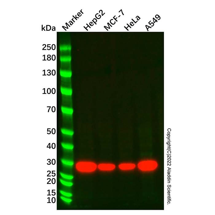
WB: HepG2, MCF-7, HeLa and A549 whole cell lysates Flow Cytometry: MCF-7 cells IF/ICC: MCF-7 cells
偶联
Unconjugated
克隆类型
单克隆抗体
Format
Whole IgG
轻链亚型
kappa
纯化方法
Protein A purified
浓度
1.0 mg/mL
分子类型
抗体
产品线
生物产品
储存与运输
储存缓冲液
PBS, pH 7.4
储存条件
-20°C储存,避免反复冻融
运输条件
超低温运输
稳定性与储存
短期储存2-8℃(1-2周);长期储存-20℃(24个月);收货后建议分装,避免反复冻融.
图片
NQO1 Mouse mAb (Ab224768) - Western Blot All lanes: NQO1 Mouse Antibody (Ab224768) at 1/1000 dilution Samples: Lysates at 20 µg per lane Secondary: Goat Anti-Mouse IgG H&L (HRP) (Ab179001) at 1/20000 dilution
Predicted band size: 31 kDa Observed band size: 30 kDa Exposure time: 30.0 s
NQO1 Mouse mAb (Ab224768) - Flow Cytometry Overlay histogram showing MCF-7 cells stained with NQO1 Mouse mAb (Ab224768) (Red) at 1.0 μg/mL for 1 hour at 4°C. The Goat Anti-Mouse IgG H&L (PE) (Ab179006) was used as the secondary antibody. Unlabeled sample (Black) was used as a negative control. Mouse IgG2b-kappa, anti-E8L (Ab210600) was used as an isotype control (Blue).
NQO1 Mouse mAb (Ab224768) - IF IF analysis of NQO1 (Green) in MCF-7 cells. The cells were fixed with 4% PFA for 15 minutes and then 0.3% Triton X-100 for 15 minutes at RT. Cells were stained with NQO1 Mouse mAb (Ab224768) at 5.0 μg/mL in blocking buffer for 2 hour at RT and then incubated with Goat Anti-Mouse IgG H&L (FITC) (Ab179003) at a dilution of 1/1000 for 1 hour at room temperature in the dark. Cells were counterstained with DAPI (Blue). Images were taken on the confocal laser scanning microscope.

 ZJ25F0724123


 危险品化学品经营许可证(带存储)