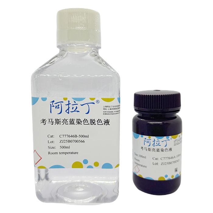
考马斯亮蓝染色试剂盒采用了最经典的考马斯亮蓝染色和脱色方法,以考马斯亮蓝R250为染料,可用于SDS-PAGE或非变性PAGE等蛋白电泳凝胶的常规染色和脱色,或Western转膜后PAGE胶上残余蛋白的检测。
C777646
| Component
| 1Kit | Storage |
C777646A
| 考马斯亮蓝染色液
| 100 mL
| RT. Store in the dark.
|
C777646B
| 考马斯亮蓝染色脱色液
| 500 mL
| RT. |
使用说明:
1、常规染色脱色方法
1.1 电泳结束后,取凝胶放入适量考马斯亮蓝染色液中,确保染色液可以充分覆盖凝胶。
1.2 置于水平摇床或侧摆摇床上缓慢摇动,室温染色1h或更长时间。
注:具体的染色时间取决于凝胶的厚度和染色时的温度。凝胶较厚,温度较低,则染色时间宜适当延长。凝胶较薄,温度较高,则染色时间可以适当缩短。通常染色至凝胶的颜色和染色液的颜色非常接近,在染色液中几乎看不清凝胶时,可以认为已染色充分。
1.3 倒出染色液。染色液可以回收重复使用至少2-3次。
1.4 加入适量考马斯亮蓝染色脱色液,确保脱色液可以充分覆盖凝胶。
1.5 置于水平摇床或侧摆摇床上缓慢摇动,室温脱色4-24h。期间更换脱色液2-4次,直至蓝色背景基本上全部被脱去,并且蛋白条带染色效果达到预期。通常蛋白条带在脱色1-2h后即可出现。注:脱色时间过长也会导致蛋白条带的颜色变浅。
1.6 完成脱色后,可把凝胶保存在水中,用于后续的拍照等。保存在水中的凝胶会发生溶涨。如需避免溶涨,可以把胶保存在含20%甘油的水中。长期保存可以制备干胶。
2、快速染色脱色方法
2.1 电泳结束后,取胶放入适量考马斯亮蓝染色液中,微波炉加热至接近沸腾或刚刚沸腾,立即停止加热。通常大于10%的胶比较坚韧,在煮沸时不易破损;对于小于10%的胶,宜尽量避免煮沸,以免出现胶碎裂的情况。
2.2 随后在染色液温度较高的情况下,在室温摇床上摇动5-10min。
2.3 倒出染色液。染色液可以回收重复使用至少2-3次。
2.4 加入适量考马斯亮蓝染色脱色液,确保染色液可以充分覆盖凝胶。
2.5 微波炉加热至接近沸腾或刚刚沸腾,立即停止加热。
2.6 随后在脱色液温度较高的情况下,在摇床上摇动5-10min。此时通常可以观察到比较清楚的蛋白条带。
2.7 更换新鲜的脱色液,重复步骤5和步骤6,直至蓝色背景基本上全部被脱去,蛋白条带染色效果达到预期。
2.8 完成脱色后,可以把凝胶保存在水中,用于后续的拍照等。保存在水中的凝胶会发生溶涨。如需避免溶涨,可以把胶保存在含20%甘油的水中。长期保存可以制备干胶。
注意事项:
为了您的自身安全,使用试剂前,请做好防护,如穿实验服,带手套等。
This Coomassie Brilliant Blue staining kit employs the classic Coomassie Brilliant Blue staining and destaining method, using Coomassie Brilliant Blue R-250 as the dye. It is suitable for routine staining and destaining of protein electrophoresis gels such as SDS-PAGE or native PAGE, and can also be used for the detection of residual proteins on the PAGE gel after Western blot transfer.
| Component No. | Component Name | Appearance | 1Kit | Storage |
| C777646A | Coomassie Brilliant Blue Staining Solution | Liquid | 100mL | Room Temperature.Store in the dark. |
| C777646B | Coomassie Brilliant Blue Destaining Solution | Liquid | 500mL | Room Temperature |
Instructions:
1. Routine Staining and Destaining Method
1.1 After electrophoresis, place the gel into an appropriate amount of Coomassie Brilliant Blue Staining Solution, ensuring the solution fully covers the gel.
1.2 Place on a horizontal shaker or side-to-side rocker and shake gently at room temperature for 1 h or longer.
Note: The specific staining time depends on the gel thickness and the staining temperature. Thicker gels and lower temperatures require a longer staining time; thinner gels and higher temperatures allow a shorter staining time. Generally, staining is considered complete when the gel color closely matches the staining solution, and the gel is barely visible in the solution.
1.3 Pour out the staining solution. The staining solution can be recovered and reused at least 2-3 times.
1.4 Add an appropriate amount of Coomassie Brilliant Blue Destaining Solution, ensuring it fully covers the gel.
1.5 Place on a horizontal shaker or side-to-side rocker and shake gently at room temperature for 4-24 h. During this period, replace the destaining solution 2-4 times until the blue background is essentially completely removed and the protein bands show the desired staining. Protein bands usually appear after 1-2 h of destaining. Note: An excessively long destaining time may also lighten the protein bands.
1.6 After destaining is complete, the gel can be stored in water for subsequent imaging, etc. Gels stored in water will swell. To avoid swelling, the gel can be stored in water containing 20% glycerol. For long-term storage, dried gels can be prepared.
2. Rapid Staining and Destaining Method
2.1 After electrophoresis, place the gel into an appropriate amount of Coomassie Brilliant Blue Staining Solution and heat in a microwave oven until near boiling or just boiling, then stop heating immediately. Gels with a concentration greater than 10% are generally tougher and less prone to damage when boiled; for gels below 10%, boiling should be avoided as much as possible to prevent the gel from cracking.
2.2 While the staining solution is still hot, shake on a room-temperature shaker for 5-10 min.
2.3 Pour out the staining solution. The staining solution can be recovered and reused at least 2-3 times.
2.4 Add an appropriate amount of Coomassie Brilliant Blue Destaining Solution, ensuring it fully covers the gel.
2.5 Heat in a microwave oven until near boiling or just boiling, then stop heating immediately.
2.6 While the destaining solution is still hot, shake on a shaker for 5-10 min. Clearly visible protein bands are usually observed at this point.
2.7 Replace with fresh destaining solution and repeat Steps 5 and 6 until the blue background is essentially completely removed and the protein bands show the desired staining.
2.8 After destaining is complete, the gel can be stored in water for subsequent imaging, etc. Gels stored in water will swell. To avoid swelling, the gel can be stored in water containing 20% glycerol. For long-term storage, dried gels can be prepared.
Precautions:
For your own safety, take proper protective measures before using the reagent, such as wearing a lab coat and gloves.

 ZJ25F0723939


 危险品化学品经营许可证(带存储)