是否无菌:是
复溶(Reconstitution):Reconstitute in Ultrapure water or 10mM PBS (pH 7.4).
脂肪间充质干细胞(ADSCs)是一类源自脂肪组织的多能干细胞,具有自我更新和多向分化潜能。它们可以分化为多种细胞类型,包括脂肪细胞、成骨细胞、软骨细胞、心肌细胞、神经细胞等。ADSCs 还具有低免疫原性、可塑性强、旁分泌多种生物活性因子等特性,使其在临床应用中具有广阔的前景。脂肪间充质干细胞外泌体(Adipose-Derived Mesenchymal Stem Cells Exosomes, ADSCs-Exos)是近年来再生医学和组织修复领域的研究热点。ADSCs 凋亡过程中释放的细胞外囊泡(ApoEVs)在皮肤损伤修复中表现出色。
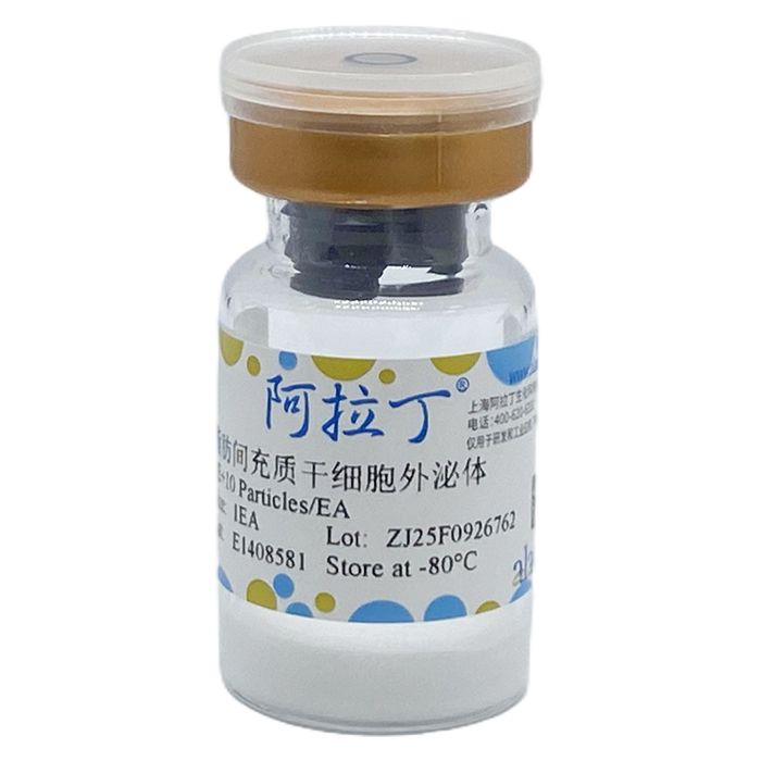
该产品由脂肪间充质干细胞培养获得的上清液,经分离纯化得到的外泌体,可用于实验对照、直接进行负载和靶向改造、组织修复等研究。
产品优势:
严格的质控标准:多项质控分析,有严格的浓度、纯度等不同质控标准。
完整的表征结果:外泌体原料产品按照MISEV2018指南,从电镜形态、粒径大小及颗粒分布、WB三阳一阴蛋白标志物三个维度进行表征。
广泛的应用场景:可用于实验流程及功能实验的对照;可用于靶向改造、装载分子等工程化改造,以及外泌体治疗产品的开发。
产品规格:
名称
| 脂肪间充质干细胞外泌体
| 规格
| 1E+10 Particles/瓶
| 外观
| 白色粉末
| pH
| 7.0 | 无菌检测
| 无菌生长
| 总蛋白浓度
| (BCA)
| 纯度
| ≥90% |
|
案例展示:
1. NTA检测外泌体的粒径分布和颗粒浓度:
将得到的外泌体震荡分散均匀,用PBS稀释至合适倍数,标记好样本名称、稀释倍数、完成样本的稀释配制;测样之前,先测试稀释液:移液枪吸取200μL稀释液上机检测,确认组件、仪器
都处于正常运行;稀释液测量完成,测试结果正常后开始测试样本,取稀释后的200μL样本上机检测;待计数颗粒数达到100个以上时停止测试,导出数据结果,完成样本检测。

2. TEM观察外泌体的形态:
重悬外泌体至50-100μL 2% PFA中,将5μL混合悬液加到Formvar carbon载样铜网上;也可将5-10μL混合悬液滴加到封口膜上,将铜网Formvar膜面朝下放在悬液上。每个样品准备2-3个铜网;将100μL PBS 加到封口膜上。用镊子将铜网 (Formvar 膜面朝下) 放在PBS液滴上清洗 (在所有步骤中,都应保持Formvar膜面湿润,而另一面干燥);将铜网放在50μL 1%戊二醛液滴上5 min;将铜网放在100μL ddH₂O洗涤8次,每次洗2min;将铜网放在50μL草酸双氧铀液滴上 (pH 7.0),5min;将铜网放在50甲基纤维素液滴上10min,冰上操作;铜网放到样品台顶端的不锈钢环上,用滤纸吸去多余液体;空气中干燥5-10min;将铜网放在样品盒内,80kV下拍摄电镜照片。

3. Western Blot 检测外泌体标志物 (三阳一阴):
将分离纯化得到的外泌体加入裂解液 (E778170),裂解后吸取上清液,根据测定蛋白浓度稀释样本至合适浓度;裂解液加入 4×LDS上样缓冲液 (T466588),95°C煮5min,待降至室温后,瞬离;加样:将15μL样本全部加到泳道中,并在样本首尾加上marker;电泳:190V,70min;提前10min用甲醇活化PVDF膜;转膜:转膜液需要提前预冷,275mA,70min;封闭:配制1% BSA的TBST溶液,室温封闭1h;孵育一抗:4°C,摇床上过夜,抗体用1% BSA的TBST溶液稀释;洗膜:1xTBST 洗三次,10 min/次;孵育二抗:抗体用1% BSA的TBST溶液稀释,室温摇床1h;洗膜:1xTBST洗三次,10min/次;曝光拍照。

注意事项和免责声明:
本品仅限于专业人员的科学研究使用,不得用于临床诊断或治疗,不得用于食品或药品。
Adipose-derived mesenchymal stem cells (ADSCs) are a type of pluripotent stem cells derived from adipose tissue, with the potential of self-renewal and multi-directional differentiation. They can differentiate into various cell types, including adipocytes, osteoblasts, chondrocytes, cardiomyocytes, and nerve cells. ADSCs also have characteristics such as low immunogenicity, strong plasticity, and paracrine secretion of a variety of bioactive factors, which endow them with broad prospects in clinical applications. Adipose-Derived Mesenchymal Stem Cells Exosomes (ADSCs-Exos) have been a research hotspot in the fields of regenerative medicine and tissue repair in recent years. The extracellular vesicles (ApoEVs) released during the apoptosis of ADSCs have shown excellent performance in skin damage repair.
This product is exosomes isolated and purified from the supernatant obtained by culturing adipose-derived mesenchymal stem cells, and can be used in studies such as experimental control, direct loading and targeted modification, and tissue repair.
Product Advantages:
Strict quality control standards: Multiple quality control analyses are conducted, with strict standards for various aspects such as concentration and purity.
Comprehensive characterization results: Exosome raw material products are characterized in three dimensions in accordance with the MISEV2018 guidelines, including electron microscope morphology, particle size and particle distribution, and WB three-positive and one-negative protein markers.
Wide range of application scenarios: It can be used as a control in experimental procedures and functional experiments; it can be applied in engineering modifications such as targeted modification and molecular loading, as well as the development of exosome therapeutic products.
Product Specifications:
Name
| ADSCs Exosomes
| Specifications
| 1E+10 Particles/EA
| Appearance
| White Powder
| pH
| 7.0 | Aseptic Testing
| Aseptic
| Total Protein Concentration
| (BCA)
| Purity
| ≥90% |
|
Case Presentation:
1. NTA detection of exosome particle size distribution and particle concentration:
Shake and disperse the obtained exosomes evenly, dilute them to an appropriate multiple with PBS, mark the sample name and dilution multiple, and complete the dilution preparation of the sample; before testing the sample, test the diluent first: absorb 200μL of diluent with a pipette for on-machine detection, and confirm that the components and instruments are in normal operation; after the measurement of the diluent is completed and the test result is normal, start testing the sample, take 200μL of the diluted sample for on-machine detection; stop the test when the number of counted particles reaches more than 100, export the data results, and complete the sample detection.
2. Observation of exosome morphology by TEM:
Resuspend exosomes in 50-100μL of 2% PFA. Add 5μL of the mixed suspension to a Formvar carbon-coated copper grid; alternatively, drop 5-10μL of the mixed suspension onto a piece of parafilm and place the copper grid with the Formvar film facing down on the suspension. Prepare 2-3 copper grids for each sample. Add 100μL of PBS onto the parafilm. Use tweezers to place the copper grid (with the Formvar film facing down) on the PBS droplets for washing (during all steps, keep the Formvar film surface moist while the other surface remains dry). Place the copper grid on 50μL of 1% glutaraldehyde droplets for 5 minutes. Wash the copper grid on 100μL of ddH₂O 8 times, 2 minutes each time. Place the copper grid on 50μL of uranyl oxalate droplets (pH 7.0) for 5 minutes. Place the copper grid on 50μL of methyl cellulose droplets for 10 minutes, operating on ice. Put the copper grid on the stainless steel ring at the top of the sample stage and blot off excess liquid with filter paper. Air-dry for 5-10 minutes. Place the copper grid in the sample box and take electron microscope photos at 80kV.
3. Western Blot Detection of Exosome Markers (Three Positive and One Negative):
Add the isolated and purified exosomes to the lysis buffer (E778170). After lysis, aspirate the supernatant and dilute the sample to an appropriate concentration according to the measured protein concentration. Add 4×LDS loading buffer (T466588) to the lysis buffer, boil at 95°C for 5 minutes, and perform a quick centrifugation after cooling to room temperature. Loading: Load all 15μL of the sample into the lane, and add markers at the start and end of the sample lanes. Electrophoresis: 190V for 70 minutes. Activate the PVDF membrane with methanol 10 minutes in advance. Membrane transfer: The transfer buffer needs to be pre-cooled in advance, 275mA for 70 minutes. Blocking: Prepare 1% BSA in TBST solution and block at room temperature for 1 hour. Primary antibody incubation: Incubate overnight at 4°C on a shaker; dilute the antibody with 1% BSA in TBST solution. Membrane washing: Wash three times with 1×TBST, 10 minutes each time. Secondary antibody incubation: Dilute the antibody with 1% BSA in TBST solution, and incubate on a shaker at room temperature for 1 hour. Membrane washing: Wash three times with 1×TBST, 10 minutes each time. Exposure and photography.
Precautions and Disclaimer:
This product is limited to scientific research use by professional personnel. It must not be used for clinical diagnosis or treatment, nor for food or drugs.

 ZJ25F0926762


 危险品化学品经营许可证(带存储)