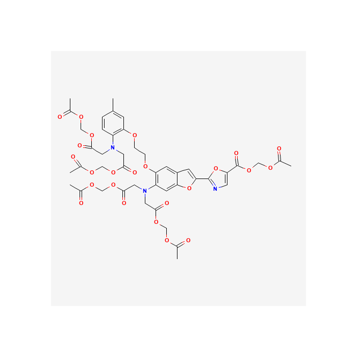
Fura-2 是细胞内钙离子(Ca²⁺)的特异性荧光指示剂,是最常用的检测细胞内钙离子浓度的荧光探针之一。
Fura-2 AM 是一种可以穿透细胞膜的荧光染料。Fura-2 AM 的荧光比较弱,最大激发波长为 369 nm,最大发射波长为 478 nm,并且其荧光不会随钙离子浓度改变而改变。Fura-2 AM 进入细胞后可以被细胞内的酯酶剪切形成 Fura-2,从而被滞留在细胞内。Fura-2 和钙离子结合后,最大激发波长为 335 nm(最大激发波长随离子浓度的不同而有所不同),最大发射波长为 505 nm。实际检测时推荐使用的激发波长为 340 nm,发射波长为 510 nm。如果做双波长检测,则推荐使用的激发波长为 340 nm 和 380 nm。
本 Fura-2 AM (钙离子荧光探针) 是配制于无水 DMSO (anhydrous DMSO) 中的储存液,浓度为 2mM。
用于细胞内钙离子检测时,Fura-2 AM 的常用浓度为 0.5-5 μM。通常用含有 0.5-5μM 的 Fura-2 AM 的适当溶液和细胞一起在 4-37℃孵育 15-60 分钟,即可完成荧光探针的装载。
使用方法
以下方法仅供参考
1. 用 HBSS 缓冲液(含钙镁)来稀释本产品,制成 1μM 的 Fura-2 AM 工作液。
2. 除去培养细胞的培养液,清洗细胞,使用 HBSS 缓冲液洗涤细胞 3 次。(如果使用含血清的培养基,血清中的酯酶会分解 AM 体,从而降低 Fura-2 AM 进入细胞的效果。另外含有酚红的培养基会使本底值略微偏高,所以加工作液之前需尽量去除培养基残留。)
* 注:本 Fura-2AM 在 4℃、冰浴等较低温度情况下会凝固而粘在离心管管底、管壁或管盖内,可以 20-25℃水浴温育片刻至全部融解后使用。
3. 加入适量的 Fura-2AM 工作液,溶液量以覆盖细胞为准。
4. 培养 15-60 分钟,然后除去 Fura-2AM 工作液。(首次孵育时间 30 分钟,看荧光效果:若细胞死亡较多,则缩短时间;若荧光强度太弱,则延长时间。)
5. 用 HBSS 缓冲液洗涤细胞 3 次,然后将细胞在 HBSS 缓冲液中培养 20-30 分钟,以确保 AM 体在细胞内的完全去酯化作用。
6. 将此细胞用于荧光钙离子检测。
7. 检测:激发波长 380 nm(游离钙)和 340 nm(结合钙),发射波长 510nm。
* 注:标记的条件因细胞种类而异,在每次实验前,请先确定最佳条件。
注意事项
1. 本 Fura-2 AM 在 4℃、冰浴等较低温度情况下会凝固而粘在离心管管底、管壁或管盖内,可以 20-25℃水浴温育片刻至全部融解后使用。
2. 为获得更好的染色效果,可选择 HEPES-BSA 缓冲液代替 HBSS 缓冲液。
3. 荧光染料均存在淬灭问题,请尽量注意避光,以减缓荧光淬灭。
4. 为了您的安全和健康,请穿实验服并戴一次性手套操作。
Fura-2 is a specific fluorescent indicator for intracellular calcium ions (Ca²⁺), and it is one of the most commonly used fluorescent probes for detecting intracellular calcium ion concentration.Fura-2 AM is a cell-permeable fluorescent dye. It exhibits weak fluorescence, with a maximum excitation wavelength of 369 nm and a maximum emission wavelength of 478 nm. Notably, its fluorescence intensity does not change with variations in calcium ion concentration. After entering cells, Fura-2 AM can be cleaved by intracellular esterases to form Fura-2, which is then trapped inside the cells. When Fura-2 binds to calcium ions, its maximum excitation wavelength is 335 nm (the maximum excitation wavelength varies with ion concentration), and the maximum emission wavelength is 505 nm. For practical detection, the recommended excitation wavelength is 340 nm and the emission wavelength is 510 nm. For dual-wavelength detection, the recommended excitation wavelengths are 340 nm and 380 nm.
This Fura-2 AM (Calcium Fluorescent Probe) is a stock solution formulated in anhydrous DMSO, with a concentration of 2 mM.
For intracellular calcium ion detection, the commonly used concentration of Fura-2 AM is 0.5–5 μM. Typically, probe loading can be achieved by incubating cells with an appropriate solution containing 0.5–5 μM Fura-2 AM at 4–37 °C for 15–60 minutes.
Usage Instructions
The following method is for reference only:
1. Dilute this product with HBSS buffer (containing calcium and magnesium) to prepare a 1 μM Fura-2AM working solution.
2. Remove the culture medium from the cultured cells, wash the cells, and rinse them 3 times with HBSS buffer. (If serum-containing medium is used, esterases in the serum will decompose the AM moiety, thereby reducing the efficiency of Fura-2AM entering the cells. Additionally, phenol red-containing medium may slightly increase the background value, so residual medium should be removed as much as possible before adding the working solution.)
*Note: This Fura-2AM may solidify and adhere to the bottom, wall, or inner cap of the centrifuge tube at low temperatures (e.g., 4°C or in an ice bath). Incubate it in a 20–25°C water bath for a short while until fully dissolved before use.
3. Add an appropriate amount of Fura-2AM working solution, sufficient to cover the cells.
4. Incubate for 15–60 minutes, then remove the Fura-2AM working solution. (For the first incubation, use 30 minutes and check the fluorescence effect: if there is excessive cell death, shorten the time; if the fluorescence intensity is too weak, prolong the time.)
5. Wash the cells 3 times with HBSS buffer, then incubate the cells in HBSS buffer for 20–30 minutes to ensure complete de-esterification of the AM moiety inside the cells.
6. Use these cells for fluorescent calcium ion detection.
7. Detection: Use excitation wavelengths of 380 nm (for free calcium) and 340 nm (for bound calcium), with an emission wavelength of 510 nm.
*Note: Labeling conditions vary by cell type. Please determine the optimal conditions before each experiment.
Precautions:
1. This Fura-2AM may solidify and adhere to the bottom, wall, or inner cap of the centrifuge tube at low temperatures (e.g., 4°C or in an ice bath). Incubate it in a 20–25°C water bath for a short while until fully dissolved before use.
2. For better staining results, HEPES-BSA buffercan be used instead of HBSS buffer.
3. All fluorescent dyes are subject to quenching; please keep the reagent away from light as much as possible to slow down fluorescence quenching.
4. For your safety and health, please wear a lab coat and disposable gloves during operation.

 ZJ26F0131150


 危险品化学品经营许可证(带存储)