
Cell counting experiments
Cell counting experiments
Cell counting is a basic technique in cell biology experiments, which is widely used to determine the growth status of cultured cells and the inoculation density in primary culture, passaged culture, freezing and resuscitation experiments, in addition, it is also an important means of determining the biological effects of culture media, serum, drugs and other substances.
Principle
The basic principle of the cell counting experiment is that cells can be counted using a cell counting plate.
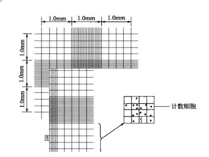
Each large square of the plate is 1 mm in length, 1 mm in width, 0.1 mm in height, 0.1 mm in volume, and can hold 0.1 μL of solution, so the number of cells per 1 mL of solution is 10,000 times the number of cells counted in each large square in the field of view. (Figure below)
Operation method
Cell counting experiments
Principle
The basic principle of the cell counting experiment is that cells can be counted using a cell counting plate. Each large square of the plate is 1 mm in length, 1 mm in width, 0.1 mm in height, 0.1 mm in volume, and can hold 0.1 μL of solution, so the number of cells per 1 mL of solution is 10,000 times the number of cells counted in each large square of the field of view. (Figure below).
Materials and Instruments
Equipment: Move The basic procedure for microscopic size measurement of cells can be divided into the following steps: Caveat 1. Before cell counting, in order to make the result more accurate, the cells should be digested sufficiently so that they can be dispersed as much as possible to prepare single cell suspension. Before adding samples, the cell suspension should be well mixed, and bubbles should be avoided when adding samples. The amount of samples added should be moderate, not overflowing the coverslip, and not too little or with bubbles. 2. When counting under the microscope, when encountering cell clusters consisting of more than 2 cells, the cells should be counted as single cells. If the cell mass accounts for more than 10% of the sample, it means that the digestion is insufficient, and the cell suspension should be re-prepared and counted. For more product details, please visit Aladdin Scientific website.
① Coverslip
② Pipette
② Straw ③ Pipette with a sharp tip
④ Glue cap
⑤ Centrifuge tube
⑥ Alcohol cotton ball
⑦ Alcohol lamp
⑧ Cell counting plate
⑨ Inverted microscope
⑩ Ultra-clean bench
⑪ CO
2
Incubator
Reagents: ① D-Hanks liquid
D-Hanks liquid
② 0.25% Trypsin solution
③ DMEM medium
A. Clean the counting plate and coverslip with 75% ethanol cotton balls, gently dry them with absorbent paper, observe the large squares in the corners of the counting plate with a 10x objective lens, and after adjusting the focal length so that the field of view is clear, cover the coverslip with a coverslip in the middle of the two grooves of the counting plate.
B. Digest the monolayers of fibroblasts that have been passaged for 7 d with 0.25% trypsin solution to produce a single-cell suspension. Make a single-cell suspension.
C. Blow gently on the cell suspension with a sharp-nosed pipette and add a small amount of the suspension to the edge of one side of the coverslip on the counting plate.
D. Under a microscope, observe the number of cells in the large squares in the corners of the counting plate with a 10x objective lens, and count the cells pressing against the center line, only those on the left side and the top, and not those on the right side and the bottom.


 危险品化学品经营许可证(带存储)