
Coumarin Core and Simple Coumarins: Substitution Patterns, Electronic Effects, and Physicochemical Behavior
Coumarin Core and Simple Coumarins: Substitution Patterns, Electronic Effects, and Physicochemical Behavior
1 Coumarin Core and Simple Coumarins
1.1 Benzo-α-pyrone Core and Atom Numbering
The molecular formula of parent coumarin is C₉H₆O₂, and its relative molecular mass is 146.14. According to the 2H-chromen-2-one numbering system, the endocyclic oxygen atom is at position 1, the lactone carbonyl carbon is at position 2, and C3 and C4 form a double bond; the hydrogen-bearing, substitutable carbons on the benzene ring are C5–C8, and the two ring-junction carbons are C4a and C8a. The coumarin core consists of a benzene ring fused to an α-pyrone ring. Within the α-pyrone ring, C2=O and C3=C4 constitute an α,β-unsaturated carbonyl moiety, while the endocyclic oxygen atom participates in the lactone structure.
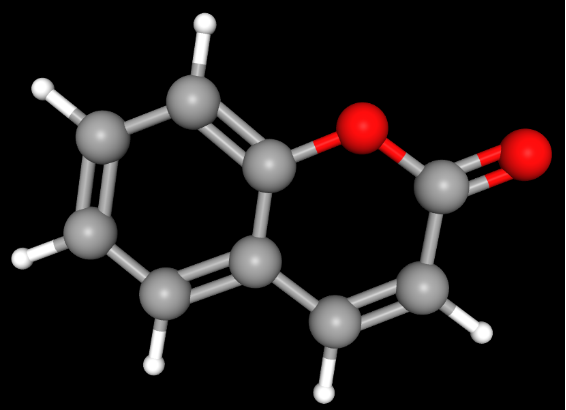
In this structure, C2=O and C3=C4 constitute an α,β-unsaturated carbonyl moiety and, together with the fused benzene ring, form an extended π system. The carbonyl group exerts a pronounced electron-withdrawing effect, whereas electron-donating or electron-withdrawing substituents on the benzene ring can redistribute electron density through the conjugated system. The spectral properties and reactivity of coumarin are jointly determined by the entire benzopyrone system.[2]
1.2 Structural Scope of Simple Coumarins
The scope of “simple coumarins” is not entirely consistent across the natural-products literature. A common approach is to classify hydroxy and methoxy derivatives that have not undergone additional ring fusion, together with their glycosides, as simple coumarins; some classification systems also include noncyclized alkyl or prenyl derivatives.[1] Accordingly, whether a structure remains a simple coumarin can be assessed by determining whether the coumarin core remains intact and whether an additional fused heterocycle has formed.
Representative Structure | Typical Compounds | Principal Structural Variable | Principal Physicochemical Effect |
Unsubstituted core | Coumarin | Reference structure | Provides the conjugated benzo-α-pyrone system |
Monohydroxy | Umbelliferone (7-hydroxycoumarin) | Position of the phenolic hydroxyl group | Alters ionization, hydrogen bonding, and excited-state electron distribution |
Vicinal dihydroxy | Esculetin (6,7-dihydroxycoumarin), daphnetin (7,8-dihydroxycoumarin) | Adjacent phenolic hydroxyl groups | Alters polarity, redox properties, and metal-coordination ability |
Hydroxy–methoxy | Scopoletin (6-methoxy-7-hydroxycoumarin), isoscopoletin (7-methoxy-6-hydroxycoumarin) | Interchange of OH/OCH₃ positions | Alters resonance electron donation and acid–base response |
Methoxy | 7-Methoxycoumarin, 6,7-dimethoxycoumarin | O-Methylation of a phenolic hydroxyl group | Eliminates phenolic hydroxyl ionization and hydrogen-bond donation at that site |
C-Prenyl | Osthenol, osthole, and related compounds | Hydrophobic carbon chain directly attached to the core | Increases hydrophobic surface area and conformational freedom |
O-Linked long-chain alkenyl | Auraptene | Phenolic oxygen forms an ether linked to a geranyl group | Both masks the phenolic hydroxyl group and substantially increases hydrophobic surface area |
O-Glycoside | Skimmin, scopolin, esculin | Glycosylation of a phenolic hydroxyl group | Increases polarity and hydrogen-bonding capacity while masking the original phenolic hydroxyl group |
In some classification systems, 4-hydroxycoumarin is also considered a simple coumarin. However, its hydroxyl group is located at C4 of the α-pyrone ring and can directly participate in tautomerism and the distinctive reactivity of the C2 carbonyl–C3–C4 oxygen-containing system; its behavior therefore cannot be described by the substitution patterns of ordinary phenolic hydroxyl groups on the benzene ring. Accordingly, the acid–base equilibria, tautomerism, and reactivity of 4-hydroxycoumarin should be discussed separately from those of simple coumarins bearing phenolic hydroxyl groups at C5–C8 of the benzene ring.[1]
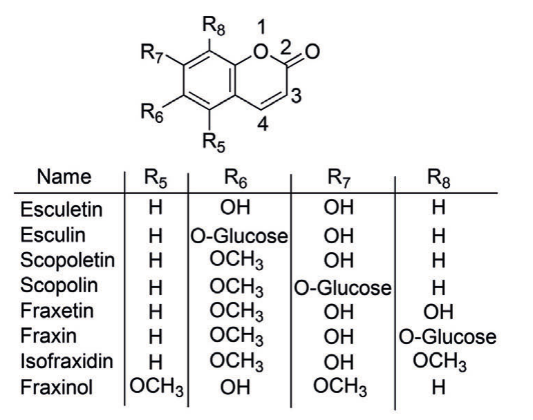
Substitution Positions of the Simple Coumarin Core and Representative Hydroxy, Methoxy, and Glycoside Derivatives
In the figure, R₅–R₈ represent the substituents at C5–C8 of the coumarin core, and the substitution patterns of esculetin, esculin, scopoletin, scopolin, fraxetin, fraxin, isofraxidin, and fraxinol are shown.[8] All of these compounds retain the benzo-α-pyrone core; their structural differences arise primarily from the number and attachment positions of hydroxy, methoxy, and O-glycosyl groups at C5–C8.
Esculin, scopolin, and fraxin are the 6-O-glucoside of esculetin, the 7-O-glucoside of scopoletin, and the 8-O-glucoside of fraxetin, respectively. Isofraxidin and fraxinol each contain one hydroxy group and two methoxy groups, but at different positions, allowing the effects of changes in oxygen-containing substituent positions on the electronic environment and intermolecular interactions of coumarins to be compared.
O-Glycosylation removes the proton-dissociation behavior of the free phenolic hydroxyl group at the glycosylated position, while introducing multiple hydroxyl groups through the glucose residue and increasing the hydrophilic surface area. The overall physicochemical properties of a glycoside also depend on the free phenolic hydroxyl groups that remain unglycosylated and on the original substitution pattern of the aglycone.
Figure source: Cropped from Sisó-Terraza P, Luis-Villarroya A, Fourcroy P, et al. Frontiers in Plant Science, 2016, 7:1711, Figure 1A.[8] Used under the Creative Commons Attribution (CC BY) license.
2 Substitution Position and the π-Electron Structure of Coumarins
2.1 Resonance and Inductive Effects
The oxygen atoms in hydroxy and methoxy groups exert two electronic effects in opposing directions. On the one hand, the electronegativity of oxygen produces an electron-withdrawing inductive effect; on the other hand, the oxygen lone pairs can conjugate with the aromatic π system and exert a resonance electron-donating effect. At positions that can couple effectively with the conjugated benzopyrone system, resonance can markedly alter the electron density of the core.
Alkyl groups do not possess the lone-pair conjugation of oxygen-containing substituents, but they can weakly donate electron density to an adjacent π system through inductive effects and hyperconjugation. Longer hydrocarbon chains, such as prenyl and geranyl groups, generally exert their more pronounced effects not by strongly altering the electronic structure of the core, but by increasing nonpolar surface area and conformational freedom.
At least two types of structural effects should be distinguished in simple coumarins:
Electronic effects primarily determine the energy levels and absorption and emission properties of the conjugated system; intermolecular-interaction effects primarily determine hydrogen bonding, solvation, partitioning, and chromatographic retention. The two types of effects often occur simultaneously, and their relative contributions depend on the substituent.
2.2 Positional Effects and Electronic Coupling
When the same substituent is located at C5, C6, C7, or C8, its resonance-transmission pathway to the C2 carbonyl group and C3=C4 double bond is not equivalent. This is why positional isomers may exhibit different spectral and acid–base properties even when they have the same molecular formula and the same numbers of hydroxy and methoxy groups.
Substituent effects are particularly evident in fluorescence properties. Unsubstituted coumarin itself exhibits weak fluorescence, whereas introducing an electron-donating group at an appropriate position or establishing a more pronounced donor–acceptor electronic structure can markedly enhance or shift absorption and fluorescence.[2] In coumarin derivatives with substantial donor–acceptor differentiation, these changes may involve intramolecular charge transfer (ICT). However, the photophysical processes of natural simple coumarins are also influenced by protonation state, solvent hydrogen bonding, and the local substitution pattern.[2]
3 Hydroxy Substitution and Ionization, Hydrogen Bonding, and Excited-State Properties
3.1 Monohydroxy Substitution and Phenolate Formation
The hydroxy group is one of the substituents that markedly affect the electronic structure of simple coumarins. A neutral phenolic hydroxyl group can participate in intermolecular hydrogen bonding; when the solution pH rises into the range of the relevant acid–base equilibrium, the phenolic hydroxyl group loses a proton to form a phenolate anion:
Coumarin–OH ⇌ Coumarin–O⁻ + H⁺
Conversion from the neutral phenol to the phenolate markedly enhances the resonance electron-donating ability of the oxygen atom; the molecule also acquires a negative charge and becomes more strongly hydrated. Consequently, the same hydroxycoumarin may exhibit different UV absorption, fluorescence, and apparent solubility behavior at different pH values.
Umbelliferone (7-hydroxycoumarin) is a representative example of this effect. Its 7-OH group is conjugated with the benzopyrone system, and its excited-state acidity is substantially greater than its ground-state acidity. Time-resolved spectroscopic studies have shown that umbelliferone can undergo rapid excited-state proton transfer (ESPT) in water, with a reported excited-state pKₐ* of approximately 0.4 and a proton-transfer rate of approximately 2 × 10¹⁰ s⁻¹.[3] This finding indicates that the 7-hydroxy group not only increases molecular polarity but also directly participates in charge redistribution and proton transfer after photoexcitation.
3.2 Vicinal Dihydroxy Groups and Catechol-Type Structures
Esculetin (6,7-dihydroxycoumarin) and daphnetin (7,8-dihydroxycoumarin) both contain two adjacent phenolic hydroxyl groups. Compared with monohydroxycoumarins, these structures provide additional hydrogen-bond donors, acceptors, and ionizable sites and form a local catechol-type moiety.
Under suitable pH and metal-ion conditions, adjacent phenolic hydroxyl groups can provide an O,O-bidentate coordination site. Metal coordination can be coupled with phenolic hydroxyl deprotonation and redox processes. In the presence of metal ions, coordination, redox processes, and protonation states may be interdependent; under alkaline conditions, the oxidative stability of catechol-type coumarins may also decrease. The degradation rates of esculetin, scopoletin, and fraxetin increase with increasing pH; under aerobic conditions, their oxidation is irreversible. Changes in pH can induce both acid–base speciation and changes in chemical stability, so these two processes must be distinguished when interpreting changes in absorption or fluorescence signals.[5]
Esculetin also exhibits pronounced fluorescent photoacid behavior. Its first ground-state pKₐ has been measured at approximately 7.3, while its corresponding excited-state acidity is substantially greater; the fluorescence lifetimes of its neutral and anionic forms also occur on different timescales.[4] This demonstrates that adding a second hydroxyl group to the coumarin core does more than simply “increase polarity”; it can further alter proton transfer, excited-state dynamics, and interactions with metals or solvents.
3.3 Nonlinear Relationship Between Hydroxy-Group Number and Water Solubility
Adding hydroxy groups generally increases hydrogen-bonding capacity and hydrophilic surface area, but actual water solubility is jointly determined by ionization state, lattice energy, molecular packing, and temperature.
In a study that directly compared umbelliferone and its derivatives under identical measurement conditions, the water solubility of the 6,7-dihydroxylated product esculetin was approximately 1.28 times that of umbelliferone.[6] This result is consistent with the expected increase in hydration capacity upon addition of a hydroxy group, but the increase was substantially smaller than that caused by glycosylation in the same study, further demonstrating competition between solid-state structure and solvation.
4 Methoxylation and Masking of Phenolic Hydroxyl Groups
4.1 O-Methylation Simultaneously Alters Electronic Effects and Intermolecular Interactions
O-Methylation of a phenolic hydroxyl group produces an aromatic methyl ether. The oxygen atom of the methoxy group can still donate electron density to the aromatic conjugated system through its lone pairs; therefore, O-methylation is not equivalent to “removing the electron-donating effect.” The more pronounced changes are as follows:
The acidic proton of the hydroxyl group is replaced by a methyl group, so the site no longer undergoes a phenol–phenolate acid–base equilibrium and no longer serves as a hydrogen-bond donor; a nonpolar methyl group is also introduced.
Methylation of the 7-OH group of umbelliferone produces herniarin (7-methoxycoumarin), providing a comparable structural pair. Experimental studies have shown that, under identical water-solubility measurement conditions, the water solubility of herniarin decreases to approximately 1/1.89 that of umbelliferone.[6] This difference cannot be attributed solely to the increase in molecular weight; it is mainly associated with masking of the 7-OH group, reduced hydrogen-bonding and ionization capacity, and the hydrophobic contribution of the methyl group.
4.2 Interchange of Hydroxy and Methoxy Positions
Scopoletin is 6-methoxy-7-hydroxycoumarin, whereas isoscopoletin is 7-methoxy-6-hydroxycoumarin. The two compounds have the same molecular formula and the same numbers of functional groups, but the positions of their hydroxy and methoxy groups are interchanged.
This positional change simultaneously alters two factors: the coupling pathway between the free phenolic hydroxyl group and the conjugated coumarin system, and the position from which the methoxy group donates electron density by resonance.
In 6,7-dimethoxycoumarin (scoparone), both phenolic hydroxyl-type sites are converted into methyl ethers. Compared with the 6,7-dihydroxy substitution pattern of esculetin, its C6 and C7 positions no longer undergo phenolic hydroxyl dissociation under ordinary acid–base conditions, and the number of hydrogen-bond donors is reduced. Its pH-dependent spectral response, hydrophilicity, and intermolecular interactions consequently change.
5 Effects of Alkyl and Prenyl Substitution on Hydrophobicity and Conformation
5.1 Hyperconjugative and Hydrophobic Contributions of Small Alkyl Groups
Small alkyl groups such as methyl can donate some electron density to a conjugated system through inductive effects and hyperconjugation, but their direct resonance-modulating ability is weaker than that of phenolate, hydroxy, and methoxy groups. Another effect of small-alkyl substitution is an increase in nonpolar surface area and a change in local spatial structure.
When a methyl group is located directly in the conjugated α-pyrone region, such as at C3 or C4, its perturbation of the local electronic structure and excited state may be greater than that of ordinary aryl-ring alkyl substitution farther from this region. When it is located at different positions on the benzene ring, the electron-transmission pathway and spatial orientation also differ.
5.2 C-Linked Prenyl Groups and Hydrophobic Surface Area
Osthenol is 7-hydroxy-8-prenylcoumarin, osthole is 7-methoxy-8-prenylcoumarin, and suberosin is 7-methoxy-6-prenylcoumarin. Osthole and suberosin share the same C7 methoxy group, while their prenyl groups are located at C8 and C6, respectively, allowing the effects of side-chain position on molecular shape and intermolecular interactions to be examined.
C-Prenylation does not itself mask a phenolic hydroxyl group; if another free phenolic hydroxyl group is present in the molecule, such as the C7-OH group in osthenol, that site can still participate in hydrogen bonding and the corresponding acid–base equilibrium. The prenyl group itself increases the nonpolar hydrocarbon surface and introduces a rotatable single bond, thereby increasing hydrophobic interactions and side-chain conformational freedom. Changing the attachment position from C6 to C8 also changes the side-chain attachment site, local spatial environment, and overall molecular shape, which may in turn affect molecular packing, solvation, and interactions with hydrophobic environments.
5.3 Differences Between O-Alkylation and C-Alkylation
O-Prenylation or O-geranylation and C-prenylation do not produce the same type of structural effect. In the former, the phenolic oxygen forms an ether bond, simultaneously producing two changes: masking of the phenolic hydroxyl group + addition of a nonpolar hydrocarbon chain.
Auraptene is 7-geranyloxycoumarin. Its long geranyl group substantially increases the nonpolar surface area, while position 7 no longer bears an ionizable free phenolic hydroxyl group. Consequently, these O-alkyl ethers generally exhibit more pronounced hydrophobic characteristics than the corresponding hydroxycoumarins.
If a C-prenylcoumarin retains a 7-OH group, phenolic hydroxyl ionization may still occur; after O-prenylation or O-geranylation, the acid–base response at that site is masked. Thus, even when the two types of structures contain hydrocarbon chains of similar length, their pH-dependent properties are not the same.
6 Effects of Glycosylation on Hydration, Ionization, and Spectral Response
6.1 Formation of O-Glycosides Alters Local Acid–Base Sites and Overall Hydration
When a phenolic hydroxyl group of a simple coumarin forms an O-glycosidic bond, the O–H bond of the original phenolic hydroxyl group disappears, and the corresponding phenolic hydroxyl proton-dissociation behavior is no longer retained at that position. At the same time, the sugar residue introduces multiple aliphatic alcohol hydroxyl groups, increasing the number of hydrogen-bonding sites, hydrophilic surface area, and molecular weight.
Glycosylation therefore produces two simultaneous structural effects:
Disappearance of the free phenolic hydroxyl group at the glycosylated position → loss of phenolic hydroxyl ionization and hydrogen-bond-donor character at that site;
Introduction of multiple hydroxyl groups by the sugar residue → increased hydration and number of hydrogen-bonding sites.
When only some of the phenolic hydroxyl groups of a polyhydroxycoumarin are glycosylated, the remaining free phenolic hydroxyl groups can still undergo the corresponding acid–base equilibria. The overall solvation properties of the glycoside are jointly determined by the sugar residue, the remaining free hydroxyl groups, the substitution pattern of the aglycone, and the solvent conditions.
6.2 Solubility Difference Between Skimmin and Umbelliferone
Chu et al. compared the solubilities of umbelliferone and its derivatives in phosphate-buffered saline at pH 7.6. Under identical measurement conditions, the measured solubility of skimmin was approximately 3.98 times that of umbelliferone, while that of esculetin was approximately 1.28 times that of umbelliferone.[6]
Skimmin is the glucoside formed at the C7 phenolic hydroxyl group of umbelliferone. The multiple hydroxyl groups introduced by the glucose residue enhance the molecule’s ability to form hydrogen bonds and hydration interactions with the aqueous phase, consistent with the increased solubility observed in this experimental system.[6]
Solubility is also affected by crystal form, temperature, solvent composition, pH, and ionic strength. The 3.98-fold difference corresponds to the specific buffer system and measurement conditions used by Chu et al. and does not indicate that skimmin maintains the same proportional difference in solubility under all aqueous conditions.
6.3 pH-Dependent Spectral Responses of Scopoletin and Scopolin
Scopoletin retains a free C7 phenolic hydroxyl group, and its protonation state changes with pH. Agati et al. found that, as the pH of phosphate buffer increased from 5.4 to 8.5, the neutral-state excitation band of scopoletin at approximately 340 nm gradually weakened, while the longer-wavelength excitation band associated with the anion at approximately 385 nm increased. Its emission band was located mainly at approximately 460 nm, with little change in peak position over this pH range.[7]
The C7 phenolic oxygen of scopolin forms an O-glucosidic bond and therefore no longer undergoes the same proton-dissociation process as the C7-OH group of scopoletin. Its fluorescence excitation spectrum has a main peak at approximately 340 nm and another absorption band at approximately 290 nm; over the pH range of 5.4–8.5, the excitation spectrum is largely unaffected by changes in pH, and the emission band is located mainly at approximately 420 nm.[7]
The spectral differences between the two compounds correspond to the different chemical states of their C7 oxygen-containing groups:
The C7-OH group of scopoletin retains a dissociable proton
→ As pH increases, the relative proportions of the neutral and anionic forms change
→ The excitation spectrum exhibits a pronounced acid–base response;
The C7-OH group of scopolin forms an O-glycosidic bond
→ The proton-dissociation site of the C7 phenolic hydroxyl group is eliminated
→ The corresponding change in the long-wavelength anionic excitation band is no longer observed over the same pH range.
7 Correspondence Between Substitution Patterns and Physicochemical Properties
7.1 Structural Variables, Electronic Effects, and Property Changes
Structural Change | Change in Electronic Structure or Intermolecular Interactions | Principal Physicochemical Outcome | Representative Compounds | Typical Observable Response |
Introduction of a single OH group on the benzene ring | Resonance electron donation; increased hydrogen bonding; phenolate formation possible | Increased polarity and pH responsiveness | Umbelliferone | UV/fluorescence changes with protonation state |
Introduction of two adjacent OH groups | Additional ionizable sites; formation of a catechol-type structure; coordination possible | Increased hydrophilicity, accompanied by enhanced oxidation and coordination behavior | Esculetin, daphnetin | pH and metal ions can markedly affect spectra and stability |
OH → OCH₃ | Retains resonance electron donation by oxygen; loses the phenolic proton and hydrogen-bond-donor ability | Weaker acid–base response and generally greater hydrophobicity | Umbelliferone → herniarin | Decreased water solubility; altered pH-dependent spectral response |
Interchange of OH/OCH₃ positions | Alters the electronic-coupling pathways between substituents and the core | The same molecular formula can still give different spectral and partitioning properties | Scopoletin, isoscopoletin | Positional isomers exhibit different electronic responses |
C-Alkyl/C-prenyl substitution | Increases nonpolar surface area and conformational freedom | Enhanced hydrophobic interactions | Osthenol, osthole, suberosin | Lower affinity for the aqueous phase; stronger tendency toward reversed-phase retention |
O-Linked long-chain alkylation | Masks the phenolic hydroxyl group and adds a long hydrocarbon chain | Weaker acid–base response and further increased hydrophobicity | Auraptene | Partitioning and chromatographic retention are strongly affected by the hydrophobic chain |
O-Glycosylation | Masks the original phenolic hydroxyl group while adding multiple aliphatic alcohol hydroxyl groups | Increased hydrophilicity and hydrogen-bonding capacity; the glycosylated site no longer undergoes phenolic hydroxyl dissociation | Skimmin, scopolin, esculin | Water solubility often increases; specific pH-dependent spectral responses of the aglycone are attenuated |
7.2 Conditions Governing the Applicability of Structural Effects
Several conditional factors cannot be ignored when interpreting structure–property relationships in simple coumarins.
① The ionized fraction of a free phenolic hydroxyl group is jointly determined by the solution pH, the corresponding pKₐ, and the medium conditions. Neutral phenols and phenolate anions differ greatly in charge, solvation, and electron-donating ability; consequently, the solubility and spectral properties of the same compound may change markedly with pH.[3–5,7]
② The number of hydroxy or glycosyl groups alone does not determine water solubility. Hydrogen bonding and hydration in the liquid phase favor dissolution, whereas intermolecular hydrogen bonding, π stacking, and lattice stability in the crystal may have the opposite effect. Experimental solubilities must therefore be compared under clearly defined temperature, pH, and medium conditions.[6]
③ Spectral changes under strongly alkaline conditions do not necessarily arise solely from phenolic hydroxyl ionization. The coumarin core contains a lactone structure, and strong base or prolonged alkaline treatment may promote lactone-ring opening; coumarins containing adjacent dihydroxy groups may also undergo oxidation. Therefore, when changes in absorption or fluorescence are observed at elevated pH, they should be evaluated in light of treatment time, reversibility, and sample stability, rather than being attributed entirely to the acid–base equilibrium between neutral phenols and phenolate anions.[4,5]
④ Substitution position is as important as substituent identity. Interchanging hydroxy and methoxy positions or changing the position of a C-prenyl group can alter the π-electron distribution, spatial shape, and intermolecular interactions without changing the molecular formula. Therefore, when comparing the structures of simple coumarins, the “functional-group identity–substitution position–protonation state” must be defined simultaneously.
8 Representative Chemicals for Research on the Coumarin Core and Simple Coumarins: Classification and Applications in Structure–Property Studies
Table 1. Coumarin Core and Monohydroxy-, Monomethoxy-, and Methyl-Substituted Simple Coumarins
Category | CAS Number | Aladdin Catalog No. | Name | Specification or Purity | Product Features and Applications |
Coumarin core | 91-64-5 | Coumarin | AR, ≥98% | Unsubstituted coumarin core that can serve as a baseline control in studies of hydroxylation, methoxylation, methylation, and side-chain substitution, enabling comparison of the conjugated electronic structure, polarity, and partitioning properties before and after substitution. | |
Methyl-substituted coumarin | 92-48-8 | 6-Methylcoumarin | ≥99% | A C6-methyl-substituted structure that can be compared with coumarin and 4-methylcoumarin to examine how methyl position affects the electronic environment of the aromatic ring, hydrophobic surface area, and molecular packing. | |
Monohydroxycoumarin | 93-35-6 | 7-Hydroxycoumarin | ≥99% | A representative C7 phenolic hydroxyl compound suitable for studies of hydroxyl ionization, hydrogen bonding, solvation, and the effects of substitution position on the electronic properties of coumarins; it also serves as an aglycone control for structural comparisons involving glycosylation and O-methylation. | |
Monohydroxycoumarin | 2442-31-1 | 8-Hydroxycoumarin | ≥99% | A C8-hydroxy positional isomer that can be compared with 6- and 7-hydroxycoumarin to investigate how hydroxy-group position affects intramolecular interactions, acid–base behavior, and conjugated electron distribution. | |
Methyl–hydroxycoumarin | 90-33-5 | 4-Methylumbelliferone (4-MU) | ≥98% | Contains both a C4 methyl group and a C7 hydroxy group and can be used to examine electronic structure, acid–base equilibria, and fluorescence responses under the combined effects of alkyl and phenolic hydroxyl substitution. | |
Monohydroxycoumarin | 6093-68-1 | 6-Hydroxycoumarin | ≥98% | A C6-hydroxy positional isomer that forms a positional-isomer series with 7- and 8-hydroxycoumarin for investigating how hydroxy-group attachment position affects electronic effects, hydrogen bonding, and physicochemical properties. | |
Monomethoxycoumarin | 531-59-9 | 7-Methoxycoumarin | ≥98% | A representative structure obtained by O-methylation of a C7 hydroxyl group; it can be compared with 7-hydroxycoumarin to examine changes in hydrogen-bond-donor ability, acid–base sites, and hydrophobicity after the phenolic hydroxyl group is methyl-masked. | |
Methyl-substituted coumarin | 607-71-6 | 4-Methylcoumarin | ≥97% | The C4 methyl group is directly adjacent to the conjugated α-pyrone moiety; comparison with 6-methylcoumarin can reveal how placing a methyl group in different electronic regions affects core properties and intermolecular interactions. |
Table 2. Polyhydroxy, Methoxy, and Hydroxy–Methoxy Simple Coumarins
Category | CAS Number | Aladdin Catalog No. | Name | Specification or Purity | Product Features and Applications |
Dimethoxycoumarin | 120-08-1 | Scoparone | Analytical standard, ≥98% | A 6,7-dimethoxy-substituted structure that can be compared with 6,7-dihydroxycoumarin to examine changes in hydrogen-bonding capacity, acid–base properties, and hydrophobicity after complete O-methylation of adjacent hydroxyl groups. | |
Hydroxy–dimethoxycoumarin | 486-21-5 | Isofraxidin | Analytical standard | Contains a free C7 hydroxy group with methoxy groups at C6 and C8 and can be used to investigate electronic effects, hydrogen bonding, and combined substitution effects when a phenolic hydroxyl group is flanked by two methoxy groups. | |
Vicinal dihydroxycoumarin | 305-01-1 | 6,7-Dihydroxycoumarin | Moligand™, ≥98% | Contains a vicinal 6,7-dihydroxy moiety and can be used to study catechol-type hydrogen bonding, metal-ion coordination, oxidation behavior, and the effects of two hydroxyl groups on coumarin polarity and electronic structure. | |
Dimethoxycoumarin | 487-06-9 | 5,7-Dimethoxycoumarin | ≥98% | Contains separated methoxy groups at C5 and C7 and can be compared as a positional isomer with 6,7-dimethoxycoumarin to study how methoxy-group position affects electron distribution, hydrophobicity, and chromatographic behavior. | |
Hydroxy–methoxycoumarin | 776-86-3 | Isoscopoletin | ≥98% | Contains a C6 hydroxy group and a C7 methoxy group and can be compared with scopoletin as a hydroxy/methoxy positional pair to study electronic and hydrogen-bonding differences arising from changes in the positions of adjacent oxygen-containing substituents. | |
Vicinal dihydroxy–methoxycoumarin | 574-84-5 | Fraxetin | ≥98% | Contains adjacent C7 and C8 hydroxy groups together with a C6 methoxy group and can be used to compare the combined physicochemical effects of vicinal-dihydroxy coordination, oxidation, and hydrogen bonding with the electronic effect of the methoxy group. | |
Hydroxy–methoxycoumarin | 92-61-5 | Scopoletin | ≥98% | Contains a C7 hydroxy group and a C6 methoxy group and can form a substitution-position and O-methylation comparison series with 7-hydroxycoumarin, 7-methoxycoumarin, and the product with CAS No. 776-86-3. | |
Vicinal dihydroxycoumarin | 486-35-1 | Daphnetin | ≥90% (HPLC) | A 7,8-vicinal-dihydroxy positional isomer that can be compared with 6,7-dihydroxycoumarin to investigate how vicinal-dihydroxy position affects intramolecular interactions, metal coordination, oxidation tendency, and polarity. |
Table 3. Prenyl- and Long-Chain Alkenyloxy-Substituted Simple Coumarins
Category | CAS Number | Aladdin Catalog No. | Name | Specification or Purity | Product Features and Applications |
Methoxy–prenylcoumarin | 484-12-8 | Osthole | Analytical standard, Moligand™, ≥99.5% | Contains a C7 methoxy group and a C8 prenyl group and can be used to study the effects of introducing a hydrophobic side chain and O-methylating a phenolic hydroxyl group on partitioning, membrane interactions, and chromatographic retention. | |
Methoxy–prenylcoumarin | 581-31-7 | Suberosin | Moligand™, ≥99% | Contains a C7 methoxy group and a C6 prenyl group and can be compared with osthole as a pair of prenyl positional isomers to study how side-chain position affects molecular shape, hydrophobic surface area, and retention behavior. | |
Geranyloxycoumarin | 495-02-3 | Auraptene | ≥98% (HPLC) | Contains a long geranyloxy chain at C7 and can be used to study how a flexible long chain affects hydrophobicity, conformational freedom, solvent partitioning, and chromatographic retention after O-alkylation masks the phenolic hydroxyl group. | |
Hydroxy–prenylcoumarin | 484-14-0 | Osthenol | ≥98% | Contains a free C7 hydroxy group and a C8 prenyl group and can be compared with osthole to investigate acid–base, hydrogen-bonding, and hydrophobic properties before and after methylation of the C7 hydroxy group while retaining the same C8 prenyl structure. |
Table 4. Simple Coumarin Glycosides
Category | CAS Number | Aladdin Catalog No. | Name | Specification or Purity | Product Features and Applications |
Hydroxy–methoxycoumarin glycoside | 531-44-2 | Scopolin | Moligand™, ≥98% | A glucoside of scopoletin that can be compared with scopoletin to examine changes in acid–base sites, hydration capacity, partitioning properties, and fluorescence response after glycosylation of the C7 phenolic hydroxyl group. | |
Dihydroxycoumarin glycoside | 486-55-5 | Daphnin | Moligand™, ≥98% | A monoglucoside of daphnetin that retains one free phenolic hydroxyl group while introducing a glycosyl group; it can be compared with daphnetin to examine the effects of selective glycosylation on hydrogen bonding, hydrophilicity, and partitioning behavior. | |
Hydroxy–methoxycoumarin glycoside | 524-30-1 | Fraxin | Moligand™, ≥98% | The 8-O-glucoside of fraxetin, retaining a free C7 hydroxy group and a C6 methoxy group; it can be used to study the effects of glycosylating a single hydroxyl group in a vicinal oxygen-containing system on hydration, acid–base properties, and partitioning. | |
Monohydroxycoumarin glycoside | 93-39-0 | Skimmin | Moligand™, ≥98% | A glucoside of 7-hydroxycoumarin that can be directly compared with 7-hydroxycoumarin to examine changes in hydrogen-bonding sites, ionization behavior, hydrophilicity, and chromatographic retention after glycoside formation at the phenolic hydroxyl group. | |
Dihydroxycoumarin glycoside | 531-75-9 | Esculin hydrate | ≥98% | A hydrated form of the esculetin glucoside that can be compared with 6,7-dihydroxycoumarin to examine hydration, intermolecular hydrogen bonding, and partitioning after monoglycosylation of a vicinal-dihydroxy system and to serve in aglycone/glycoside comparison experiments. |
Note: The products listed above are representative Aladdin products relevant to scientific research. Specific uses should be determined according to the product specification, batch certificate of analysis (COA), and the intended reaction or evaluation system. Additional information on product specifications, grades, and COAs can be retrieved from the Aladdin website using the “product name/CAS number/catalog number.”
References
[1] Zhu Z, Chen R, Zhang L. Simple phenylpropanoids: recent advances in biological activities, biosynthetic pathways, and microbial production. Natural Product Reports, 2024, 41(1): 6–24. DOI: 10.1039/D3NP00012E.
[2] Donovalová J, Cigáň M, Stankovičová H, Gašpar J, Danko M, Gáplovský A, Hrdlovič P. Spectral Properties of Substituted Coumarins in Solution and Polymer Matrices. Molecules, 2012, 17(3): 3259–3276. DOI: 10.3390/molecules17033259.
[3] Simkovitch R, Huppert D. Photoprotolytic Processes of Umbelliferone and Proposed Function in Resistance to Fungal Infection. The Journal of Physical Chemistry B, 2015, 119(46): 14683–14696. DOI: 10.1021/acs.jpcb.5b08439.
[4] Knoor L H, Du Laney G R, Jonker I B, Hoogewerf L P, Tu Y, Pham H T, Yoo J, Muyskens M A. Aesculetin Exhibits Strong Fluorescent Photoacid Character. Journal of Fluorescence, 2022, 32(1): 307–318. DOI: 10.1007/s10895-021-02842-w.
[5] Kang K, Schenkeveld W D C, Weber G, Kraemer S M. Stability of Coumarins and Determination of the Net Iron Oxidation State of Iron–Coumarin Complexes: Implications for Examining Plant Iron Acquisition Mechanisms. ACS Earth and Space Chemistry, 2023, 7(12): 2339–2352. DOI: 10.1021/acsearthspacechem.3c00199.
[6] Chu L L, Pandey R P, Lim H N, Jung H J, Thuan N H, Kim T S, Sohng J K. Synthesis of umbelliferone derivatives in Escherichia coli and their biological activities. Journal of Biological Engineering, 2017, 11: 15. DOI: 10.1186/s13036-017-0056-5.
[7] Agati G, Brunetti C, Tuccio L, Degano I, Tegli S. Retrieving the in vivo Scopoletin Fluorescence Excitation Band Allows the Non-invasive Investigation of the Plant–Pathogen Early Events in Tobacco Leaves. Frontiers in Microbiology, 2022, 13: 889878. DOI: 10.3389/fmicb.2022.889878.
[8] Sisó-Terraza P, Luis-Villarroya A, Fourcroy P, Briat J-F, Abadía A, Gaymard F, Abadía J, Álvarez-Fernández A. Accumulation and Secretion of Coumarinolignans and other Coumarins in Arabidopsis thaliana Roots in Response to Iron Deficiency at High pH. Frontiers in Plant Science, 2016, 7: 1711. DOI: 10.3389/fpls.2016.01711.
For more related articles, see below:
Will glycosylation affect the biological activity of recombinant protein?


 危险品化学品经营许可证(带存储)