
Experimental Protocol for the Russell Modified Movat Pentachrome Method for Comprehensive Visualization of Atherosclerotic Plaques and Connective Tissue Components
Experimental Protocol for the Russell Modified Movat Pentachrome Method for Comprehensive Visualization of Atherosclerotic Plaques and Connective Tissue Components
1 Overview
1.1 Purpose and Scope of Application
This protocol is used to establish a routine operating procedure for the Russell modified Movat pentachrome staining method. It is suitable for comprehensive color visualization of elastic fibers, collagen fibers, reticular fibers, proteoglycans, fibrinoid material, fibrin, and muscle tissue components in paraffin-embedded tissue sections. It is especially suitable for morphological observation of atherosclerotic plaques and complex connective tissue structures.
1.2 Principle
(1) Russell modified Movat pentachrome staining is a multi-step composite differential staining method. Different staining solutions are applied sequentially, allowing multiple tissue components to show stable and layered color differences.
(2) Weigert hematoxylin is mainly used to stain nuclei and elastic fibers, which usually appear black.
(3) Crocein scarlet-fuchsin is used to display fibrinoid material, fibrin, and some muscle tissue components. Fibrinoid material and fibrin are usually dark red, while cardiac muscle and smooth muscle are usually red.
(4) Alcian blue is used to display proteoglycans in the matrix, staining them blue-green.
(5) Alcoholic saffron is mainly used to stain collagen fibers and reticular fibers, making them appear yellow.
(6) Foam cells usually appear purple in this staining system, which is helpful for identifying different lesion structures in atherosclerosis-related sections.
1.3 Staining Quality Requirements
(1) Nuclei and elastic fibers should appear black with clear contours.
(2) Collagen fibers and reticular fibers should appear yellow with clear distribution.
(3) Proteoglycans should appear blue-green; fibrinoid material and fibrin should appear dark red; cardiac muscle and smooth muscle should appear red; foam cells should appear purple.
(4) The overall section should show rich color layering, a relatively clean background, and intact tissue structure.
2 Materials and Reagents
2.1 Sample Types
(1) Routine paraffin-embedded tissue sections.
(2) Vascular and atherosclerosis-related tissue sections.
(3) Tissue sections requiring comprehensive observation of connective tissue, matrix, and muscle tissue components.
2.2 Main Reagents
(1) Hypo solution.
(2) Alcian blue staining solution.
(3) Alkaline ethanol solution.
(4) Weigert A, Weigert B, and Weigert C.
(5) Crocein scarlet-fuchsin A and crocein scarlet-fuchsin B.
(6) Phosphotungstic acid solution.
(7) Weak acid differentiation solution.
(8) Alcoholic saffron staining solution.
(9) Bouin’s fixative.
(10) Graded ethanol.
(11) Distilled water.
(12) Xylene or deparaffinization clearing solution.
(13) Neutral balsam.
2.3 Consumables and Equipment
(1) Glass slides and coverslips.
(2) Staining jars.
(3) Forceps and absorbent paper.
(4) Paraffin microtome.
(5) Microwave oven.
(6) Microscope.
3 Staining Solution and Sample Preparation
3.1 Sectioning Requirements
(1) Paraffin sections should be prepared routinely, with a recommended thickness of approximately 5 μm.
(2) Sections should be treated for strong adhesion to prevent detachment during long and multi-step staining procedures.
(3) Before staining, sections should be fully deparaffinized and hydrated through graded ethanol.
3.2 Preparation of Weigert Hematoxylin Staining Solution
(1) Before use, mix Weigert A, Weigert B, and Weigert C at a ratio of 3:2:1.
(2) After thorough mixing, the solution is ready as Weigert hematoxylin staining solution.
(3) This staining solution should not be prepared long in advance and should be freshly prepared before use.
3.3 Preparation of Crocein Scarlet-Fuchsin Staining Solution
(1) Before use, mix crocein scarlet-fuchsin A and crocein scarlet-fuchsin B at a ratio of 4:1.
(2) After thorough mixing, the solution is ready as crocein scarlet-fuchsin staining solution.
(3) This staining solution should not be prepared long in advance and should be freshly prepared before use.
3.4 Preheating Requirements for Bouin’s Fixative
(1) Place an appropriate amount of Bouin’s fixative in a microwave oven and heat moderately for 30–60 s.
(2) Use immediately for section treatment after heating.
(3) Temperature should be controlled during treatment to avoid tissue section damage caused by overheating.
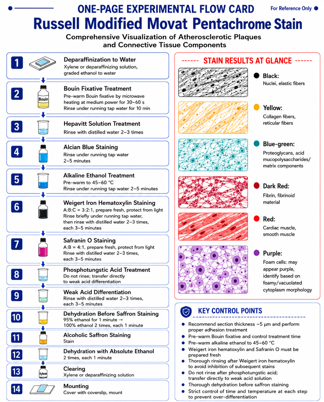
4 Experimental Procedure
4.1 Deparaffinization and Hydration
(1) Routinely deparaffinize paraffin sections with xylene or deparaffinization clearing solution.
(2) Hydrate sections to water through graded ethanol.
4.2 Bouin’s Fixative Treatment
(1) Take preheated Bouin’s fixative.
(2) Immediately place sections into the fixative and treat for 10 min.
(3) Rinse in running water for 10 min after treatment.
4.3 Hypo Solution Treatment
(1) Place sections in hypo solution for 5 min.
(2) Rinse with distilled water 2–3 times after treatment.
4.4 Alcian Blue Staining
(1) Place sections in Alcian blue staining solution and stain for 20 min.
(2) Rinse in running water for 2–5 min after staining.
4.5 Alkaline Ethanol Treatment
(1) Preheat the alkaline ethanol solution to 45–60°C using high power in a microwave oven.
(2) Place sections in the alkaline ethanol solution and treat for 10 min.
(3) Rinse in running water for 2–5 min after treatment.
4.6 Weigert Hematoxylin Staining
(1) Place sections in the freshly prepared Weigert hematoxylin staining solution.
(2) Stain in the dark for 60 min.
(3) After staining, briefly rinse with running water, then rinse with distilled water 2–3 times, 3–5 min each time.
(4) After this step, excess staining solution should be thoroughly removed to avoid affecting subsequent staining steps.
4.7 Crocein Scarlet-Fuchsin Staining
(1) Place sections in the freshly prepared crocein scarlet-fuchsin staining solution.
(2) Stain in the dark for 1 min.
(3) Rinse with distilled water 2–3 times, 3–5 min each time.
4.8 Phosphotungstic Acid and Weak Acid Differentiation Treatment
(1) Place sections in phosphotungstic acid solution for 5 min.
(2) Without intermediate rinsing, directly transfer sections into weak acid differentiation solution and treat for 5 min.
(3) Rinse with distilled water 2–3 times, 3–5 min each time.
4.9 Dehydration Before Saffron Staining
(1) Place sections in 95% ethanol for 1 min.
(2) Then place sections in 100% ethanol twice, 1 min each time.
4.10 Alcoholic Saffron Staining
(1) Place sections in alcoholic saffron staining solution and stain for 5 min.
4.11 Dehydration, Clearing, and Mounting
(1) Dehydrate sections in absolute ethanol twice, 1 min each time.
(2) Transfer sections into xylene or deparaffinization clearing solution for clearing.
(3) Apply coverslips and mount.
(4) After mounting, avoid air bubbles and ensure even mounting.
5 Staining Results
5.1 Black
(1) Nuclei.
(2) Elastic fibers.
5.2 Yellow
(1) Collagen fibers.
(2) Reticular fibers.
5.3 Blue-Green
(1) Proteoglycans.
5.4 Dark Red
(1) Fibrinoid material.
(2) Fibrin.
5.5 Red
(1) Cardiac muscle.
(2) Smooth muscle.
5.6 Purple
(1) Foam cells.
6 Key Points for Result Interpretation
6.1 Interpretation of Elastic Fibers and Nuclei
(1) When elastic fibers and nuclei appear black, their boundaries are usually clear, which is helpful for observing vascular wall structure and tissue layering.
(2) If the black background is too heavy, first check whether washing after Weigert hematoxylin staining was sufficient.
6.2 Interpretation of Collagen Fibers and Reticular Fibers
(1) Yellow collagen fibers and reticular fibers are helpful for observing fibrous stroma and structures such as the fibrous cap of plaques.
(2) If yellow staining is not distinct, phosphotungstic acid treatment, weak acid differentiation, and alcoholic saffron staining should be considered together.
6.3 Interpretation of Proteoglycans
(1) Blue-green proteoglycan staining helps identify matrix-rich regions.
(2) Insufficient Alcian blue staining or overly strong subsequent processing may weaken this color.
6.4 Interpretation of Fibrinoid Material, Fibrin, and Muscle Tissue
(1) Fibrinoid material and fibrin appear dark red, while cardiac muscle and smooth muscle appear red, which helps distinguish them from yellow collagen and blue-green matrix.
(2) If red components are generally weak, first check the crocein scarlet-fuchsin staining and subsequent differentiation steps.
6.5 Interpretation of Foam Cells
(1) Purple foam cells have good auxiliary identification value in atherosclerosis-related sections.
(2) Interpretation should be integrated with their location and surrounding lesion background.
7 Common Problems and Cause Analysis
7.1 Not All Five Colors Are Displayed After Staining
(1) The staining strength of different staining solutions varies.
(2) The distribution of corresponding tissue components in the section itself is limited.
(3) Inconsistent endpoint control in multi-step staining may result in insufficient development of some colors.
7.2 Section Detachment
(1) Adhesion treatment was not performed.
(2) Sections were not firmly attached.
(3) Repeated rinsing and long staining procedures caused mechanical section detachment.
7.3 Unsatisfactory Elastic Fiber Visualization
(1) Differentiation time for elastic fibers was not properly controlled.
(2) Elastic fiber-related differentiation is usually completed within 2–3 min. Excessive treatment may weaken visualization.
7.4 Subsequent Staining Is Inhibited
(1) Running water rinsing after Weigert hematoxylin staining was insufficient.
(2) Residual staining solution can inhibit subsequent steps, resulting in unclear overall color layering.
7.5 Deep Background or Imbalanced Overall Staining
(1) The staining procedure is complex, and the operator’s timing is inconsistent.
(2) Treatment times for different staining solutions are unstable.
(3) Tissue sections are uneven in thickness or incompletely deparaffinized.
8 Application Scope
8.1 Atherosclerosis-Related Research
(1) Used to display multiple tissue components in atherosclerotic plaques.
(2) Suitable for observing fibrous caps, proteoglycans, foam cells, and elastic fiber distribution.
8.2 Comprehensive Observation of Connective Tissue
(1) Used to simultaneously display collagen fibers, reticular fibers, and elastic fibers.
(2) Suitable for multi-component comparative observation in complex connective tissue backgrounds.
8.3 Observation of the Relationship Between Muscle Tissue and Matrix
(1) Used to display the relationship between cardiac or smooth muscle and surrounding stroma or matrix components.
9 Safety and Operating Requirements
9.1 Personal Protection
(1) Wear a laboratory coat and disposable gloves during the experiment.
(2) Bouin’s fixative, ethanol, xylene, and various staining solutions should be handled in a well-ventilated environment.
9.2 Waste Disposal
(1) Waste liquids containing fixatives, ethanol, xylene, and residual staining solutions should be collected separately.
(2) All waste liquids and contaminated consumables should be disposed of according to laboratory chemical waste handling regulations.
10 Reagent Selection for the Russell Modified Movat Pentachrome Staining Method
Cat. No. | Product Name | Grade and Purity | Corresponding Step | Use |
Russell Modified Movat Pentachrome Staining Solution | BioReagent,Biological Stain,for microscopy | Main pentachrome staining system | Directly corresponds to the core staining system of this method; suitable for comprehensive color visualization of atherosclerotic plaques and connective tissue components | |
Weigert's Hematoxylin Staining Kit | BioReagent, Biological Stain, for microscopy | Weigert hematoxylin staining | Corresponds to the staining step for nuclei and elastic fibers | |
Alcian Blue Staining Solution (pH2.5) | BioReagent,for microscopy,Biological Stain | Alcian blue staining | Corresponds to the staining step for proteoglycans/matrix components | |
Alcian Blue Staining Solution (pH 2.5) | BioReagent, Biological Stain, for microscopy | Alcian blue staining | Can be used as a direct alternative for the Alcian blue staining step | |
Alcian Blue Staining Solution (pH 1.0) | BioReagent, Biological Stain, for microscopy | Alcian blue staining | Can be used as an optimization option for Alcian blue staining under acidic conditions | |
Alkaline Ethanol Differentiation Solution | BioReagent,Biological Stain,for microscopy,1× | Alkaline ethanol treatment | Directly corresponds to the alkaline ethanol treatment step in the protocol | |
Neutral Formalin Buffered Solution | 10% | Fixation | Can be used as an option for routine tissue fixation | |
Carnoy's Fluid | BioReagent, ready-to-use | Fixation | Can be used as an alternative fixation system for tissue pretreatment | |
Carnoy Fixative Ⅱ | BioReagent, ready-to-use | Fixation | Can serve as an alternative Carnoy fixation system | |
Water | for biotechnology nuclease-free, sterile | Rinsing/solution preparation | Can be used as a standardized experimental water option | |
Xylene | Premium-Grade Reagents, ≥99%, xylene isomer and ethyl benzene | Deparaffinization/clearing | Corresponds to paraffin section deparaffinization and post-staining clearing steps | |
Xylene | ACS, ≥98.5%, isomers plus ethylbenzene | Deparaffinization/clearing | Suitable for standardized deparaffinization and clearing | |
Xylene | Anhydrous Grade, ≥98%, mixture of isomers | Deparaffinization/clearing | Corresponds to routine clearing treatment | |
Environment Friendly Wax Impregnation Dewaxing Transparent Solution | BioReagent,Suitable for Immunohistochemistry(IHC),for microscopy | Deparaffinization/clearing | Can replace xylene for deparaffinization or clearing steps | |
dewaxing liquid |
| Deparaffinization | Can be used for paraffin section deparaffinization | |
Neutral gum | FMP | Mounting | Corresponds to the neutral gum mounting step |
For more related articles, please see below:
[1] Connective tissue observation experiment
[2] Morphological observation experiments on loose connective tissue


 危险品化学品经营许可证(带存储)