
Oxalate Metabolism Research Methods: Analysis of Oxalate Production, Degradation, Transport, and Kidney Stone Mechanisms
Oxalate Metabolism Research Methods: Analysis of Oxalate Production, Degradation, Transport, and Kidney Stone Mechanisms
Oxalate metabolism research is mainly used to analyze mechanisms involving oxalate production, intestinal absorption, microbial degradation, renal excretion, and calcium oxalate crystal formation. Common applications include urinary oxalate detection, hyperoxaluria, calcium oxalate kidney stones, renal tubular injury, and gut-kidney axis regulation.
Keywords: oxalate metabolism; oxalate detection; urinary oxalate; calcium oxalate crystals; kidney stones; hyperoxaluria; oxalate decarboxylase; oxalate oxidase; AGXT; GRHPR; HOGA1; SLC26A6; KIM-1; sodium oxalate; calcium oxalate monohydrate; renal tubular injury
1 Research Positioning of Oxalate Metabolism
1.1 Sources of Oxalate
(1) Endogenous production
In vivo oxalate is mainly produced from metabolic precursors such as glyoxylate, glycolate, hydroxyproline, and ascorbic acid. The liver is an important site of endogenous oxalate production. Glyoxylate can be diverted through transaminase, reductase, or oxidase pathways. When glyoxylate clearance decreases or oxidative conversion increases, glyoxylate is more readily converted into oxalate, resulting in increased oxalate burden.
(2) Exogenous intake
Dietary oxalate comes from spinach, beetroot, nuts, tea, cocoa, certain legumes, and high-oxalate plant foods. Intake amount, food processing methods, dietary calcium status, and intestinal binding capacity jointly affect oxalate absorption. A high-oxalate diet does not necessarily cause hyperoxaluria directly; the key factors are the proportion of free oxalate in the intestine, microbial degradation capacity, and renal excretion capacity.
(3) Gut microbiota degradation
Some gut microorganisms can use oxalate as a carbon source or participate in oxalate degradation. Oxalobacter formigenes is commonly used in studies of oxalate-degrading bacteria. Some strains of Lactobacillus and Bifidobacterium may also show oxalate-degrading capacity, but there are substantial differences among strains. Verification requires integration of in vitro oxalate degradation assays, functional gene detection, and microbial abundance quantification.
1.2 Research Significance
(1) Calcium oxalate kidney stones
Oxalate binds calcium ions to form calcium oxalate, which is an important component of most calcium stones. Even a mild increase in urinary oxalate can significantly increase calcium oxalate supersaturation. Therefore, urinary oxalate detection, calcium oxalate crystallization models, and renal tubular injury evaluation are core components of stone mechanism research.
(2) Primary hyperoxaluria
Primary hyperoxaluria is associated with abnormalities in genes such as AGXT, GRHPR, and HOGA1, which affect glyoxylate transamination, glyoxylate reduction, and hydroxyproline metabolism-related processes, respectively. Research in this field focuses on excessive oxalate production, renal calcium oxalate deposition, renal functional injury, and systemic oxalate deposition.
(3) Gut-kidney axis regulation
Intestinal oxalate absorption, microbial oxalate degradation, and renal oxalate excretion together form the gut-kidney axis of oxalate metabolism. Antibiotic use, inflammatory bowel disease, fat malabsorption, changes in gut microbiota, and abnormal intestinal epithelial transport may all affect oxalate burden and kidney stone risk.
Table 1 Main Targets and Questions in Oxalate Metabolism Research
Research target | Key question | Common samples | Recommended detection direction |
Endogenous oxalate production | Whether glyoxylate, glycolate, and hydroxyproline are diverted toward oxalate | Liver tissue, serum, cell lysate | Oxalate, glyoxylate, glycolate, related enzyme activities |
Intestinal oxalate absorption | Whether oxalate is excessively absorbed or microbial degradation is insufficient | Feces, intestinal contents, intestinal epithelial cells | Oxalate-degrading bacteria, SLC26A6, fecal oxalate |
Renal oxalate excretion | Whether urinary oxalate is elevated and whether supersaturation occurs | Urine, kidney tissue | Urinary oxalate, urinary calcium, urinary citrate, calcium oxalate crystals |
Calcium oxalate crystallization | Whether crystals nucleate, aggregate, and adhere | Urine, in vitro crystallization systems, renal tubular cells | Crystal number, morphology, cell injury indicators |
Genetic hyperoxaluria | Whether genetic defects exist in oxalate production pathways | Blood, urine, hepatocyte models | AGXT, GRHPR, HOGA1, metabolite profiles |
2 Oxalate Metabolic Pathways and Key Regulatory Nodes
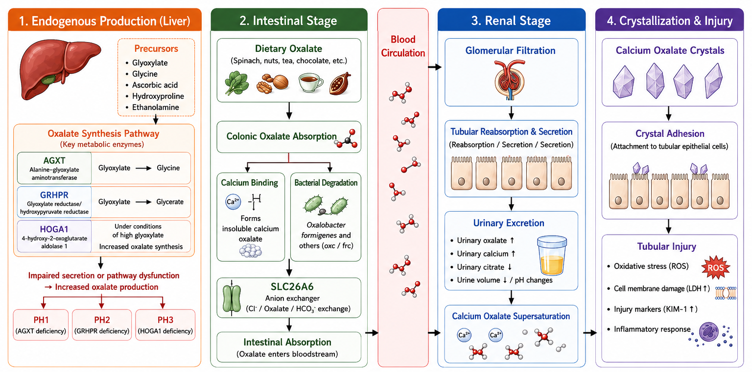
Figure 1. Mechanistic overview of oxalate metabolism
2.1 Glyoxylate Metabolic Diversion
(1) AGXT pathway
AGXT encodes alanine-glyoxylate aminotransferase, which mainly converts glyoxylate into glycine. When this pathway is impaired, glyoxylate is more easily oxidized to oxalate, which is an important mechanism of primary hyperoxaluria type 1. In experiments, AGXT expression, gene silencing, glyoxylate accumulation, and oxalate production should be analyzed together.
(2) GRHPR pathway
GRHPR encodes glyoxylate/hydroxypyruvate reductase, which can reduce glyoxylate to glycolate. GRHPR deficiency changes glyoxylate clearance capacity and shifts glyoxylate toward oxalate production. Studies often detect oxalate, glycolate, and glyoxylate simultaneously to determine whether metabolic diversion is abnormal.
(3) HOGA1-related pathway
HOGA1 participates in hydroxyproline metabolism. Functional abnormalities can increase glyoxylate-related precursors and promote oxalate production. Because hydroxyproline originates from collagen metabolism, this pathway is often related to liver metabolism, collagen turnover, and primary hyperoxaluria type 3 research.
2.2 Oxalate Oxidation and Oxalate Degradation
(1) Oxalate oxidase
Oxalate oxidase can catalyze the oxidation of oxalate to produce CO₂ and H₂O₂. It is commonly used for enzymatic oxalate detection, oxalate consumption assays, and oxalate quantification system development. When used in detection systems, the linear range of H₂O₂-coupled colorimetric or fluorescent readouts and sample background interference should be considered.
(2) Oxalate decarboxylase
Oxalate decarboxylase can catalyze the decarboxylation of oxalate to generate formate and CO₂. It is a more direct functional enzyme in oxalate degradation research. This enzyme is suitable for oxalate clearance, microbial oxalate degradation pathway studies, oxalate-degrading enzyme engineering, and in vitro oxalate consumption models.
(3) Microbial oxalate degradation pathways
Some oxalate-degrading bacteria depend on functional enzymes such as oxalyl-CoA decarboxylase and formyl-CoA transferase to utilize oxalate. Compared with detecting microbial composition alone, detection of functional genes such as oxc and frc better reflects the oxalate degradation potential of a strain or microbial community.
2.3 Intestinal and Renal Transport
(1) Intestinal absorption
Intestinal oxalate absorption is affected by free oxalate concentration, calcium binding, intestinal pH, the ability of fatty acids to bind calcium ions, and the status of the intestinal epithelial barrier. During fat malabsorption, fatty acids bind calcium and reduce calcium-mediated precipitation of oxalate in the intestinal lumen. As free oxalate increases, enteric hyperoxaluria is more likely to occur.
(2) SLC26A6 transporter
Anion transporters such as SLC26A6 participate in the exchange of oxalate, chloride, bicarbonate, and other anions. Changes in SLC26A6 expression in the intestine and renal tubules affect oxalate absorption and excretion. Studies should combine transporter expression, siRNA intervention, transmembrane oxalate flux, and urinary oxalate changes.
(3) Renal excretion
The kidney is the main organ for oxalate excretion. Increased urinary oxalate, reduced urine volume, increased urinary calcium, and decreased urinary citrate all increase the risk of calcium oxalate crystallization. Renal tubular epithelial cells can also be stimulated by soluble oxalate or calcium oxalate crystals, resulting in oxidative stress, inflammatory responses, and cell injury.
Table 2 Key Pathways and Detection Indicators in Oxalate Metabolism
Pathway node | Representative molecule | Main function | Recommended detection indicators |
Glyoxylate transamination | AGXT | Converts glyoxylate into glycine | AGXT expression, glyoxylate, glycine, oxalate |
Glyoxylate reduction | GRHPR | Converts glyoxylate into glycolate | GRHPR expression, glycolate, glyoxylate, oxalate |
Hydroxyproline metabolism | HOGA1 | Affects conversion of hydroxyproline to glyoxylate-related precursors | HOGA1 expression, hydroxyproline, oxalate |
Oxalate oxidation | Oxalate oxidase | Oxidizes oxalate and generates H₂O₂ | Oxalate oxidase activity, oxalate consumption, H₂O₂ readout |
Oxalate decarboxylation | Oxalate decarboxylase | Converts oxalate into formate and CO₂ | Oxalate decarboxylase activity, oxalate reduction, formate production |
Intestinal transport | SLC26A6 | Regulates intestinal oxalate transport | SLC26A6 expression, intestinal luminal oxalate, fecal oxalate |
Microbial degradation | Oxalobacter formigenes, etc. | Degrades intestinal luminal oxalate | Abundance of oxalate-degrading bacteria, oxc/frc functional genes, in vitro degradation rate |
3 Oxalate Detection Methods
3.1 Enzymatic Detection
(1) Detection principle
Enzymatic oxalate detection usually uses oxalate oxidase or related coupled systems to convert oxalate into detectable products, followed by colorimetric or fluorescence-based quantification. This method is relatively simple and suitable for batch detection of urine, cell culture medium, or certain tissue extract samples.
(2) Applicable scenarios
Enzymatic methods are suitable for routine screening, treatment group comparisons, and high-throughput experiments. They are convenient for urinary oxalate detection in animal models, renal tubular cell oxalate treatment systems, oxalate degradation experiments, and drug intervention screening.
(3) Limiting factors
Ascorbic acid, uric acid, reducing substances, color background, and matrix interference in samples may affect readings. Urine samples also require attention to pH, precipitation, dilution ratio, and storage conditions. For low-concentration samples or complex matrices, validation by standard addition recovery is recommended.
3.2 Ion Chromatography
(1) Method characteristics
Ion chromatography can separate oxalate from other anions and is suitable for oxalate detection in urine, drinking water, culture medium, food extracts, and environmental samples. Compared with enzymatic methods, ion chromatography offers stronger anion separation capacity and is suitable for complex matrix analysis.
(2) Sample processing
Urine and culture media usually require dilution, filtration, and, when necessary, protein precipitation. Tissue samples require extraction, centrifugation, and purification. If calcium ion levels are high in the sample, calcium oxalate precipitation may lead to falsely low free oxalate detection. Therefore, acidification, redissolution, or chelation procedures should be standardized in the methodology.
(3) Result advantages
Ion chromatography can simultaneously detect some inorganic anions, helping analyze the relationship between oxalate and other ions in urine or culture systems. For calcium oxalate crystallization risk studies, combined information on urinary calcium, citrate, magnesium ions, and pH is more informative.
3.3 HPLC and LC-MS/MS
(1) HPLC detection
HPLC often analyzes oxalate through derivatization or specific detection methods and can be used for oxalate quantification in relatively complex samples. Method development focuses on derivatization stability, peak separation, matrix effects, and standard curve range.
(2) LC-MS/MS detection
LC-MS/MS provides high specificity and sensitivity and is suitable for plasma, urine, tissue extracts, and metabolomics research. This method can simultaneously detect metabolites such as oxalate, glyoxylate, glycolate, and hydroxyproline, making it suitable for oxalate metabolic pathway analysis.
(3) Methodological control
LC-MS/MS requires attention to internal standard selection, ion suppression, sample pretreatment, lower limit of quantification, and stability. Because oxalate has a small molecular weight and strong polarity, method development is challenging in terms of retention, separation, and matrix interference control.
3.4 Calcium Oxalate Crystallization Models
(1) Calcium oxalate nucleation
In vitro crystallization models commonly mix calcium salts with oxalate salts to observe crystal nucleation rate, turbidity changes, and crystal number. This method is suitable for screening candidates that inhibit calcium oxalate nucleation and for comparing crystallization backgrounds in different oxalate systems, such as sodium oxalate and potassium oxalate.
(2) Crystal growth and aggregation
After crystal formation, particle size, aggregation degree, and morphological changes can be further observed. Citrate, magnesium ions, urinary proteins, renal tubular injury molecules, and candidate interventions may all affect crystal growth and aggregation.
(3) Cell adhesion and injury
Calcium oxalate crystals can adhere to the surface of renal tubular epithelial cells and induce oxidative stress, membrane damage, and inflammatory responses. Common HK-2 and MDCK cell models are used to detect crystal adhesion, ROS, LDH release, cell viability, and KIM-1-related injury indicators.
Table 3 Comparison of Oxalate Detection Methods
Method | Applicable samples | Advantages | Limitations | Applicable scenarios |
Enzymatic method | Urine, culture medium, tissue extracts | Simple operation, suitable for batch detection | Susceptible to reducing substances and color interference | Routine screening, animal models, cell experiments |
Ion chromatography | Urine, food, culture medium, water samples | Strong anion separation capacity | Higher instrument requirements; pretreatment must be standardized | Urinary oxalate, dietary oxalate, complex anion samples |
HPLC | Urine, blood, tissue extracts | Allows relatively specific quantification | Derivatization and method development requirements are high | Method validation, complex sample analysis |
LC-MS/MS | Plasma, urine, tissue, metabolomics samples | High sensitivity and specificity; multi-analyte detection | High cost; matrix effects must be controlled | Mechanistic research, inherited metabolic diseases, metabolic pathway analysis |
Calcium oxalate crystallization model | Artificial urine, buffers, cell systems | Direct observation of crystallization risk | Still differs from the in vivo environment | Crystallization inhibitor screening, kidney stone mechanism research |
4 Oxalate Metabolism and Disease Mechanisms
4.1 Kidney Stone Formation
(1) Urinary supersaturation
The core condition for calcium oxalate stone formation is supersaturation of calcium ions and oxalate in urine. Increased urinary oxalate, increased urinary calcium, reduced urine volume, and decreased urinary citrate all promote crystallization. Compared with urinary calcium, a small increase in urinary oxalate often has a stronger effect on calcium oxalate supersaturation.
(2) Crystal adhesion
After calcium oxalate crystals form, their adhesion to renal tubular epithelium determines whether they can be retained and continue to grow. Renal tubular injury, oxidative stress, and changes in cell-surface glycoproteins can all enhance crystal adhesion.
(3) Renal tubular injury
Both soluble oxalate and calcium oxalate crystals can induce renal tubular epithelial cell injury, but their mechanistic emphases differ. Sodium oxalate is more suitable for modeling soluble oxalate load, whereas calcium oxalate monohydrate is more suitable for modeling crystal adhesion, crystal stimulation, and stone-related local injury.
4.2 Primary Hyperoxaluria
(1) PH1
PH1 is usually associated with AGXT functional defects, causing glyoxylate to be inefficiently converted into glycine and increasing oxalate production. Research should focus on AGXT expression, glyoxylate metabolism, urinary oxalate, renal calcium deposition, and renal functional injury.
(2) PH2
PH2 is related to abnormal GRHPR function. When the glyoxylate reduction pathway is blocked, oxalate production increases. This model is suitable for simultaneous detection of glycolate, glyoxylate, and oxalate to determine the direction of metabolic diversion.
(3) PH3
PH3 is associated with HOGA1 and involves abnormal hydroxyproline metabolism. In addition to oxalate, hydroxyproline metabolites, hepatic metabolic flux, and renal calcium oxalate deposition can also be examined.
4.3 Enteric Hyperoxaluria
(1) Fat malabsorption
During fat malabsorption, unabsorbed fatty acids bind calcium and reduce the opportunity for calcium to form insoluble precipitates with oxalate, resulting in increased free oxalate and easier intestinal absorption. This mechanism is commonly seen in inflammatory bowel disease, short bowel syndrome, pancreatic insufficiency, and post-gastrointestinal surgery states.
(2) Microbiota changes
Antibiotic use, dietary structure changes, and intestinal diseases may reduce the abundance of oxalate-degrading bacteria and decrease intestinal oxalate clearance capacity. Such studies should combine fecal oxalate, microbial composition, oxalate-degrading function, and urinary oxalate changes.
(3) Transport abnormalities
Changes in SLC26A6 expression or function may affect the balance between intestinal oxalate secretion and absorption. If only elevated urinary oxalate is detected, it is not possible to distinguish increased intake, insufficient microbial degradation, or transport abnormalities. Therefore, intestinal samples, transporter expression, and urinary excretion data should be analyzed together.
4.4 Chronic Kidney Disease and Systemic Oxalate Burden
(1) Reduced renal excretion
Declining renal function reduces oxalate excretion capacity, leading to increased blood oxalate. In chronic kidney disease models, oxalate burden may interact with renal tubular injury, calcium salt deposition, and oxidative stress.
(2) Oxalate deposition
When blood oxalate remains elevated, calcium oxalate can deposit in the kidney and other tissues. Studies of systemic oxalate deposition require combined analysis of blood oxalate, urinary oxalate, tissue calcium deposition, and renal function indicators.
(3) Injury markers
KIM-1, LDH, and ROS can be used to evaluate oxalate- or calcium oxalate crystal-induced cell injury. KIM-1 is more specific to renal tubular injury, LDH reflects disruption of cell membrane integrity, and ROS is suitable for evaluating the involvement of oxidative stress.
Table 4 Relationships Between Oxalate Metabolic Abnormalities and Disease Mechanisms
Disease direction | Key mechanism | Recommended indicators | Mechanistic interpretation |
Calcium oxalate kidney stones | Increased urinary oxalate, calcium oxalate supersaturation, crystal adhesion | Urinary oxalate, urinary calcium, urinary citrate, crystal number | Oxalate load and urinary inhibitory factors should be analyzed together |
Primary hyperoxaluria | Abnormal AGXT, GRHPR, or HOGA1 | Oxalate, glyoxylate, glycolate, related genes | Clarify the source of abnormal oxalate production |
Enteric hyperoxaluria | Increased intestinal absorption, insufficient microbial degradation | Fecal oxalate, urinary oxalate, microbiota, SLC26A6 | Distinguish intake, absorption, and microbial degradation factors |
Chronic kidney disease | Reduced oxalate excretion, increased blood oxalate | Blood oxalate, urinary oxalate, renal function, calcium deposition | Determine whether oxalate burden is caused by excretory dysfunction |
Renal tubular injury | Oxalate or crystals induce oxidative stress and cell injury | KIM-1, LDH, ROS, histopathology | Distinguish soluble oxalate stimulation from crystal stimulation |
5 Experimental Design and Result Interpretation
5.1 Sample Selection
(1) Urine samples
Urine is the most commonly used sample in oxalate metabolism research. A 24-hour urine sample is more suitable for evaluating total excretion, while spot urine requires creatinine correction. Urine testing should record urine volume, pH, storage conditions, and whether precipitation is present.
(2) Blood samples
Blood oxalate is suitable for studies of chronic kidney disease, primary hyperoxaluria, and systemic oxalate burden. Blood oxalate concentrations are low and easily affected by pretreatment and contamination, so high-specificity methods such as LC-MS/MS are usually more suitable.
(3) Tissue and cell samples
Liver tissue is suitable for studying endogenous oxalate production, kidney tissue for calcium oxalate deposition and renal tubular injury, and intestinal samples for oxalate absorption and microbial degradation. In cell models, soluble oxalate stimulation using sodium oxalate should be distinguished from calcium oxalate crystal stimulation because the two induce different injury mechanisms.
5.2 Control Settings
(1) Dietary control
Oxalate metabolism is highly sensitive to diet. Animal experiments should control oxalate intake, calcium intake, vitamin C intake, water intake, and salt intake. If a high-oxalate diet is being studied, controls with equivalent energy, calcium, and mineral background should be included.
(2) Positive models
Common high-oxalate models can be established using ethylene glycol, hydroxyproline, high-oxalate diets, or genetic models. Different models have different mechanisms: ethylene glycol is more oriented toward endogenous oxalate production and kidney injury, while dietary oxalate is more oriented toward intestinal absorption and urinary oxalate elevation.
(3) Methodological blanks
Oxalate detection should include reagent blanks, matrix blanks, standard curves, and spike recovery. If precipitation exists in the sample, it should be clearly stated whether total oxalate, soluble oxalate in the supernatant, or free oxalate is being measured.
5.3 Combined Indicators
(1) Metabolite combinations
Measuring oxalate alone cannot explain its metabolic source. According to the research objective, it is recommended to simultaneously detect precursors or related metabolites such as glyoxylate, glycolate, glycine, hydroxyproline, and ascorbic acid.
(2) Urinary risk combinations
Kidney stone research should simultaneously detect urinary oxalate, urinary calcium, urinary citrate, urinary magnesium, uric acid, urine pH, and urine volume. Calcium oxalate crystallization risk is determined by multiple factors and should not be judged only by urinary oxalate.
(3) Injury and oxidative stress combinations
Oxalate or calcium oxalate crystals can induce renal tubular injury. KIM-1, LDH, ROS, and histopathological changes are recommended in combination to distinguish metabolic abnormalities, crystallization stimulation, and cellular injury outcomes.
Table 5 Key Points in Experimental Design for Oxalate Metabolism Research
Research objective | Recommended samples | Core indicators | Key control points |
Evaluation of urinary oxalate excretion | 24-hour urine, spot urine | Oxalate, creatinine, urine volume | Standardize sampling time, storage conditions, and dilution ratio |
Endogenous production mechanism | Liver tissue, hepatocytes, blood | Oxalate, glyoxylate, glycolate, AGXT/GRHPR/HOGA1 | Distinguish metabolic production from excretion changes |
Enteric absorption research | Feces, intestinal contents, intestinal tissue | Fecal oxalate, microbiota, SLC26A6 | Control dietary oxalate, calcium intake, and antibiotic effects |
Calcium oxalate crystallization model | Artificial urine, urine, cell systems | Crystal number, particle size, cell adhesion | Control pH, calcium/oxalate concentration, and incubation time |
Kidney injury mechanism | Kidney tissue, renal tubular cells | KIM-1, LDH, ROS, histopathology | Distinguish oxalate stimulation from crystal stimulation |
Intervention effect evaluation | Urine, blood, kidney tissue, feces | Oxalate, urinary calcium, kidney injury, microbiota | Analyze metabolism, crystallization, and tissue injury simultaneously |
6 Product Selection for Oxalate Metabolism Research
Table 6 Product Selection for Oxalate Metabolism Research
Cat. No. | Product Name | Grade/Specification | Product category | Application positioning |
Oxalic acid | PrimorTrace™, Purifed, ≥99.999% metals basis | Oxalic acid standard / model substrate | Used for high-purity oxalate standard systems, oxalate detection with low trace-metal interference, and method validation | |
Oxalic acid | PrimorTrace™, Anhydrous Grade, ≥99.999% metals basis | Oxalic acid standard / model substrate | Used for oxalate metabolism and detection method development requiring strict impurity control | |
Oxalic acid | AR, ≥98% | Oxalic acid standard / model substrate | Used for routine oxalate models, oxalate solution preparation, and methodological controls | |
Oxalic acid dihydrate | GR, ≥99.8% | Oxalic acid standard / model substrate | Used for oxalate quantification, oxalate treatment systems, and routine laboratory standard preparation | |
Oxalic acid dihydrate | Standard for GC, ≥99.6% | Oxalic acid methodological standard | Used for chromatographic method development, standard curves, and quantitative quality control | |
Oxalic acid dihydrate | UltraBio™, ≥99%(RT) | Oxalate substrate for biological experiments | Used for oxalate treatment systems related to cells or biological samples | |
Oxalate standard solution | 1000ppm in water | Oxalate quantitative standard solution | Used for ion chromatography, HPLC, or oxalate detection standard curves | |
Oxalic acid anion standard solution | 1mg/ml (1,000ppm) | Oxalate quantitative standard solution | Used for oxalate anion quantification, method calibration, and quality control | |
Oxalic acid anion standard solution | 0.1mg/ml (100ppm) | Oxalate quantitative standard solution | Used for low-concentration oxalate standard curves and sample detection quality control | |
Oxalic acid standard solution | c(1/2H2C2O4)=0.1mol/L | Oxalic acid standard solution | Used for titration, standard curves, and calibration of oxalate detection systems | |
Sodium carbonate/Oxalic Acid concentrate | Na2CO3 360mM and H2C2O4 0.5mM in water, IC eluent concentrate (100x) | Ion chromatography methodological reagent | Used as eluent or for method development in ion chromatography detection systems | |
Sodium oxalate | ≥99% | Soluble oxalate model reagent | Used for soluble oxalate stimulation of renal tubular cells and oxalate treatment models | |
Sodium oxalate | analytical standard, ≥99.96% | Oxalate analytical standard | Used as a standard material in urinary oxalate, culture medium oxalate, and method validation | |
Sodium oxalate | PrimorTrace™, ≥99.99% metals basis | High-purity oxalate model reagent | Used in oxalate stimulation, crystallization, and detection systems sensitive to metal impurities | |
Sodium oxalate | AR | Soluble oxalate model reagent | Used for routine oxalate treatment, in vitro oxalate stimulation, and experimental controls | |
Sodium oxalate solution | 0.05M | Sodium oxalate standard solution | Used for volumetric analysis, method validation, and quantitative quality control | |
Potassium oxalate monohydrate | AR, ≥99.8% | Oxalate system reagent | Used for oxalate ion systems, crystallization models, and methodological controls | |
Potassium oxalate monohydrate | ACS, ≥99.5% | Oxalate system reagent | Used for oxalate model construction and analytical detection systems | |
Potassium oxalate monohydrate | ≥99.98% metals basis | High-purity oxalate system reagent | Used for calcium oxalate crystallization and detection method research sensitive to metal impurities | |
Potassium oxalate monohydrate | BioReagent, ≥98.5% | Oxalate reagent for biological experiments | Used for oxalate treatment and oxalate model construction involving biological samples | |
Calcium oxalate | ≥99.9% metals basis | Calcium oxalate crystallization model material | Used for calcium oxalate deposition, crystallization models, and kidney stone mechanism research | |
Calcium oxalate monohydrate | ≥97% | Calcium oxalate monohydrate model | Used for COM crystal adhesion, renal tubular injury, and stone formation mechanism research | |
Calcium oxalate monohydrate | PrimorTrace™, ≥99.99% metals basis | High-purity calcium oxalate monohydrate model | Used for high-purity calcium oxalate crystal models, crystallization intervention, and cell injury research | |
Calcium oxalate hydrate | CP, ≥98% | Calcium oxalate hydrate model | Used for calcium oxalate crystal formation, deposition, and in vitro crystallization evaluation | |
Oxalate Oxidase (OXO) | ActiBioPure™, Bioactive, High Performance, EnzymoPure™, ≥0.5 U/mg powder | Oxalate detection/degradation enzyme | Used for enzymatic oxalate detection, oxalate consumption experiments, and oxalate oxidation reaction system establishment | |
Oxalate Decarboxylase | Bioactive,Recombinant,ActiBioPure™,High Performance,EnzymoPure™,≥1 U/mg enzyme powder; ≥5 U/mg protein | Oxalate-degrading enzyme | Used for oxalate decarboxylation degradation, oxalate clearance, mechanisms of oxalate-degrading bacteria, and enzyme engineering research | |
Oxalic acid decarboxylase (OXDC) | Bioactive, Recombinant, ActiBioPure™, High Performance, EnzymoPure™, ≥90%(SDS-PAGE), ≥70 U/mg protein | Oxalate-degrading enzyme | Used for oxalate degradation pathways, OxdC functional validation, and oxalate metabolism mechanism research | |
AGXT Human Pre-designed siRNA Set A |
| Glyoxylate transamination pathway validation | Used for AGXT knockdown to validate glyoxylate diversion to glycine and PH1-related oxalate production mechanisms | |
AGXT2 Human Pre-designed siRNA Set A |
| Glyoxylate metabolism-related gene validation | Used for AGXT2-related glyoxylate metabolism, oxalate production regulation, and gene function validation | |
GRHPR Human Pre-designed siRNA Set A |
| Glyoxylate reduction pathway validation | Used for GRHPR knockdown to analyze glyoxylate reduction, glycolate production, and PH2-related abnormal oxalate metabolism | |
GRHPR Mouse mAb | KD Validation | GRHPR pathway protein detection | Used for GRHPR protein expression detection and knockdown model validation | |
GRHPR Mouse mAb | See COA | GRHPR pathway protein detection | Used for GRHPR expression, localization, and glyoxylate reduction pathway analysis | |
GRHPR Mouse mAb | KD Validation | GRHPR pathway protein detection | Used for protein validation in GRHPR knockdown experiments | |
HOGA1 Human Pre-designed siRNA Set A |
| Hydroxyproline metabolism pathway validation | Used for HOGA1 knockdown to study abnormal hydroxyproline metabolism and PH3-related oxalate production mechanisms | |
SLC26A6 Human Pre-designed siRNA Set A |
| Oxalate transport function validation | Used to validate the effects of SLC26A6 on oxalate transmembrane transport, gut-kidney axis regulation, and renal tubular oxalate load | |
Recombinant Human TIM-1/KIM-1/HAVCR Protein | Animal Free,Carrier Free,His Tag,PBS Only,≥95%(SDS-PAGE) | Renal tubular injury marker | Used for KIM-1/HAVCR-related renal tubular injury mechanism research and positive controls | |
Rat Kidney Injury Molecule 1 (Kim-1) ELISA Kit | BioReagent | Renal tubular injury detection | Used for KIM-1 detection in rat hyperoxaluria, calcium oxalate deposition, and renal tubular injury models | |
Mouse Kidney Injury Molecule 1 (Kim-1) ELISA Kit | BioReagent | Renal tubular injury detection | Used for KIM-1 detection in mouse oxalate metabolic abnormality, renal crystallization, and kidney injury models | |
Lactate Dehydrogenase (LDH) Cytotoxicity Assay Kit (DNPH, Micro Method) | BioReagent | Oxalate/crystal cell injury evaluation | Used for evaluating renal tubular cell oxalate injury, calcium oxalate crystal toxicity, and membrane integrity | |
LDH Cytotoxicity Assay Kit with WST-8 | BioReagent,ready-to-use,for IP | Oxalate/crystal cell injury evaluation | Used for rapid detection of oxalate-induced injury in cell models | |
Lactate Dehydrogenase (LDH) Cytotoxicity Assay Kit (DNPH, Colorimetric Method) | BioReagent | Oxalate/crystal cell injury evaluation | Used for colorimetric detection of cell injury after calcium oxalate crystal stimulation | |
Mitochondrial Reactive Oxygen Species (ROS) Production Rate Assay Kit (Fluorometric Method) | BioReagent | Oxidative stress detection | Used to evaluate mitochondrial ROS generation and oxidative injury induced by oxalate or calcium oxalate crystals |
7 FAQ
7.1 Is 24-hour urine or spot urine more suitable for urinary oxalate detection?
For evaluating total oxalate excretion, 24-hour urine is more suitable. Spot urine can be used for preliminary screening or high-throughput detection in animal experiments, but creatinine correction should be performed. For kidney stone risk research, urine volume, urine pH, urinary calcium, and urinary citrate should also be recorded.
7.2 Why is oxalate detection easily affected by sample storage?
Oxalate can form precipitates with calcium. Sample pH, temperature, storage time, and freeze-thaw processes can all affect measurable oxalate levels. If precipitation occurs in urine samples, it should be clearly stated whether total oxalate or soluble oxalate in the supernatant is being detected, and the same processing method should be maintained throughout the study.
7.3 What is the difference between oxalate oxidase and oxalate decarboxylase?
Oxalate oxidase mainly catalyzes the oxidation of oxalate to generate CO₂ and H₂O₂ and is commonly used for enzymatic oxalate detection and oxalate consumption experiments. Oxalate decarboxylase catalyzes the decarboxylation of oxalate to produce formate and CO₂, making it more suitable for oxalate degradation, oxalate clearance, and oxalate-degrading bacterial mechanism studies. Both are related to oxalate metabolism, but their application scenarios differ.
7.4 Can sodium oxalate and calcium oxalate monohydrate replace each other in cell experiments?
They should not be used interchangeably. Sodium oxalate mainly models soluble oxalate load and is suitable for studying oxalate transport, soluble oxalate stimulation, and cellular metabolic responses. Calcium oxalate monohydrate mainly models crystal adhesion, crystal stimulation, and stone-related injury. The mechanisms represented by the two models are different.
7.5 Can calcium oxalate crystallization experiments replace animal stone models?
Not completely. In vitro crystallization experiments are suitable for analyzing crystal nucleation, growth, aggregation, and candidate inhibitor effects. Animal models also reflect the urinary environment, renal tubular injury, metabolic regulation, and tissue deposition. The two types of models should be used according to the research stage.
7.6 Is hyperoxaluria always caused by eating too much oxalate?
Not necessarily. Hyperoxaluria may result from increased dietary intake, enhanced intestinal absorption, insufficient microbial degradation, excessive endogenous oxalate production, or abnormal renal excretion. Dietary records, fecal oxalate, blood oxalate, metabolic precursors, and genetic pathway analysis are needed to identify the source.
7.7 What should be mainly examined for SLC26A6 in oxalate metabolism research?
SLC26A6 is mainly used to analyze intestinal and renal tubular oxalate transport. Studies may detect changes in its expression, or use siRNA knockdown to validate its effects on oxalate transmembrane transport, intracellular oxalate load, and urinary oxalate changes.
7.8 Why should KIM-1, LDH, and ROS be detected in oxalate metabolism research?
KIM-1 reflects renal tubular injury, LDH reflects cell membrane injury, and ROS reflects oxidative stress. Oxalate or calcium oxalate crystals not only alter metabolic burden but also cause renal tubular cell injury. These indicators help determine whether abnormal oxalate metabolism has translated into cellular or tissue injury.
Oxalate metabolism research needs to address production, degradation, transport, excretion, and crystallization processes at the same time. Urinary oxalate detection is suitable for evaluating oxalate burden. Oxalate oxidase and oxalate decarboxylase can be used for detection or degradation mechanism analysis. AGXT, GRHPR, HOGA1, and SLC26A6 help explain metabolic and transport sources. Calcium oxalate crystal models and injury indicators such as KIM-1, LDH, and ROS help connect metabolic abnormalities with kidney stone mechanisms.


 危险品化学品经营许可证(带存储)