
Potential Mechanisms of p-Cymene in Problem-Prone Skin: Membrane Perturbation, Inflammatory Signaling Modulation, and Localized Delivery
Potential Mechanisms of p-Cymene in Problem-Prone Skin: Membrane Perturbation, Inflammatory Signaling Modulation, and Localized Delivery
1 Chemical Structure and Raw Material Characteristics of p-Cymene
1.1 p-Cymene Is an Aromatic Monoterpene Hydrocarbon
The English name is p-cymene, and its chemical name is 1-isopropyl-4-methylbenzene. Its molecular formula is C₁₀H₁₄, its relative molecular mass is 134.22, and its CAS number is 99-87-6.
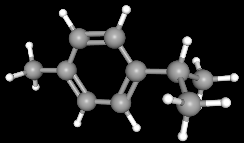
Its simplified structure is:
p-CH₃—C₆H₄—CH(CH₃)₂
The methyl and isopropyl groups are located at the para positions of the benzene ring, hence the name “p-cymene.” It contains no polar functional groups such as hydroxyl, carboxyl, or amino groups and is generally classified as an aromatic monoterpene hydrocarbon. It may also be described as a monoterpene-derived aromatic hydrocarbon.[1]
p-Cymene has the following properties that are closely related to its use in formulations:
Raw Material Property | Influence on Its Mode of Action |
Strong hydrophobicity | Readily partitions into oil phases, sebum, and phospholipid membranes |
Low water solubility | Makes it difficult to establish a uniform free concentration in aqueous systems |
Small molecular size | Allows redistribution among oil phases, stratum corneum lipids, and biological membranes |
Volatility | May result in concentration loss during storage and after application |
Absence of a phenolic hydroxyl group | Direct disruption of bacterial ion homeostasis and the proton motive force is generally weaker than that caused by phenolic monoterpenes such as carvacrol |
1.2 p-Cymene Is Different from o-Cymen-5-ol
p-Cymene can easily be confused with o-cymen-5-ol, but the two compounds differ substantially in both structure and function.
Comparison Item | p-Cymene | o-Cymen-5-ol |
English name | p-Cymene | O-Cymen-5-ol |
Key functional group | No phenolic hydroxyl group | Contains a phenolic hydroxyl group |
Main physical characteristic | Hydrophobic aromatic hydrocarbon | Hydrophobic phenolic compound |
Direct antimicrobial activity | Generally weak and markedly affected by environmental conditions | Generally stronger |
Main research focus | Membrane partitioning, antimicrobial synergy, anti-inflammatory effects, and delivery | Antimicrobial activity and preservation |
2 How p-Cymene Acts on Bacterial Membranes
2.1 Hydrophobic Partitioning Alters Membrane Structure
Bacterial cell membranes are composed primarily of phospholipid bilayers. The hydrophilic head groups of the phospholipids face the aqueous phase, whereas the hydrophobic fatty acid chains are located within the membrane interior.
Because p-cymene lacks polar functional groups and has low affinity for the aqueous phase, once it reaches the vicinity of bacteria, it tends to transfer from the aqueous phase into the hydrophobic region of the phospholipid bilayer.
In liposome and Bacillus cereus models, p-cymene showed a strong tendency to partition into lipid membranes and caused pronounced membrane expansion. In the study, the membrane expansion caused by p-cymene was even greater than that caused by carvacrol.[2]
This membrane expansion can be explained by the entry of p-cymene between the phospholipid fatty acid chains, where it occupies part of the hydrophobic space and alters interactions among the fatty acid chains.
2.2 Membrane Expansion Does Not Necessarily Mean Collapse of the Bacterial Energy System
The bacterial membrane does more than separate the cell from its surroundings. It also performs the following functions:
① maintaining differences in ion concentrations across the membrane;
② generating the membrane potential;
③ establishing the proton motive force;
④ driving adenosine triphosphate synthesis;
⑤ mediating nutrient transport.
Adenosine triphosphate (ATP) is an important energy source for bacterial metabolism and growth. After mild alterations in membrane structure, bacteria may still maintain basic functions through ion pumps, membrane lipid rearrangement, and metabolic regulation.
In the same study, although p-cymene caused pronounced membrane expansion, it did not significantly affect the transmembrane pH gradient or ATP levels, and its effect on membrane potential was also weaker than that of carvacrol.[2]
2.3 The Phenolic Hydroxyl Group Determines the Difference Between the Membrane Effects of p-Cymene and Carvacrol
p-Cymene and carvacrol have similar aromatic carbon skeletons, but carvacrol contains a phenolic hydroxyl group, whereas p-cymene does not contain a hydroxyl group. Both compounds can enter the hydrophobic membrane phase and alter membrane structure, but they differ in their effects on bacterial transmembrane ion gradients, energy metabolism, and growth. The molecular structure of carvacrol is illustrated below.
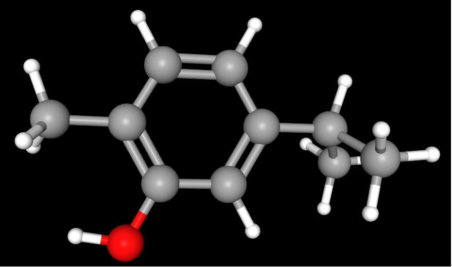
The original study used egg-yolk-derived L-α-phosphatidylethanolamine to prepare unilamellar liposomes for evaluating membrane-phase partitioning and membrane expansion. Glucose-energized vegetative cells of Bacillus cereus IFR-NL94-25 were used to measure membrane potential, the transmembrane pH gradient, and the ATP pool, while growth inhibition experiments were conducted in brain heart infusion medium.[2].
Structure and Effect | p-Cymene | Carvacrol |
Phenolic hydroxyl group | Absent | Present |
Partitioning into lipid membranes | Strong affinity for the hydrophobic membrane phase | Clear affinity for the hydrophobic membrane phase |
Liposome membrane expansion | Causes pronounced membrane expansion; the maximum extent of expansion is greater than that caused by carvacrol, but a higher membrane loading is required | Causes pronounced membrane expansion; reaches its maximum effect at a lower membrane loading |
Effect on membrane potential Δψ | Can reduce membrane potential, but a higher concentration is required to produce the same decrease | Produces a more pronounced reduction in membrane potential |
Effect on the transmembrane pH gradient ΔpH | No significant effect observed within the range of 0.5–2 mmol/L | Can eliminate the transmembrane pH gradient at 1 mmol/L |
Effect on the ATP pool | No significant change in intracellular or extracellular ATP levels at 1 and 2.4 mmol/L | Can reduce the intracellular ATP pool |
Effect on the growth of Bacillus cereus | No growth inhibition observed within the concentration range tested in the study | Shows clear growth-inhibitory activity |
The strong membrane partitioning and membrane-expanding capacity of p-cymene mainly reflect its effects on phospholipid organization and the physical state of the membrane and do not directly indicate potent antimicrobial activity. The phenolic hydroxyl group of carvacrol can participate in proton and monovalent cation exchange, thereby reducing the transmembrane pH gradient and membrane potential and consequently weakening the proton motive force and ATP synthesis. This proton-exchange process is a mechanistic model proposed on the basis of changes in ion flux and cellular energy status.[2]
p-Cymene primarily causes physical perturbation of the membrane. In addition to perturbing membrane structure, carvacrol can disrupt ion homeostasis and energy metabolism through its phenolic hydroxyl group and therefore exhibits more pronounced direct antimicrobial activity.
3 Antimicrobial Combination Effects and Potential Synergistic Mechanisms of p-Cymene
Note: The figure below was prepared on the basis of existing studies on microbial membranes, inflammatory signaling, and delivery systems and is intended to illustrate the potential pathways through which p-cymene may act in problem-prone skin. Some of the pathways shown represent mechanistic hypotheses proposed on the basis of current evidence and have not yet been directly validated in acne-related skin models or human topical-use studies.

3.1 p-Cymene Enhances the Effects of Other Components Through Membrane Perturbation
After entering the bacterial membrane, p-cymene may increase the spacing between phospholipid molecules and alter membrane fluidity and permeability. Under these conditions, antimicrobial components containing phenolic hydroxyl groups, such as carvacrol, may partition more readily into the membrane phase and approach their sites of action.
This potential synergy arises from the interaction of different stages of action:
① p-cymene primarily affects the physical state of the membrane;
② carvacrol further affects membrane permeability, ion balance, and energy metabolism.
3.2 Synergistic Effects Are Influenced by Environmental Conditions
In a study using unpasteurized apple juice, 1.25 mmol/L p-cymene reduced Escherichia coli O157:H7 to below the detection limit within 1–2 days. A combination of 0.25 mmol/L p-cymene and 0.5 mmol/L carvacrol also produced a pronounced antimicrobial effect.[3]
These results indicate that p-cymene can exert direct antimicrobial activity under specific environmental conditions. When combined with carvacrol, it can also reduce the individual concentrations required for the two components to achieve antimicrobial effects in the apple juice model. However, the apple juice system has the following characteristics:
① a pH of approximately 3.2;
② water activity and nutrient composition that differ from those of the skin;
③ Escherichia coli O157:H7 as the test organism;
④ experimentally controlled temperature and exposure time.
These conditions differ substantially from those on the surface of human skin or within hair follicles. Therefore, effective concentrations identified in food systems cannot be directly used as addition levels in skincare formulations, nor can they directly demonstrate an equivalent effect against Cutibacterium acnes.
3.3 Antimicrobial Synergy Must Be Evaluated Together with Skin Tolerability
p-Cymene acts on bacterial membranes through lipid partitioning, but this mechanism does not provide clear recognition of pathogenic bacteria. When local concentrations increase, skin commensal microorganisms and host cell membranes may also be affected.
A synergistic system with value for skincare applications should satisfy three conditions:
Evaluation Level | Key Indicator |
Microbial effect | Reduced growth, biofilm formation, or inflammatory-stimulatory capacity of the target microorganism |
Combination efficiency | Reduced effective concentration of the primary antimicrobial component |
Skin tolerability | Effects on keratinocytes, barrier lipids, and commensal microorganisms remain within an acceptable range |
If antimicrobial activity and cell membrane damage are enhanced simultaneously, the result is more consistent with an additive, nonselective membrane effect than with antimicrobial synergy that offers a clear advantage for skin applications.
4 How p-Cymene Influences NF-κB and MAPK Signaling
4.1 NF-κB and MAPK Participate in the Amplification of Inflammatory Signaling
Nuclear factor-kappa B (NF-κB) is an important transcription factor that regulates the transcription of inflammatory genes. In unstimulated cells, NF-κB is usually bound to inhibitor of nuclear factor-kappa B alpha (IκBα) and remains primarily in the cytoplasm.
Stimuli such as lipopolysaccharide (LPS) can induce IκBα phosphorylation and subsequent degradation, allowing NF-κB to enter the nucleus and promote the expression of pro-inflammatory genes.
The mitogen-activated protein kinase (MAPK) pathway includes several branches, such as extracellular signal-regulated kinase 1/2, p38, and c-Jun N-terminal kinase.
Among these:
① extracellular signal-regulated kinase 1/2 is abbreviated as ERK1/2;
② c-Jun N-terminal kinase is abbreviated as JNK.
The principal inflammatory signaling process can be represented as follows:
LPS and other stimuli → IκBα phosphorylation/degradation → NF-κB nuclear translocation
→ERK1/2, p38, and JNK phosphorylation → enhanced transcriptional regulation by AP-1 and other factors
↓
increased expression of pro-inflammatory mediators such as TNF-α, IL-1β, and IL-6
Activator protein-1 (AP-1) is an important downstream transcriptional regulator of MAPK signaling. NF-κB and MAPK do not function entirely independently and can jointly regulate the expression of multiple inflammatory mediators.
4.2 p-Cymene Reduces LPS-Induced Signaling Activation
In LPS-stimulated RAW 264.7 murine macrophages, p-cymene reduced the production of tumor necrosis factor-alpha, interleukin-1 beta, and interleukin-6 and downregulated the corresponding messenger RNA expression.[4]
Among these:
① tumor necrosis factor-alpha is abbreviated as TNF-α;
② interleukin-1 beta is abbreviated as IL-1β;
③ interleukin-6 is abbreviated as IL-6.
The study also showed that p-cymene inhibited LPS-induced phosphorylation of IκBα, ERK1/2, p38, and JNK.[4]
In a mouse model of LPS-induced acute lung injury, p-cymene also reduced pro-inflammatory cytokine levels, inflammatory cell infiltration, and tissue edema and inhibited the phosphorylation of IκBα and MAPK-related proteins.[5]
4.3 The Direct Molecular Targets Through Which p-Cymene Modulates the NF-κB/MAPK Pathways Remain Unclear
The above results indicate that p-cymene treatment is associated with reduced LPS-induced phosphorylation of IκBα and MAPK-related proteins and decreased production of pro-inflammatory cytokines.[4,5] However, these findings primarily reflect changes in pathway activation and do not establish whether p-cymene directly binds to or inhibits any particular protein.
Current studies have not confirmed whether p-cymene directly acts on the following molecules:
① Toll-like receptor 4 (TLR4);
② the IκB kinase complex (IKK);
③ ERK1/2, p38, or JNK;
④ the NF-κB protein itself.
At present, it can be concluded that p-cymene intervention is associated with reduced activation of NF-κB and MAPK signaling. Its upstream site of action and direct molecular targets remain to be determined.
5 Relationship Between p-Cymene and Problem-Prone Skin
5.1 Problem-Prone Skin Is Jointly Driven by the Follicular Environment, Microorganisms, and Inflammation
Excessive sebum secretion, abnormal keratinization of the follicular opening, and sebum retention can alter the lipid composition, redox state, and nutrient conditions within the follicle. In this environment, certain microbial strains may exhibit stronger adhesion, biofilm formation, or inflammatory-stimulatory capacity.
The microorganism formerly known as Propionibacterium acnes is now named Cutibacterium acnes and is a resident microorganism of sebum-rich skin regions. The difference between acne-affected and healthy skin does not involve only changes in the total abundance of this bacterium but also differences in the proportions of particular strains and genetic lineages.[6]
The interactions in problem-prone skin can be represented as follows:
Sebum retention and abnormal keratinization → altered follicular environment → changes in microbial composition or metabolism → increased microbial-derived stimulation → NF-κB/MAPK activation → release of inflammatory mediators → follicular wall edema and further abnormalities in keratinization
On the basis of existing microbial membrane studies and LPS inflammatory models, a potential framework can be proposed for the action of p-cymene in problem-prone skin. However, this framework has not yet been directly validated in Cutibacterium acnes-related skin models or human topical-use studies:
Microbial membrane effects and antimicrobial combination effects ← p-Cymene → NF-κB/MAPK-related inflammatory signaling
The effect on the left may theoretically reduce the growth or activity of certain microorganisms, whereas the effect on the right may weaken inflammatory signaling output from host cells. Together, these effects may influence the cycle of “microbial stimulation–inflammatory amplification.”
5.2 Microbiome Effects Depend on Strain Sensitivity and Local Exposure
p-Cymene enters microbial membranes primarily through hydrophobic partitioning.[2] There is currently no evidence that it can specifically recognize acne-associated strains or selectively preserve skin commensal microorganisms. Different microorganisms may vary in their sensitivity to p-cymene, and these differences may be related to cell envelope structure, membrane lipid composition, biofilm status, and stress-adaptation capacity. However, this does not mean that p-cymene has clear microbiome selectivity.
Its actual effects on the skin microbiome depend on strain sensitivity, the free concentration at the target site, exposure duration, combination components, and the condition of the hair follicle and skin barrier. If the local concentration is too low, the intended effect may not be achieved. In contrast, extensive or sustained exposure to high concentrations may simultaneously affect problem-associated microorganisms, commensal microorganisms, and skin cells.
Therefore, when developing a topical p-cymene system for problem-prone skin, the research focus should be on controlling concentration and follicular delivery so that its action is concentrated as much as possible within the follicular sebum environment requiring intervention, while minimizing exposure in nontarget areas.
6 How Delivery Control Influences the Local Effects of p-Cymene
p-Cymene is hydrophobic and volatile. After incorporation into a formulation, it continuously partitions among the oil phase, micelles or carriers, the skin surface, stratum corneum lipids, and follicular sebum. The total amount added to the formulation does not directly represent the concentration that microbial membranes or skin cells actually encounter.
The primary role of a delivery system is to control three processes:
Release from the formulation → partitioning into the skin and hair follicles → establishment of local exposure at the target site
6.1 Total Formulation Content Differs from Exposure at the Target Site
After p-cymene is incorporated into an emulsion, gel, or particulate carrier, it may be present in oil droplets, micelles, polymer matrices, packaging headspace, stratum corneum lipids, and follicular sebum. p-Cymene located in different compartments does not have the same bioavailability.
When evaluating delivery performance, the following three levels can be distinguished:
Evaluation Level | Meaning |
Total formulation content | The total amount of p-cymene present in the formulation |
Releasable fraction | The proportion that can be released from the oil phase, inclusion complex, or particulate carrier, together with its release rate |
Local concentration at the target site | The concentration of p-cymene that partitions into the stratum corneum, follicular sebum, or the vicinity of cells and is available to participate in biological activity |
If p-cymene remains primarily within the formulation oil phase or carrier, the bioavailable amount at the target site may be insufficient. If it is released too rapidly, a short-lived concentration peak may form on the skin surface, followed by a decline caused by volatilization, diffusion, and tissue partitioning.
Therefore, formulation studies should measure not only total content but also release rate, volatilization loss, distribution across skin layers, and follicular deposition.
6.2 β-Cyclodextrin Inclusion Alters Dispersion and Duration of Action
β-Cyclodextrin (β-CD) is a cyclic oligosaccharide with a hydrophilic outer surface and a relatively hydrophobic internal cavity. p-Cymene can enter this hydrophobic cavity and form a dynamic, noncovalent inclusion equilibrium:
p-Cymene + β-CD ⇌ p-Cymene/β-CD inclusion complex
Inclusion may improve the apparent dispersion of p-cymene in aqueous systems and influence its volatilization, dissociation, and release behavior by altering the free–included equilibrium.
In oral administration experiments in mice, the analgesic effect of the p-cymene/β-CD inclusion complex lasted longer than that of free p-cymene, and its anti-inflammatory effect in a carrageenan-induced paw edema model was also maintained for a longer period.[7] This study supports the conclusion that β-CD inclusion can alter the in vivo exposure and duration of action of p-cymene, but it did not evaluate topical release on human skin, follicular distribution, or local tolerability.
Topical inclusion systems require further measurement of:
① inclusion efficiency and drug loading;
② stability of the inclusion complex within the formulation;
③ dissociation and release rate after application;
④ volatilization loss of p-cymene;
⑤ distribution within the stratum corneum and hair follicles.
6.3 PLGA Carriers Regulate the Release of p-Cymene
Poly(lactic-co-glycolic acid) (PLGA) can encapsulate p-cymene within polymeric nanoparticles. Noncovalent interactions between the carrier and the encapsulated molecule influence particle stability and release behavior.
Related research has shown that carvacrol can form strong and directionally defined hydrogen bonds with PLGA. Because p-cymene lacks a hydroxyl group, its interactions with PLGA consist mainly of dispersion forces and hydrophobic interactions. In the experiments, carvacrol showed relatively rapid initial release, whereas p-cymene exhibited delayed release and increased release in the presence of Staphylococcus aureus.[8]
These results indicate that the release of p-cymene is determined not only by molecular hydrophobicity but also by the following factors:
Influencing Factor | Significance for Release Behavior |
Interactions between p-cymene and PLGA | Affect molecular retention and migration within the polymer matrix |
PLGA molecular weight and lactic acid/glycolic acid ratio | Affect polymer hydration, degradation, and diffusion |
Particle size and internal structure | Affect diffusion distance and surface area |
External medium and microbial environment | May alter particle structure or p-cymene release |
The study used PLGA nanoparticles, in vitro release experiments, and a Staphylococcus aureus system and therefore cannot directly determine the release rate in topical skin formulations. For skin-related studies, particle stability in the formulation, initial release, sustained release, skin deposition, and cellular tolerability must also be evaluated.
The objective of polymer encapsulation is not simply to prolong the release period but to establish relatively stable and reproducible local exposure to p-cymene at the target site.
6.4 General Research Basis for Enhancing Follicular Deposition with Particulate Carriers
Sebum secretion, Cutibacterium acnes colonization, and acne-related inflammation primarily involve the pilosebaceous unit. For these conditions, local exposure within the hair follicle is more meaningful for research than the average concentration across the entire skin surface.
Particulate carriers can deposit at follicular openings. A classic study using dye particles with a diameter of approximately 320 nm found that, under massage conditions, particles penetrated more deeply into porcine hair follicles than the same dye in nonparticulate form. In human skin experiments, the particulate form remained detectable in the follicles for approximately 10 days, compared with approximately 4 days for the nonparticulate form. Without massage, no clear difference in follicular penetration was observed between the two formulations.[9]
This study demonstrated the feasibility of particle-assisted follicular deposition and retention, but the study material consisted of model dye particles rather than p-cymene. A follicular delivery system for p-cymene would need to confirm:
① whether the particles and the encapsulated p-cymene enter the hair follicles;
② whether p-cymene can be released from the carrier within the follicles;
③ whether the release site is close to sebum and areas of microbial colonization;
④ whether the local follicular concentration reaches the intended activity range;
⑤ whether nontarget exposure of the epidermis, commensal microorganisms, and skin cells is reduced.
Follicular deposition also does not necessarily mean that p-cymene has reached the deeper follicle or sebaceous gland. Particle location, the site of active-component release, and local biological effects must be measured separately.
There is currently a lack of direct evidence regarding topical p-cymene delivery systems in humans. Its use in formulations requires simultaneous confirmation of local concentration, antimicrobial synergy, inflammatory markers, effects on commensal microorganisms, and skin tolerability before it can be determined whether the delivery technology genuinely improves its functional efficiency.
7 Classification and Research Applications of Chemicals Related to p-Cymene Antimicrobial Synergy, Inflammatory Pathways, and Delivery Research
Note: The tables below include the core p-cymene raw material, structural and functional reference compounds, inflammatory pathway research reagents, and delivery-related materials. These products are intended primarily for chemical analysis, in vitro experiments, mechanistic research, or laboratory formulation studies. Some products are extended reference compounds or candidate materials selected according to the research rationale of this article and have not necessarily been directly validated in studies involving p-cymene, Cutibacterium acnes, or topical application to human skin.
Table 1 Core p-Cymene Raw Material, Structural References, and Antimicrobial Synergistic Components
Category | CAS No. | Aladdin Catalog No. | Name | Specification or Purity | Product Features and Applications |
Core aromatic monoterpene raw material | 99-87-6 | 4-Isopropyltoluene | ≥98% | Core raw material for p-cymene research; used to investigate aromatic monoterpene structure, bacterial membrane partitioning, changes in membrane fluidity, antimicrobial synergy, and inflammatory signaling modulation. | |
Cymene positional isomer reference | 527-84-4 | o-Cymene | ≥98% (GC) | Used to compare the effects of methyl and isopropyl substitution positions on hydrophobic partitioning, lipid membrane interactions, and antimicrobial activity. | |
Cymene positional isomer reference | 535-77-3 | m-Cymene | ≥98% | Used for structural identification, chromatographic analysis, membrane partitioning, and antimicrobial structure–activity relationship studies of cymene positional isomers. | |
Nonaromatic monoterpene skeleton reference | 99-85-4 | γ-Terpinene | ≥95% (GC) | Used as a structural reference for the conversion of nonaromatic monoterpenes into aromatic monoterpenes and for studies of oxidative stability and lipid membrane partitioning. | |
Cyclic monoterpene hydrophobicity reference | 138-86-3 | Dipentene | ≥95% | Used to compare cyclic nonaromatic monoterpenes with p-cymene in terms of hydrophobic partitioning, membrane perturbation, volatility, and oxidation behavior. | |
Phenolic monoterpene antimicrobial synergistic component | 499-75-2 | Carvacrol | Moligand™, ≥99% | Phenolic structural reference related to p-cymene; used to investigate phenolic hydroxyl structure–activity relationships, membrane potential, transmembrane acid–base gradients, adenosine triphosphate changes, and antimicrobial synergy. | |
Phenolic monoterpene antimicrobial synergistic component | 89-83-8 | Thymol | Moligand™, ≥99% (GC) | Phenolic hydroxyl-containing monoterpene; used in combination with p-cymene to investigate bacterial membrane permeability, ion homeostasis, the proton motive force, and biofilm inhibition. | |
Phenolic hydroxyl antimicrobial structural reference | 3228-02-2 | 4-Isopropyl-3-methylphenol | ≥99% (HPLC) | Used to compare differences in direct antimicrobial activity, preservation, membrane permeability, and microbial sensitivity following the introduction of a phenolic hydroxyl group into the cymene skeleton. |
Table 2 Bacterial Membrane Models, Inflammation Inducers, and Signaling Pathway Reference Reagents
Category | CAS No. | Aladdin Catalog No. | Name | Specification or Purity | Product Features and Applications |
Defined phospholipid membrane model material | 63-89-8 | 1,2-Dipalmitoyl-sn-glycero-3-phosphocholine (DPPC) | Moligand™, ≥99% | Used to construct liposome or phospholipid bilayer models for investigating p-cymene membrane partitioning, membrane expansion, fluidity, and permeability changes. | |
Inflammatory model inducer | 93572-42-0 | Lipopolysaccharide (LPS) | From Escherichia coli O55:B5, purified by trichloroacetic acid extraction | Used to establish inflammatory models in macrophages and related immune cells and to examine the effects of p-cymene on pro-inflammatory cytokines, nuclear factor-kappa B, and mitogen-activated protein kinase signaling. | |
p38 pathway reference inhibitor | 152121-47-6 | SB-203580, p38 MAPK Inhibitor | Moligand™, ≥98% (HPLC) | Used as a reference inhibitor for blocking the p38 mitogen-activated protein kinase pathway and for analyzing the relationship between the effects of p-cymene, p38 phosphorylation, and inflammatory mediator expression. | |
Nuclear factor-kappa B pathway reference inhibitor | 19542-67-7 | BAY 11-7082, IκBα Phosphorylation Inhibitor | Moligand™, ≥98% | Used to inhibit IκBα phosphorylation and as a tool-compound reference for blocking NF-κB signaling, allowing investigation of changes in pro-inflammatory cytokine transcription and expression following p-cymene treatment. | |
Extracellular signal-regulated kinase pathway reference inhibitor | 109511-58-2 | U0126, MEK1/2 Inhibitor | Moligand™, ≥98% | Used to block MEK1/2–ERK1/2 signaling and to analyze the effects of p-cymene on extracellular signal-regulated kinase phosphorylation and inflammatory responses. | |
c-Jun N-terminal kinase pathway reference inhibitor | 129-56-6 | SP600125 [Anthra[1,9-cd]pyrazol-6(2H)-one] | Moligand™, ≥98% | Used as a JNK pathway tool-inhibitor reference for analyzing related kinase phosphorylation, transcriptional regulation, and inflammatory mediator expression. |
Table 3 Materials for p-Cymene Inclusion, Controlled Release, and Localized Delivery
Category | CAS No. | Aladdin Catalog No. | Name | Specification or Purity | Product Features and Applications |
Host–guest inclusion material | 7585-39-9 | β-Cyclodextrin | ≥98% | Used to form host–guest inclusion systems with p-cymene, improve aqueous-phase dispersion, reduce volatilization loss, and regulate the release of the free component. | |
Water-soluble cyclodextrin inclusion material | 128446-35-5 | Cyclodextrin HPB | PharmPure™, USP | Water-soluble cyclodextrin derivative; used to investigate p-cymene solubilization, inclusion stability, free concentration, and release kinetics. | |
Biodegradable controlled-release carrier | 26780-50-7 | Resomer® RG 505, Poly(D,L-lactide-co-glycolide) (PLGA) | Ester end-capped, Mw 54,000–69,000 | Used to prepare biodegradable nanoparticles or microcapsules for investigating p-cymene encapsulation, initial release, sustained release, skin deposition, and follicular delivery. | |
Cationic polymer encapsulation material | 9012-76-4 | Chitosan | Medium viscosity, 200–400 mPa·s | Used to prepare cationic microparticles, nanoparticles, composite films, or hydrogels for investigating adhesion, encapsulation, controlled release, and local follicular retention. | |
Ionic gelation and microcapsule material | 9005-38-3 | Sodium Alginate, from Brown Algae | Medium viscosity | Used to prepare ionically gelled microcapsules, hydrogels, and composite carriers for investigating p-cymene encapsulation, diffusion-controlled release, and local retention. | |
Particle emulsification and suspension-stabilizing material | 9002-89-5 | Mowiol® PVA-124 Poly(vinyl alcohol) (PVA) | Viscosity: 54–66 mPa·s | Used for emulsification and suspension stabilization during polymer particle preparation and for regulating particle size, encapsulation efficiency, surface properties, and release behavior. | |
Micellar and thermoresponsive gel material | 9003-11-6 | K434429 | Kolliphor® P 407 | Oxyethylene content: 71.5–74.9% | Used in micellar solubilization, thermoresponsive gels, and localized controlled-release systems to investigate p-cymene dispersion, release, skin retention, and application rheology. |
Phospholipid vesicle and lipid carrier material | 8002-43-5 | Phospholipids from Sunflower, Non-GMO | Natural, with ≥60% phosphatidylcholine | Used to construct liposomes, vesicles, and lipid nanocarriers for investigating p-cymene encapsulation, phospholipid membrane interactions, release, and localized delivery. | |
Lipid bilayer-stabilizing component | 57-88-5 | Cholesterol, from Lanolin | PharmPure™, JP, BP, European Pharmacopoeia (Ph. Eur.), NF, ultrapure grade | Used to regulate liposomal bilayer organization, fluidity, and leakage and to evaluate the structural stability, encapsulation retention, and release behavior of p-cymene carriers. |
Note: The products listed above are representative Aladdin products related to scientific research and formulation studies. Additional product specifications, grades, and certificates of analysis may be found on the Aladdin website by searching by product name, CAS number, or catalog number.
References
[1] National Center for Biotechnology Information. PubChem Compound Summary for CID 7463, p-Cymene. PubChem Database. Accessed July 23, 2026.
[2] Ultee A, Bennik M H J, Moezelaar R. The phenolic hydroxyl group of carvacrol is essential for action against the food-borne pathogen Bacillus cereus. Applied and Environmental Microbiology, 2002, 68(4): 1561-1568. DOI: 10.1128/AEM.68.4.1561-1568.2002.
[3] Kiskó G, Roller S. Carvacrol and p-cymene inactivate Escherichia coli O157:H7 in apple juice. BMC Microbiology, 2005, 5: 36. DOI: 10.1186/1471-2180-5-36.
[4] Zhong W, Chi G, Jiang L, Soromou L W, Chen N, Huo M, Guo W, Deng X, Feng H. p-Cymene modulates in vitro and in vivo cytokine production by inhibiting MAPK and NF-κB activation. Inflammation, 2013, 36(3): 529-537. DOI: 10.1007/s10753-012-9574-y.
[5] Xie G, Chen N, Soromou L W, Liu F, Xiong Y, Wu Q, Li H, Feng H, Liu G. p-Cymene protects mice against lipopolysaccharide-induced acute lung injury by inhibiting inflammatory cell activation. Molecules, 2012, 17(7): 8159-8173. DOI: 10.3390/molecules17078159.
[6] Fitz-Gibbon S, Tomida S, Chiu B H, Nguyen L, Du C, Liu M, et al. Propionibacterium acnes strain populations in the human skin microbiome associated with acne. Journal of Investigative Dermatology, 2013, 133(9): 2152-2160. DOI: 10.1038/jid.2013.21.
[7] Quintans J S S, Menezes P P, Santos M R V, Bonjardim L R, Almeida J R G S, Gelain D P, Araújo A A S, Quintans-Júnior L J. Improvement of p-cymene antinociceptive and anti-inflammatory effects by inclusion in β-cyclodextrin. Phytomedicine, 2013, 20(5): 436-440. DOI: 10.1016/j.phymed.2012.12.009.
[8] Romero-Montero A, Medel E, Gutiérrez-Flores J, García-Aguirre I A, Vargas R, Del Prado-Audelo M L. The crucial role of noncovalent interactions in PLGA nanoparticles: impact on carvacrol/p-cymene release, biological activity, and antibiofilm properties. ACS Omega, 2026, 11(1): 1505-1520. DOI: 10.1021/acsomega.5c09202.
[9] Lademann J, Richter H, Teichmann A, Otberg N, Blume-Peytavi U, Luengo J, Weiß B, Schaefer U F, Lehr C M, Wepf R, Sterry W. Nanoparticles—an efficient carrier for drug delivery into the hair follicles. European Journal of Pharmaceutics and Biopharmaceutics, 2007, 66(2): 159-164. DOI: 10.1016/j.ejpb.2006.10.019.
For more related articles, see below:
Sources, Biological Functions, Isolation/Characterization, and Emerging Applications of Exosomes
Applicability Comparison of Vitronectin, Laminin and Fibronectin in Cell Culture Coating Systems
Ferulic Acid: Structural Features, Preparation Routes, and Key Points for Research Applications
Role of Sphingolipid Metabolic Remodeling in Apoptotic and Inflammatory Signaling
How to decipher the whitening code?
Niacin (Vitamin B3): Structural Features, Metabolic Roles, and Application Landscape
The "Six Key Checkpoints" of Skin Lightening
Tyrosinase: a key regulatory enzyme in melanin synthesis and its biological and applied significance
Enzymatic Hydroxylation Mechanism of Salicylate Conversion to Catechol


 危险品化学品经营许可证(带存储)