
Role of ENTPD3 as an Islet β-Cell Marker and in Insulin Secretion
Role of ENTPD3 as an Islet β-Cell Marker and in Insulin Secretion
ENTPD3 regulates islet purinergic signaling by hydrolyzing extracellular ATP and ADP, and can be used for live identification, sorting, and secretion function studies of adult islet β cells.
Keywords: ENTPD3; NTPDase3; CD39L3; extracellular ATP; extracellular ADP; purinergic signaling; islet β cells; insulin secretion; cell surface marker; diabetes
1 Molecular Features of ENTPD3 and Its Positioning in the ENTPD Family
1.1 Molecular Structure of ENTPD3
ENTPD3 encodes ectonucleoside triphosphate diphosphohydrolase 3 (NTPDase3), also known as CD39L3. ENTPD3 is anchored to the plasma membrane by N-terminal and C-terminal transmembrane regions, with its main catalytic domain facing the extracellular space, allowing it to directly contact and hydrolyze extracellular nucleotides. Its extracellular region contains multiple conserved apyrase conserved regions, and N-glycosylation, disulfide bond formation, and transmembrane region conformation jointly influence protein folding, cell surface localization, and catalytic activity.
1.2 Functional Positioning in the ENTPD Family
The ENTPD family includes ENTPD1-ENTPD8. ENTPD1, ENTPD2, ENTPD3, and ENTPD8 are mainly localized to the plasma membrane, with their catalytic domains facing the extracellular space. ENTPD4-ENTPD7 are mainly distributed in the Golgi apparatus, endoplasmic reticulum, and other intracellular membranous structures, where they participate in nucleotide metabolism within organelle lumens.
Table 1. Functional Differences Among Major Plasma Membrane-Type ENTPD Family Members
Family Member | Common Name | Substrate Hydrolysis Features | Main Functional Positioning |
ENTPD1 | NTPDase1/CD39 | Efficiently hydrolyzes ATP and ADP | Nucleotide metabolism in vascular homeostasis, platelet regulation, and immune microenvironments |
ENTPD2 | NTPDase2/CD39L1 | Relatively prominent hydrolysis of nucleoside triphosphates such as ATP | Maintains local ADP levels and regulates vascular and stromal signaling |
ENTPD3 | NTPDase3/CD39L3 | Hydrolyzes ATP, ADP, and multiple NTPs and NDPs | Regulation of purinergic signaling in neural, endocrine, and islet environments |
ENTPD8 | NTPDase8 | Hydrolyzes multiple extracellular NTPs and NDPs | Nucleotide metabolism in the liver and digestive system |
1.3 Differences Between ENTPD3 and ENTPD1/CD39
Both ENTPD3 and ENTPD1 can sequentially hydrolyze ATP and ADP into AMP, but their tissue distribution and physiological functions differ. ENTPD1 broadly participates in platelet responses, vascular homeostasis, and adenosine generation in the immune microenvironment. ENTPD3 is more prominent in neural and endocrine tissues and has value as a surface marker in adult islet β cells.
2 ENTPD3-Mediated Hydrolysis of Extracellular ATP and ADP
2.1 Stepwise Hydrolysis of Extracellular Nucleotides
ENTPD3 can remove terminal phosphate groups from nucleoside triphosphates and nucleoside diphosphates, thereby stepwise regulating extracellular ATP, ADP, and AMP levels.
(1) ATP or UTP is first hydrolyzed to ADP or UDP.
(2) ADP or UDP is further hydrolyzed to AMP or UMP.
(3) ENTPD3 cannot directly convert AMP to adenosine; AMP requires further hydrolysis by extracellular 5′-nucleotidases such as CD73.
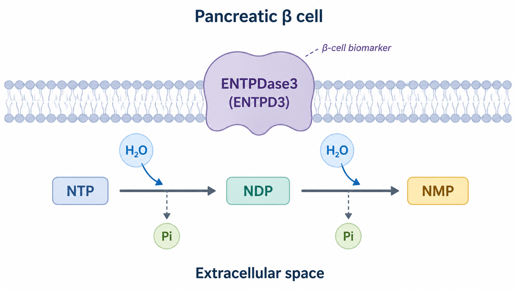
Figure 1. Schematic of ENTPD3 Localization on the Pancreatic β-Cell Surface and Stepwise Hydrolysis of Extracellular Nucleotides
2.2 Factors Affecting ENTPD3 Enzymatic Activity
ENTPD3 catalytic reactions depend on divalent cations such as Ca²⁺ or Mg²⁺. Its activity is also affected by substrate concentration, local pH, glycosylation status, and cell membrane localization. Decreased cell surface expression, abnormal glycosylation, or altered membrane structure may all reduce effective nucleotide hydrolysis. In cell systems, intracellular ATP release caused by cell rupture, nonspecific phosphatase activity, and contributions from other ectonucleotidases such as ENTPD1 and ENTPD2 should also be excluded.
2.3 Regulation of P2 and P1 Receptor Signaling by ENTPD3
Extracellular ATP and ADP can activate P2X ion channel receptors and P2Y G protein-coupled receptors, while adenosine formed from further AMP conversion can act on P1 receptors. ENTPD3 regulates ligand input to P2 receptors and adenosine receptors by altering the concentration, ratio, and duration of ATP, ADP, and AMP, rather than simply terminating all purinergic signaling.
3 Tissue Distribution and Physiological Functions of ENTPD3
3.1 Expression in the Nervous System
ENTPD3 is expressed in some central and peripheral neurons. It can regulate nucleotide levels in extrasynaptic and neuroendocrine microenvironments and influence sensory information transmission, neuronal excitability, energy intake regulation, and neuroendocrine output. The specific effects depend on brain region, neuronal subtype, and local P2 receptor composition.
3.2 Expression in Pancreatic Endocrine Cells
ENTPD3 is highly expressed in islet endocrine cells and is an important nucleotidase that regulates local extracellular ATP in islets. In adult human islets, ENTPD3 shows strong selectivity on the β-cell surface and can be used to identify insulin-positive cells and enrich mature β cells. Its islet distribution shows species differences, and expression patterns in mice or rats cannot be directly used to define human β-cell specificity. Antibody epitopes, tissue processing methods, and detection platforms can also affect the ENTPD3-positive range.
3.3 Functions in Other Tissues
ENTPD3 can also be expressed in gastrointestinal epithelium, sensory neural structures, and some endocrine-related tissues. By regulating extracellular nucleotide levels, it affects cell secretion, sensation, and intercellular signaling. ENTPD3 positivity in tissues outside the pancreas should not be interpreted as a β-cell feature. Its marker value should be limited to islet- or pancreas-derived cells.
4 Islet β-Cell Purinergic Signaling and Regulation of Insulin Secretion
4.1 Dual Roles of ATP in Insulin Secretion
After glucose enters β cells and is metabolized, the intracellular ATP/ADP ratio increases, causing ATP-sensitive K⁺ channels to close. This then induces membrane depolarization, opening of voltage-dependent Ca²⁺ channels, and exocytosis of insulin granules. During granule release, ATP and other nucleotides can also be co-released extracellularly. Therefore, intracellular ATP mainly serves as a metabolic coupling signal, while extracellular ATP acts as an autocrine and paracrine mediator regulating β cells and neighboring islet cells.
4.2 Effects of P2 Receptors on Insulin Secretion
β cells express multiple P2X and P2Y receptors. Opening of P2X receptors can cause cation influx and alter membrane potential and intracellular Ca²⁺. Some P2Y receptors can regulate insulin secretion through phospholipase C, IP₃, and Ca²⁺ release. Different receptor subtypes may produce promoting or inhibitory effects. The final outcome is influenced by glucose concentration, ATP or ADP level, receptor expression profile, and stimulation duration. Extracellular ATP should not be uniformly interpreted as an insulin secretion-promoting factor.
4.3 ENTPD3 Controls the Duration of Nucleotide Signaling
ENTPD3 hydrolyzes ATP and ADP co-released from insulin granules, limiting the intensity and duration of local P2 receptor stimulation. In some β-cell models, reducing ENTPD3 expression or activity can slow ATP hydrolysis and enhance glucose-stimulated insulin secretion. However, excessive extracellular ATP accumulation or long-term P2 receptor activation may also increase Ca²⁺ load, secretory stress, and cellular stress.
4.4 Purinergic Communication Among Islet Cells
Islets consist of β cells, α cells, δ cells, endothelial cells, immune cells, and nerve terminals. Extracellular nucleotides can transmit metabolic and secretory information among different cells. By limiting the diffusion distance and action duration of nucleotides, ENTPD3 not only regulates β cells themselves but may also affect glucagon, somatostatin, and local blood flow-related signals.
5 Research Value of ENTPD3 as an Islet β-Cell Surface Marker
5.1 Surface Identification of Adult Islet β Cells
ENTPD3 is localized on the cell surface and can be recognized by antibodies without fixation or permeabilization. Its selective expression in adult human islet β cells enables researchers to identify β cells through flow cytometry, live-cell imaging, and tissue co-staining. It can also be used to detect residual β cells in diabetic donors. ENTPD3 positivity only indicates that cells retain the corresponding surface protein and does not independently prove intact secretory function.
5.2 Maturation Evaluation of Stem Cell-Derived β-Like Cells
β-like cells generated from directed differentiation of human pluripotent stem cells usually contain subpopulations with different maturation states. ENTPD3 expression increases in more mature stem cell-derived β-like cells and can be used to enrich candidate cells with higher insulin storage, processing, and glucose responsiveness. Its expression still needs to be evaluated together with maturation or functional indicators such as MAFA, UCN3, IAPP, and PCSK1, and cannot be directly equated with complete maturation.
5.3 Live β-Cell Sorting and Downstream Analysis
ENTPD3 antibodies recognizing extracellular epitopes can be used to sort live β cells without relying on intracellular insulin staining, enabling single-cell transcriptomics, proteomics, calcium imaging, glucose-stimulated insulin secretion, drug screening, and transplantation function evaluation. Antibody binding may affect ENTPD3 enzymatic activity or nearby receptor structures, so whether the antibody used has blocking effects should be confirmed before long-term culture and functional experiments.
5.4 Application Boundaries of ENTPD3 as a Marker
(1) ENTPD3 positivity should be validated together with β-cell indicators such as INS, IAPP, PDX1, NKX6.1, and PCSK1.
(2) Cell maturity, culture conditions, tissue digestion, and membrane protein internalization may alter ENTPD3 surface levels.
(3) Epitope recognition, affinity, and cross-reactivity of different antibodies can affect positive cell proportion and sorting purity.
(4) ENTPD3 positivity cannot replace glucose-stimulated insulin secretion, C-peptide release, and dynamic secretion evaluation.
6 ENTPD3 Activity Abnormality and Diabetes-Related Mechanisms
6.1 Research on Residual β Cells in Type 1 Diabetes
Type 1 diabetes is mainly characterized by immune-mediated β-cell injury and reduced β-cell number. Residual β cells may also show dedifferentiation or decreased secretory function. ENTPD3 can be used to localize and isolate residual human β cells and analyze their number, maturation state, immune proximity, and post-stimulation secretion function. Reduced ENTPD3 signal may reflect β-cell loss or decreased single-cell surface expression. Therefore, tissue-positive area, positive cell proportion, and single-cell expression level should be interpreted separately.
6.2 Changes in Purinergic Signaling in Type 2 Diabetes
Hyperglycemia, lipotoxicity, oxidative stress, and endoplasmic reticulum stress associated with type 2 diabetes can alter β-cell identity, nucleotide release, and P2 receptor expression. Changes in ENTPD3 expression or activity may alter Ca²⁺ signaling and insulin release by affecting ATP and ADP clearance. However, ENTPD3 abnormality should not currently be regarded as an independent pathogenic factor or clinical diagnostic marker for type 2 diabetes.
6.3 Interpretation of ENTPD3 Expression Changes
Reduced ENTPD3 detection signal may be caused by decreased β-cell number, cell dedifferentiation, reduced maturation, membrane protein internalization, or tissue processing damage. Immunohistochemistry reflects spatial distribution in tissue, flow cytometry reflects surface positivity in recoverable cells, and transcriptomic detection reflects ENTPD3 mRNA level. These three types of results correspond to different detection levels and cannot be directly substituted for one another.
6.4 Limitations of ENTPD3 as a Therapeutic Target
ENTPD3 inhibition may temporarily prolong extracellular ATP signaling and enhance insulin release in some models. However, persistent P2 receptor stimulation may increase Ca²⁺ load, oxidative stress, and secretory exhaustion. ENTPD3 is also expressed in neural and other endocrine tissues, so systemic inhibition may cause extra-islet effects. Therefore, ENTPD3 is currently more suitable as a β-cell marker and mechanistic research target.
7 ENTPD3 Detection, Cell Identification, and Functional Evaluation
7.1 ENTPD3 Expression and Localization Detection
(1) Flow cytometry
ENTPD3 antibodies recognizing extracellular epitopes can be used to detect live-cell surface expression and can be combined with live/dead dyes and other cell surface markers. Islet digestion conditions may damage extracellular epitopes, so enzyme type, digestion time, and staining temperature need to be optimized.
(2) Immunofluorescence and immunohistochemistry
ENTPD3 can be co-stained with insulin, glucagon, somatostatin, or pancreatic polypeptide to analyze its cell type and spatial distribution in islets.
(3) WB and gene expression detection
WB can evaluate total ENTPD3 protein and molecular weight changes. qPCR and transcriptomic analysis can detect ENTPD3 transcription level, but none of these methods can directly prove that the protein is localized on the live-cell surface.
7.2 Extracellular Nucleotidase Activity Detection
ENTPD3 enzymatic activity can be evaluated by measuring ATP or ADP consumption, inorganic phosphate release, and AMP generation. ATP luminescence assays are suitable for monitoring substrate consumption, malachite green assays can detect inorganic phosphate, and HPLC or mass spectrometry can simultaneously distinguish ATP, ADP, and AMP. In cell systems, ENTPD3 knockdown, blockade, or overexpression groups should be included, and cell viability and membrane integrity should be examined to avoid misinterpreting ATP changes caused by cell death as extracellular hydrolytic activity.
7.3 β-Cell Identity, Maturity, and Functional Evaluation
Evaluation Level | Representative Indicators | Main Methods | Result Significance |
Surface identity | ENTPD3 | Flow cytometry, live-cell imaging, tissue staining | Identifies and sorts candidate adult or mature β cells |
β-cell identity | INS, IAPP, PCSK1, PDX1, NKX6.1 | qPCR, WB, immunostaining | Confirms insulin synthesis and β-cell lineage |
Maturation state | MAFA, UCN3, SLC2A2, and mitochondrial metabolism | Expression detection, metabolic analysis | Determines glucose responsiveness and secretory maturity |
Secretory function | Insulin, C-peptide | Static GSIS, dynamic perifusion | Evaluates secretion capacity after glucose stimulation |
Purinergic signaling | Extracellular ATP, intracellular Ca²⁺, and P2 receptor responses | Luminescence assay, calcium imaging, pharmacological intervention | Analyzes coupling between nucleotide signaling and secretion |
ENTPD3 enzymatic activity | ATP/ADP hydrolysis and AMP generation | Inorganic phosphate detection, HPLC, mass spectrometry | Confirms actual catalytic function of ENTPD3 |
7.4 ENTPD3 Functional Validation
ENTPD3 functional research can reduce its expression or activity through siRNA, CRISPR, or blocking antibodies and simultaneously detect extracellular ATP dynamics, P2 receptor responses, intracellular Ca²⁺, and glucose-stimulated insulin secretion. Gene rescue should re-express ENTPD3 with normal membrane localization and catalytic activity. A soluble recombinant catalytic domain is suitable for enzymology, substrate selectivity, and inhibitor screening, but cannot fully replace the cellular function of membrane-type ENTPD3.
8 Products Related to ENTPD3 and Islet β-Cell Research
Cat. No. | Product Name | Grade & Purity | Research Stage | Main Application |
ENTPD3 Human Pre-designed siRNA Set A |
| ENTPD3 gene intervention | Knocks down ENTPD3 and analyzes changes in extracellular ATP hydrolysis, Ca²⁺ signaling, and insulin secretion | |
ENTPD1 Human Pre-designed siRNA Set A |
| ENTPD family control | Compared with the ENTPD3 knockdown group to distinguish the contributions of ENTPD1 and ENTPD3 to extracellular nucleotide metabolism | |
ENTPD2 Human Pre-designed siRNA Set A |
| ENTPD family control | Evaluates the effect of ENTPD2 on ATP and ADP hydrolysis and assists in determining the specificity of ENTPD3-related phenotypes | |
Recombinant Human ENTPD1 Protein | ≥90%(SDS-PAGE) | Extracellular nucleotidase control | Establishes a control system for ENTPD1-mediated ATP and ADP hydrolysis | |
NTPDase-IN-1 |
| NTPDase pharmacological intervention | Inhibits NTPDase activity and evaluates the relationship between extracellular nucleotide hydrolysis and cell function | |
NTPDase-IN-2 |
| NTPDase pharmacological intervention | Used for NTPDase-dependent ATP and ADP metabolism research | |
NTPDase-IN-3 |
| NTPDase pharmacological intervention | Validates the effect of extracellular nucleotide hydrolysis on P2 receptor signaling | |
ATP Determination Kit | BioReagent, ready-to-use, for chemiluminescence | Extracellular ATP detection | Measures ATP changes in culture supernatants or enzyme reaction systems | |
Enhanced ATP Assay Kit | BioReagent, for chemiluminescence | Low-level ATP detection | Evaluates ATP accumulation in culture supernatants after ENTPD3 knockdown or inhibition | |
Adenosine 5′-diphosphate (ADP) | Moligand™, ≥95%(HPLC) | ENTPD3 substrate research | Serves as a nucleoside diphosphate substrate to evaluate hydrolysis of ADP to AMP | |
Malachite Green Phosphate Detection Kit | BioReagent, sterile | ENTPD3 enzymatic activity detection | Measures inorganic phosphate released from ATP or ADP hydrolysis | |
Recombinant Insulin Antibody | Recombinant, ExactAb™, validated, 1.154 mg/mL | β-cell identity detection | Detects insulin expression and assists in confirming β-cell identity | |
Recombinant Insulin Antibody | Recombinant, ExactAb™, validated, see COA | β-cell identity detection | Used for insulin expression and islet β-cell-related research | |
PDX1 Human Pre-designed siRNA Set A |
| β-cell identity validation | Analyzes the role of PDX1 in β-cell identity maintenance and insulin expression | |
NKX6-1 Human Pre-designed siRNA Set A |
| β-cell lineage research | Evaluates the regulation of β-cell lineage and secretion function by NKX6.1 | |
MAFA Human Pre-designed siRNA Set A |
| β-cell maturation research | Analyzes the relationship between MAFA and glucose-stimulated insulin secretion | |
Recombinant Human UCN3 Protein | ≥90%(SDS-PAGE) | β-cell maturation research | Used for mature β-cell-related function and signaling research | |
UCN3 Human Pre-designed siRNA Set A |
| β-cell maturation research | Validates the relationship between UCN3 and β-cell maturation and glucose responsiveness | |
Calcium Green 1 AM (Calcium Ion Fluorescent Probe) | BioReagent, biological stain, suitable for fluorescence analysis, for microscopy, ≥96% | Ca²⁺ signal detection | Monitors intracellular Ca²⁺ dynamics after ATP, ADP, or glucose stimulation | |
5-BDBD | Moligand™, ≥99%(HPLC) | P2X4 functional validation | Determines whether ENTPD3-related extracellular ATP effects involve P2X4 | |
A 438079 | Moligand™, ≥98% | P2X7 functional validation | Analyzes the role of P2X7 in high extracellular ATP, Ca²⁺ load, and cellular stress | |
MRS 2179 | Moligand™, ≥98% | P2Y1 functional validation | Analyzes ADP-mediated P2Y1 signaling and changes in insulin secretion | |
BPTU | Moligand™, ≥98%(HPLC) | P2Y1 functional validation | Used with ADP stimulation to evaluate the contribution of P2Y1 to Ca²⁺ and secretion signaling | |
2-ThioUTP tetrasodium salt | ≥98%(HPLC) | P2Y2 functional validation | Activates P2Y2 to study Ca²⁺ and secretion responses after nucleotide stimulation | |
AR-C 118925XX | Moligand™, ≥97%(HPLC) | P2Y2 functional validation | Blocks P2Y2 and validates receptor dependence of ATP- or UTP-related signaling | |
Recombinant P2Y6 Antibody | Recombinant, ExactAb™, validated, 0.2 mg/mL | P2 receptor expression detection | Detects P2Y6 expression and assists in analyzing purinergic receptor composition in islet cells |
ENTPD3 connects extracellular nucleotide metabolism, P2 receptor signaling, and insulin secretion, and provides a surface marker for live identification of adult β cells and mature β-like cells. Related research needs to jointly analyze ENTPD3 expression and enzymatic activity, β-cell identity, and glucose-stimulated secretion function.
For more related articles, please see below:
[1] Autophosphorylation experiments on insulin receptor stimulated by insulin


 危险品化学品经营许可证(带存储)